Clinically Occult Diffuse Large B-Cell Lymphoma of the Middle Turbinate Identified Using
18 F-Fluorodeoxyglucose Positron Emission
Tomography/Computed
Tomography: A Case Report
양전자방출전산화단층촬영에 의해 발견된 임상증상이 없는 중비갑개의 미만성 거대 B세포 림프종: 증례 보고
Dong Hyeon Kim, MD
1, Sang Kwon Lee, MD
1* , Mi Jeong Kim, MD
1, Jae Hyuck Yi, MD
1, Mi Sun Choi, MD
2Departments of 1Radiology, 2Pathology, Keimyung University, Dongsan Hospital, Daegu, Korea
We report a case of clinically occult diffuse large B-cell lymphoma (DLBCL) of the middle turbinate (MT) identified by 18F-fluorodeoxyglucose positron emission tomography/computed tomography (18F-FDG PET/CT) in a 71-year-old man along with imaging findings. DLBCL was presented with a hypermetabolic right MT [maximum standardized uptake values (SUVmax) = 8.8 gm/dL] on 18F-FDG PET/CT, while rhinologic examination was normal. CT showed nothing but slightly more intense enhancement of the right MT compared with the opposite side. The disease progressed during next 7 months until follow-up CT demonstrated solidly enhancing mass occupying entire right nasal cavity which was intensely hypermetabolic (SUVmax = 12.8 gm/dL). Surgical biopsy con- firmed the diagnosis. Follow-up CT and 18F-FDG PET/CT performed after chemotherapy demon- strated complete resolution of DLBCL of the right nasal cavity including the right MT. This is thought to be the first case report in the literature concerning clinically occult DLBCL presenting as a hypermetabolic MT on 18F-FDG PET/CT.
Index terms Turbinate; Lymphoma, Large B-Cell, Diffuse; PET-CT
Received July 19, 2018 Revised October 16, 2018 Accepted October 29, 2018
*Corresponding author Sang Kwon Lee, MD Department of Radiology, Keimyung University, Dongsan Hospital, 56 Dalseong-ro, Jung-gu, Daegu 41931, Korea.
Tel 82-53-250-7767 Fax 82-53-250-7766 E-mail [email protected] This is an Open Access article distributed under the terms of the Creative Commons Attribu- tion Non-Commercial License (https://creativecommons.org/
licenses/by-nc/4.0) which permits unrestricted non-commercial use, distribution, and reproduc- tion in any medium, provided the original work is properly cited.
ORCID iDs Sang Kwon Lee https://
orcid.org/0000-0001-6987-6068 Dong Hyeon Kim
https://
orcid.org/0000-0002-4600-8062
서론
비부비동의 비호지킨 림프종은 서양에서는 모든 비호지킨 림프종의 1.5%를 차지하는 드문 형 태의 림프절외 림프종이지만 아시아에서는 위장관 다음으로 흔하다(1). 비부비동에서 발생하는 비호지킨 림프종의 주된 아형으로는 B세포 림프종과 natural killer (이하 NK)/T세포 림프종이 있 으며(2), 서양에서의 비부비동 비호지킨 림프종은 대부분 B세포 표현형을 가지며 미만성 거대 B세 포 림프종(diffuse large B-cell lymphoma; 이하 DLBCL)이 대부분을 차지한다. 반면에 아시아에 서는 비강에 병변이 잘 침범하는 NK/T세포 림프종이 B세포 표현형의 림프종보다 더 흔하다. 많은 연구에서 양전자방출전산화단층촬영(
18F-fluorodeoxyglucose positron emission tomography/
computed tomography; 이하
18F-FDG PET/CT)이 림프종의 병기결정과 치료에 대한 반응평가에 유용함이 입증되었다. 현재까지
18F-FDG PET/CT에서 발견된 중비갑개의 DLBCL은 보고된 바 없 다. 저자들은 71세 남자에서
18F-FDG PET/CT에 의해 확인된 임상증상이 없는 중비갑개의 DLBCL 1예를 영상소견 및 병리소견과 함께 보고하고자 한다.
증례 보고
71세의 남자가
18F-FDG PET/CT에서 우연히 발견된 중비갑개의 과대사성 병변의 추가적인 평 가를 위해 영상의학과로 의뢰되었다. 환자는 14개월 전에 간세포암으로 부분적 간절제술을 시행 받았고, 그 당시에 시행한
18F-FDG PET/CT에서는 비부비동 부위에서 비정상적인 FDG 섭취를 보 이지 않았다(Fig. 1A). 최근 간세포암의 전이여부를 확인하기 위해 시행한 추적
18F-FDG PET/CT (Discovery STE, GE Healthcare, Milwaukee, WI, USA)에서 우측 중비갑개에 과대사성 병변 [maximum standardized uptake values (이하 SUV
max) = 8.8 gm/dL]이 발견되었으며(Fig. 1A), FDG 섭취의 이상은 다른 신체 부위에서는 발견되지 않았다. 비과적 검사에서 중비갑개는 형태학 적으로 정상이었다. Sensation 64 스캐너(Siemens Healthcare, Erlangen, Germany)를 이용하 여 시행한 비부비동 조영증강 전 CT에서 양측 중비갑개는 정상 크기, 형태 및 감쇠값을 나타내었 으며, 조영증강 후 CT에서는 반대편과 비교하여 우측 중비갑개가 약간 더 조영증강이 잘 되었다 (Fig. 1B). 비과적 검사에서 형태학적으로 정상적인 중비갑개를 보였기 때문에 생검을 시행하지 않았다. 7개월 후 비강 출혈을 주소로 내원하여 시행한
18F-FDG PET/CT에서 우측 비강 전체를 차 지하는 과대사성 병변(SUV
max= 12.8 gm/dL)이 관찰되었다(Fig. 1A). 종괴는 조영증강 전 CT에서 근육과 같은 정도의 감쇠값을 보였고, 조영증강 후 CT에서는 중등도의 조영증강을 보였으며(Fig.
1C), 우측 상악동의 내벽, 우측 중비갑개, 하비갑개 및 우측 사골동의 골미란 및 개조(remodeling) 가 관찰되었다. 외과적 생검에 의해 얻은 표본에 대한 조직학적 검사 및 면역조직화학염색에서 DLBCL로 진단되었다(Fig. 1E).
Cyclophosphamide, doxorubicin, vincristine, prednisolone, rituximab (이하 R-CHOP) 화
학요법 후 시행한
18F-FDG PET/CT (Fig. 1A) 및 CT (Fig. 1D)에서 우측 비강 및 중비갑개의 DLBCL
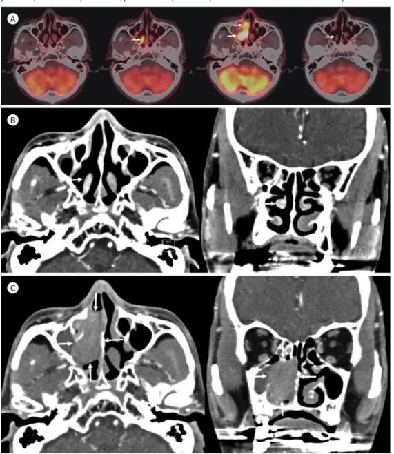
Fig. 1. A 71-year-old man with DLBCL of the MT detected using 18F-FDG PET/CT that was performed for me- tastasis work-up of HCC.
A. 18F-FDG PET/CT performed 14 months prior when he had undergone partial hepatectomy reveals no ab- normal uptake in the sinonasal area (first image). 18F-FDG PET/CT performed for metastasis work-up of HCC demonstrates hypermetabolic right MT (SUVmax = 8.8 gm/dL) (arrow) (second image). No abnormal uptake is noted at other sinonasal areas. 18F-FDG PET/CT obtained 7 months thereafter shows an intensely hypermet- abolic mass (SUVmax = 12.8 gm/dL) (arrows) occupying the right nasal cavity (third image). 18F-FDG PET/CT after R-CHOP chemotherapy shows complete resolution of DLBCL of the right nasal cavity with normal ap- pearing MT (arrow) (fourth image).
B. Contrast-enhanced axial (first image) and coronal (second image) CT for further evaluation of hypermeta- bolic MT on 18F-FDG PET/CT show morphologically normal right MT with slightly more intense enhancement compared to that on the opposite side (arrows).
C. Follow-up contrast-enhanced axial CT (first image) obtained 7 months thereafter shows a mass with moderate enhancement in the right nasal cavity (arrows). Bony erosion and remodeling of the medial wall of the right maxillary sinus, right middle and inferior turbinates, and right ethmoid sinus are also noted on the coronal CT (arrows) (second image).
CT = computed tomography, DLBCL = diffuse large B-cell lymphoma, 18F-FDG = 18F-fluorodeoxyglucose, HCC = hepatocellular carcinoma, MT = middle turbinate, PET = positron emission tomography, R-CHOP = cyclophos- phamide, doxorubicin, vincristine, prednisolone, rituximab, SUVmax = maximum standardized uptake values
A
B
C
고찰
비부비동 비호지킨 림프종은 증상이 양성 염증성 질환과 쉽게 혼동될 수 있기 때문에 임상적 진 단에 어려움이 있다. CT 또는 자기공명(이하 MR) 영상에서 림프종은 비강의 벽을 따라 확산되는 침윤성 종양 또는 인접한 골미란 및 개조(remodeling)가 동반된 부비동 벽의 종괴로 나타날 수 있다. 림프종은 조영증강 전 CT에서 근육과 비슷한 감쇠값을, T1 강조영상에서는 근육과 등신호강 도를, T2 강조영상에서는 근육과 등신호강도 또는 고신호강도를 나타내며, 중등도의 조영증강을 보인다.
Yasumoto 등(3)은 상악동의 DLBCL 7예의 CT 및 MR 영상소견을 분석하였으며 7예 중 5예에 서 직경 4~6 cm의 종괴로 보였고, 2예에서는 점막의 비후로 관찰되었다. 상악동 주변 연부조직 비
Fig. 1. A 71-year-old man with DLBCL of the MT detected using 18F-FDG PET/CT that was performed for me- tastasis work-up of HCC.D. Follow-up axial (first image) and coronal (second image) CT after R-CHOP chemotherapy shows complete resolution of the mass of the right nasal cavity with normal-appearing MT (arrows).
E. Histologic findings of the specimen obtained by surgical biopsy of the mass of the right nasal cavity dem- onstrates monotonous proliferation of medium to large lymphoid cells (first image; hematoxylin and eosin stain, × 200) with positivity for CD20 (second image; immunostaining, × 200).
CT = computed tomography, DLBCL = diffuse large B-cell lymphoma, 18F-FDG = 18F-fluorodeoxyglucose, HCC = hepatocellular carcinoma, MT = middle turbinate, PET = positron emission tomography, R-CHOP = cyclophos- phamide, doxorubicin, vincristine, prednisolone, rituximab
D
E
악동 주변 연부조직 침윤을 동반하였다. 따라서 종괴 및 점막비후에 동반된 연부조직의 침윤이 상 악동의 악성 림프종을 시사하는 중요한 소견으로 보고하였다.
Jang 등(4)은 병리학적으로 확진된 비강의 NK/T세포 림프종 21예의 CT 소견을 분석하였는데, 이들 중 9예에서는 광범위한 점막 비후로만 관찰되었고 10예에서는 작은 종괴를 동반한 광범위한 점막 비후, 2예에서는 큰 종괴를 동반한 광범위한 점막 비후로 관찰되어 전례에서 광범위한 점막 비후를 동반하였다고 보고하였다. 이러한 결과는 Yasumoto 등(3)이 보고한 DLBCL의 영상소견 과 대조된다. 비강 병변은 21예 중 20예(95%)에서 주변 조직으로의 침범을 보였는데, 피하조직이 16예(76%), 안와 9예(43%), 부비동 17예(81%), 구강인두 및 비인두가 각각 1예(10%)였다. 골변화 는 미란이 11예(52%), 파괴가 3예(14%)였다. 따라서 비강형 NK/T세포 림프종이 비강에 광범위한 점막비후를 동반하고 인접한 연부조직, 특히 피하조직으로의 침범을 잘하는 침윤성 병변이라고 보고하였다(4).
비갑개에서 발생하는 림프종에 관한 연구는 드물다. Wang 등(5)은 하비갑개에 발생한 종양 34예를 조사하였는데, 비호지킨 림프종이 6예(17.6%)였으며, 하비갑개의 진행성의 일측성 또는 양측성 비대로 나타났다. Lee 등(6)은 12세 여자에서 인접 구조물의 침범이 없는 우측 하비갑개에 서 생긴 NK/T세포 림프종 1예를 보고하였으며, 비내시경 검사에서 우측 하비갑개 점막의 궤양성 변화와 비대로 인해 우측 비강이 좁아진 소견으로 관찰되었다. CT에서는 뼈 구조와 부비동은 정 상 소견이면서 우측 하비갑개의 점막비대 소견을 보였으며, PET/CT에서는 우측 하비갑개의 FDG 섭취 증가로 관찰되었다. Lingan과 Roasa(7)는 하비갑개에 생긴 희귀하고 공격적인 형태의 림프 절외 미만성 거대 B세포 림프종인 혈관 내 림프종 1예를 보고하였는데, 비내시경 검사에서 보랏빛 의 우측 하비갑개 비대가 관찰되었으며, CT에서도 우측 하비갑개의 비대소견이 관찰되었다.
18
F-FDG PET/CT에서 관찰되는 중비갑개의 과대사성 병변에 관한 보고는 극히 드물며(8, 9), 골 모세포종과 감염된 수포성갑개 각각 1예에 관한 증례 보고가 있으나, 중비갑개의 림프종에 관한 보고는 없다.
본 증례의 최초 조영증강 후 CT에서 과대사성의 우측 중비갑개가 좌측 중비갑개에 비해 약간 더 강한 조영증강을 보였으나, 이는 생리적 순환변화(cyclic change)에 의한 소견과 큰 차이가 없 는 것으로 생각되었다. 따라서 본 증례는 임상증상이 없고 비내시경 및 CT에서 뚜렷한 해부학적 변화를 초래하지 않으며,
18F-FDG PET/CT에서 중비갑개의 과대사성 병변으로 발현한 중비갑개 의 DLBCL의 첫 번째 증례로 생각된다. 비록 초기 PET/CT에서 FDG 섭취가 증가된 중비갑개에 대 한 조직학적 확진이 이루어지지 않았으나 7개월 후에 동측 비강의 종괴에 대한 조직학적 검사에 서 DLBCL로 확진되었고, DLBCL에 대한 R-CHOP 치료 후 추적 PET/CT에서 비강종괴의 완전관 해와 더불어 과대사성 중비갑개의 소견이 소실되었다는 점, 과거의 PET/CT에서는 과대사성 중비 갑개의 소견이 없었다는 점 등을 감안하면 초기 중비갑개의 과대사성 병변을 DLBCL로 진단하는 데 논란의 여지가 없을 것으로 생각된다.
중비갑개 점막의 염증성 질환에서도 과대사성 중비갑개의 소견을 보일 수 있으나, 이 경우
SUV
max값이 3.0 gm/dL 이하인 경우가 대부분이고(10), 일부 급성염증성 질환에서는 높은 SUV
max값을 보일 수 있으나(9), 임상적 소견 및 CT 소견이 감별에 도움이 될 것으로 생각된다.
요약하면, 이 보고는 임상증상 없이
18F-FDG PET/CT에서 과대사성 중비갑개로 발현한 DLBCL 에 관한 첫 번째 증례로써, 형태학적으로 정상이지만 과대사성의 중비갑개의 감별진단에 DLBCL 이 포함되어야 할 것으로 생각된다.
Conflicts of Interest
The authors have no potential conflicts of interest to disclose.
REFERENCES
1. Woo JS, Kim JM, Lee SH, Chae SW, Hwang SJ, Lee HM. Clinical analysis of extranodal non-Hodgkin’s lym- phoma in the sinonasal tract. Eur Arch Otorhinolaryngol 2004;261:197-201
2. Oprea C, Cainap C, Azoulay R, Assaf E, Jabbour E, Koscielny S, et al. Primary diffuse large B-cell non-Hodg- kin lymphoma of the paranasal sinuses: a report of 14 cases. Br J Haematol 2005;131:468-471
3. Yasumoto M, Taura S, Shibuya H, Honda M. Primary malignant lymphoma of the maxillary sinus: CT and MRI. Neuroradiology 2000;42:285-289
4. Jang YM, Lee JH, Lee DH, Choi CG, Suh DC, Lee HK, et al. Imaging features of nasal-type natural killler/t-cell lymphomas: frequent involvement of skin and subcutaneous tissue. J Korean Radiol Soc 2006;54:161-165 5. Wang Q, Xiao S, Qin Y. Tumors originated from the inferior nasal turbinates: clinical features in 34 patients.
Lin Chung Er Bi Yan Hou Tou Jing Wai Ke Za Zhi 2014;28:1050-1052
6. Lee JY, Jang YD, Kim HK. The primary role of the otolaryngologist in managing pediatric sinonasal malig- nancies: an extranodal NK/T-cell lymphoma originating from the inferior turbinate mucosa of the nasal cav- ity. J Pediatr Hematol Oncol 2008;30:401-404
7. Lingan MB, Roasa FV. Intravascular lymphoma of the inferior turbinate: an unusual rhinologic presentation of a rare neoplasm. Philipp J Otolaryngol Head Neck Surg 2007;22:24-26
8. Jeong YJ, Sohn MH, Lim ST, Kim DW, Jeong HJ, Jang KY, et al. Osteoblastoma in the nasal cavity: F-18 FDG PET/CT and Tc-99m MDP 3-phase bone scan findings with pathologic correlation. Clin Nucl Med 2011;36:214- 217
9. Arens AI, Verbist BM, Hendrickx BW, De Geus-Oei LF, Oyen WJ. False positive 18F-FDG PET/CT due to inflamed concha bullosa. Clin Nucl Med 2012;37:509-510
10. Purohit BS, Ailianou A, Dulguerov N, Becker CD, Ratib O, Becker M. FDG-PET/CT pitfalls in oncological head and neck imaging. Insights Imaging 2014;5:585-602
양전자방출전산화단층촬영에 의해 발견된 임상증상이 없는 중비갑개의 미만성 거대 B세포 림프종: 증례 보고
김동현
1· 이상권
1* · 김미정
1· 이재혁
1· 최미선
2저자들은 71세 남자의 양전자방출전산화단층촬영(
18F-fluorodeoxyglucose positron emis- sion tomography computed tomography; 이하
18F-FDG PET/CT)에서 발견된 임상증상이 없는 중비갑개의 미만성 거대 B세포 림프종(diffuse large B-cell lymphoma; 이하 DLBCL) 1예를 영상소견과 함께 보고하고자 한다. DLBCL은 비과적 검사가 정상임에도 불구하고,
18
F-FDG PET/CT에서 과대사성[maximum standardized uptake values (이하 SUV
max) = 8.8 gm/dL]의 우측 중비갑개로 발현하였으며, CT에서는 반대측과 비교하여, 약간 더 조영증 강되는 우측 중비갑개 이외에 이상소견을 보이지 않았다. DLBCL은 7개월 후 추적 CT 및
18F- FDG PET/CT에서 우측 비강을 차지하는 조영증강을 보이는 과대사성(SUV
max= 12.8 gm/
dL) 종괴로 발견될 때까지 진행하였으며, 외과적 생검에 의해 확진되었다. 항암요법 후 시행 한 추적 CT 및
18F-FDG PET/CT에서 중비갑개를 포함한 비강의 DLBCL은 완전관해를 보였 다. 이 보고는 임상증상 없이
18F-FDG PET/CT에서 과대사성 중비갑개로 발현한 DLBCL에 관 한 첫 증례 보고로 생각된다.
계명대학교 동산의료원 1영상의학과, 2병리과