351
Copyrights © 2014 The Korean Society of Radiology
INTRODUCTION
Pancreatic neuroendocrine tumors and their hepatic metasta- ses have an inconsistent appearance with only a small percent- age of lesions appearing as cystic masses in computed tomogra- phy (CT) and magnetic resonance imaging (MRI). They can therefore be mistaken for benign or infectious lesions, leading to a false diagnosis with delayed or inadequate treatments. We re- ported a patient with an upper abdominal pain that lasted for several months, caused by a huge cystic neuroendocrine carci- noma of the liver that was mistakenly interpreted as a compli- cated or hydatid cyst. We presented the findings of the initial CT and the subsequently performed MRI under the application of hepatocyte specific contrast material, which led to distinct dif- ferential diagnoses that had a major impact on the therapeutic strategy.
CASE REPORT
A 50-year-old male patient complained of an epigastric pain that he was having for a week. Hematology laboratory data were within the normal range. Contrast enhanced CT from an exter- nal institution showed a 15 mm hypodense mass in the head of the pancreas when compared with the uninvolved pancreas (Fig.
1A). The pancreatic mass was interpreted as ductal adenocarci- noma or neuroendocrine tumor. Additionally, a CT scan showed a 10 cm hypodense mass in the right hepatic lobe with internal thin septa (Fig. 1D), and this was interpreted as a simple cyst of the right hepatic lobe. The patient was referred to our center for surgical therapy. At our center, for further preoperative evalua- tion, MRI of the liver was performed with unenhanced T1- and T2-weighted images with fat suppression, contrast enhanced se- quences using Gd-EOB-DTPA (Primovist, Bayer Schering Phar-
Case Report
pISSN 1738-2637 / eISSN 2288-2928 J Korean Soc Radiol 2014;70(5):351-354 http://dx.doi.org/10.3348/jksr.2014.70.5.351
Received January 1, 2014; Accepted February 7, 2014 Corresponding author: Seung Joon Choi, MD Department of Radiology, Gachon University Gil Hospital, 21 Namdong-daero 774beon-gil, Namdong-gu, Incheon 405-760, Korea.
Tel. 82-32-460-3060 Fax. 82-32-460-3065 E-mail: [email protected]
This is an Open Access article distributed under the terms of the Creative Commons Attribution Non-Commercial License (http://creativecommons.org/licenses/by-nc/3.0) which permits unrestricted non-commercial use, distri- bution, and reproduction in any medium, provided the original work is properly cited.
Pancreatic neuroendocrine tumors and their hepatic metastases have an inconsis- tent appearance with only a small percentage of lesions appearing as cystic masses in computed tomography (CT) and magnetic resonance imaging (MRI). Therefore, they can be mistaken as benign or infectious lesions, which can lead to a false diag- nosis with delayed or inadequate treatment. We reported a patient with upper ab- dominal pain that lasted for several months, caused by a huge cystic neuroendo- crine carcinoma of the liver. This was mistakenly interpreted as a complicated or hydatid cyst, and the findings in the CT and MRI was presented.
Index terms
Pancreatic Neuroendocrine Tumor Hepatic Metastasis
Cystic Mass
Well-Differentiated Pancreatic Neuroendocrine Tumor with Solitary Hepatic Metastasis Presenting as a Benign Cystic Mass: A Case
Report
양성낭성종괴로 오인된 고분화췌장신경내분비종양의 단발성 간전이 1예:
증례 보고
Su-Joa Ahn, MD, Seung Joon Choi, MD, Hyung Sik Kim, MD, Jeong Ho Kim, MD, Hye-Young Choi, MD
Department of Radiology, Gachon University Gil Hospital, Incheon, Korea
Well-Differentiated Pancreatic Neuroendocrine Tumor with Solitary Hepatic Metastasis Presenting as a Benign Cystic Mass
352
J Korean Soc Radiol 2014;70(5):351-354 jksronline.orgverse gradient echo sequences, the wall and septa appeared hy- pointense, while the liquid content showed subtle hyperintensity (Fig. 1E). On T2-weighted transverse turbo spin echo sequenc- es, the wall and septa were hyperintense, while the liquid con- tent showed subtle hyperintensity (Fig. 1F). On T1-weighted image with contrast enhancement, no Gd-EOB-DTPA uptake is present in wall and septations of the mass, while the cyst content remained hypointense (Fig. 1G). Based on these findings, a complicated or hydatid cystic tumor was preferred as the differ- ma, Berlin, Germany) with early dynamic acquisition, and de-
layed imaging in the hepatobiliary phase after 20 minutes (1.5 T;
AVANTO; Siemens Medical Systems, Erlangen, Germany). On T1-weighted MR image, pancreatic mass showed hypointensity compared with the uninvolved pancreas (Fig. 1B). T1-weighted image after application of Gd-EOB-DTPA shows subtle en- hancement of the mass in the arterial phase (Fig. 1C).
On MRI, the lesion in the right liver lobe, measuring 10.0 × 6.0 cm, showed a thin wall and septa. On T1-weighted trans-
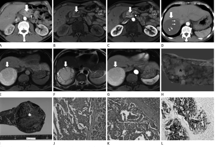
Fig. 1. A 50-year-old man presented with a week’s history of epigastric pain.
A. Transverse CT scan shows a 15 mm hypodense mass in head of pancreas compared with the normal pancreas parenchyma (arrow).
B. T1-weighted MR image shows a 15 mm hypointense mass in head of pancreas compared with the normal pancreas parenchyma (arrow).
C. T1-weighted image after application of Gd-EOB-DTPA shows subtle enhancement of the mass in the arterial phase (arrow).
D. Transverse CT scan shows a 10 cm hypodense mass (thick arrow) in the right hepatic lobe with suspicious internal thin septa (thin arrows).
E. T1-weighted MR image shows a 10 cm hyperintense mass in the right hepatic lobe with suspicious internal thin septa (arrow).
F. T2-weighted MR image shows hyperintense mass including internal multiple septations (arrow).
G. On T1-weighted image after contrast media injection, no Gd-EOB-DTPA uptake of the mass is present (arrow).
H. A photograph of the resected pancreatic specimen shows the 23 mm irregular margined yellowish mass (asterisk) in head of pancreas.
I. A photograph of the resected hepatic specimen shows the large liver mass (asterisk) possessing a capsule, containing hemorrhages.
J. Histology of the neuroendocrine carcinoma in the pancreas. Tumor cells featuring abundant cytoplasm, prominent nucleoli; frequent mitotic rate shows the organoid, nesting, trabecular and palisading pattern (H&E, × 200).
K, L. Histology of the neuroendocrine carcinoma in the liver. Tumor cells featuring abundant cytoplasm, prominent nucleoli; frequent mitotic rate shows the organoid, nesting, trabecular and palisading pattern (H&E, × 200) (K). Positive immunohistochemical stain for synaptophysin (synaptophysin, × 200) (L).
I E A
J F B
K G C
L H D
Su-Joa Ahn, et al
353
jksronline.org J Korean Soc Radiol 2014;70(5):351-354
tense compared to the surrounding normal liver parenchyma on T1-weighted image, and are hyperintense on T2-weighted images. On dynamic gadolinium enhancement phases, metasta- ses typically showed early enhancement (7). Cystic appearance of neuroendocrine carcinoma (NEC) metastases is rare. The low prevalence of NEC and the unusual, large solitary cystic lesion in the liver made this differential diagnosis quite unlikely, which initially resulted in the interpretation as an echinococcal cyst based on the monophasic contrast enhanced CT (8). Despite newer chemotherapeutic and immunologic agents, surgical therapy remains the most efficient approach to metastatic dis- ease and offers the longest lasting benefits. Surgical debulking of the hepatic disease improves the chance of survival (9).
In this article, we described the CT and MRI findings on cys- tic appearance of hepatic metastases from PNETs, which was mistakenly interpreted as complicated or hydatid cyst. Because of variability of neuroendocrine tumor appearance in imaging, percutaneous biopsy should be considered for atypical imaging to avoid delayed diagnosis and wrong treatment.
REFERENCES
1. Gumbs AA, Moore PS, Falconi M, Bassi C, Beghelli S, Mod- lin I, et al. Review of the clinical, histological, and molecu- lar aspects of pancreatic endocrine neoplasms. J Surg On- col 2002;81:45-53; discussion 54
2. Franko J, Feng W, Yip L, Genovese E, Moser AJ. Non-func- tional neuroendocrine carcinoma of the pancreas: inci- dence, tumor biology, and outcomes in 2,158 patients. J Gastrointest Surg 2010;14:541-548
3. Bosman FT, Cameiro F, Hruban RH. WHO Classification of Tumours of the Digestive System, 4th ed. Lyon: Interna- tional Agency for Research on Cancer (IARC), 2010:13-14, 64-68
4. O’Toole D, Ruszniewski P. Chemoembolization and other ablative therapies for liver metastases of gastrointestinal endocrine tumours. Best Pract Res Clin Gastroenterol 2005;
19:585-594
5. Oberg K, Eriksson B. Endocrine tumours of the pancreas.
Best Pract Res Clin Gastroenterol 2005;19:753-781 6. Sarmiento JM, Farnell MB, Que FG, Nagorney DM. Pancre-
aticoduodenectomy for islet cell tumors of the head of ential diagnosis.
Pancreatectomy and a right hemihepatectomy (Segments VI–
VII) were performed. Resected pancreatic specimen shows irreg- ular, margined, and yellowish tumor in the head of the pancreas, measuring 2.3 × 1.5 × 1.2 cm, without common bile duct inva- sion (Fig. 1H). Resected hepatic specimen shows the large liver mass possessing a capsule, containing hemorrhages (Fig. 1I).
Histology of the pancreatic tumor reveals tumor cells featuring abundant cytoplasm andprominent nucleoli; frequent mitotic rate shows the organoid, nesting, trabecular, and palisading pat- tern (Fig. 1J). Hepatic tumor shows similar histologic features with the pancreatic mass, and partially cystic neoplasm infiltrates the adjacent liver parenchyma with microcystic and trabecular growth pattern (Fig. 1K). There was an expression of CK7, syn- aptophysin, chromogranin, and CK20 (Fig. 1L). These findings resulted in the diagnosis of a well differentiated neuroendocrine tumor of pancreatic origin with hepatic metastasis (1).
DISCUSSION
Pancreatic neuroendocrine tumors (PNETs) are rare neo- plasms that affect only 1 in 300000 individuals in the United States annually, and comprise 1% to 2% of all pancreatic tumors (1, 2). The major categories of gastroenteropancreatic neuroen- docrine tumors defined by the WHO are well-differentiated neuroendocrine tumors, which show benign behavior or uncer- tain malignant potential, well-differentiated neuroendocrine carcinomas, which are characterized by low-grade malignancy, and poorly differentiated (usually small cell) neuroendocrine carcinomas of high-grade malignancy (3).
Most malignant PNETs metastasize to the liver where they de- rive their blood supply from hepatic artery branches (75–80%) (4). Hepatic metastasis occurs in 30–85% of PNETs, and is po- tentially and completely resectable only in 7–15% of the patients (5, 6). Because of the varying presentation of the patients with metastatic hepatic neuroendocrine tumors and the rarity of this disease, it is often difficult to define the ideal treatment approach based upon the current literatures. In patients with isolated he- patic diseases, an increasing number of published findings report the improved survival rates in the patients undergoing complete resection (6).
Hepatic metastases are most frequently hypointense or isoin-
Well-Differentiated Pancreatic Neuroendocrine Tumor with Solitary Hepatic Metastasis Presenting as a Benign Cystic Mass
354
J Korean Soc Radiol 2014;70(5):351-354 jksronline.organ echinococcus cyst of the liver in CT-MRI findings with hepatocyte specific contrast material. J Gastrointestin Liv- er Dis 2011;20:439-442
9. Musunuru S, Chen H, Rajpal S, Stephani N, McDermott JC, Holen K, et al. Metastatic neuroendocrine hepatic tumors:
resection improves survival. Arch Surg 2006;141:1000- 1004; discussion 1005
the pancreas: long-term survival analysis. World J Surg 2002;26:1267-1271
7. Debray MP, Geoffroy O, Laissy JP, Lebtahi R, Silbermann- Hoffman O, Henry-Feugeas MC, et al. Imaging appearanc- es of metastases from neuroendocrine tumours of the pancreas. Br J Radiol 2001;74:1065-1070
8. Krohn M, Grieser C, Weichert W, Pascher A, Denecke T.
Well-differentiated neuroendocrine carcinoma mimicking
양성낭성종괴로 오인된 고분화췌장신경내분비종양의 단발성 간전이 1예: 증례 보고
안수좌 · 최승준 · 김형식 · 김정호 · 최혜영
췌장신경내분비종양과 그들의 간전이는 극히 일부에서 전산화단층촬영이나 자기공명영상에서 낭성종괴로 보이는데 이로 인해 양성낭성종괴나 염증성종괴로 오인되어 치료가 늦어지거나 부적절한 처치를 할 수 있다. 저자들은 수개월 동안 상복 부 통증을 주소로 내원한 환자가 시행한 전산화단층촬영과 자기공명영상에서 복합낭종이나 포충낭으로 오인된 낭성 변화 를 동반한 췌장신경내분비종양의 간전이를 경험하였기에 영상 소견을 간단한 문헌 고찰과 함께 보고하고자 한다.
가천대학교 의학전문대학원 길병원 영상의학과