INTRODUCTION
Histiocytic sarcoma (HS), a rare hematopoietic neoplasm with aggressive characteristics and poor outcomes, can pre- sent as localized disease confined to the skin, lymph nodes, and intestinal tract, or as disseminated disease (1, 2). These histiocytic and dendritic cell neoplasms arise from antigen- processing phagocytes and antigen-presenting dendritic cells, cells derived from hematopoietic or mesenchymal stem cells (1). HS has often been mistaken for some types of lymphomas, due to their overlapping features and inadequate phenotyp- ic evaluation, particularly immunohistochemical and cyto- genetic data (2, 3). Current HS diagnosis is based on histo- logical and immunohistochemical evidence of histiocytic dif- ferentiation and the exclusion of epithelial, melanocytic, and lymphoid phenotypes (4). Using both immunohistochem- istry and cytogenetics, it is now generally accepted that most neoplasms diagnosed as HS were actually B- or T-cell, non- Hodgkin lymphomas (1, 4).
HS is a malignancy characterized by the proliferation of histiocytes with positive expression of the macrophage asso- ciated antigen CD68; negative expression of the T-cell asso- ciated and Langerhans cell antigen CD1a, S-100 protein, and
the dendritic cell-associated antigens CD21 and CD35; and also the absence of Birbeck granules, junctions, desmosomes and cytoplasmic projections on electron microscopy (1-6).
Although several cases of HS have been described (4-12), pri- mary bone marrow (BM) involvement is rare. We describe here a patient with HS showing primary BM involvement.
CASE REPORT
A 63-yr-old male complaining of weight loss (10 kg in 6 months), fever of unknown origin and general weakness was referred to our hospital for evaluation. Previous medical his- tory, family history and physical examination showed nothing remarkable. At presentation, white blood cell (WBC) count was 13.2×10³/mL, hemoglobin was 9.4 g/dL and platelet count was 466×103/mL. Prothrombin time and activated par- tial thromboplastin time were near normal range. His serum protein concentration was 7.5 mg/dL, albumin was 1.4 mg/
dL, aspartate aminotransferase was 53 IU/L, alanine amino- transferase was 53 IU/L, alkaline phosphatase was 496 IU/L,
g-GT was 66 IU/L, total bilirubin was 0.7 mg/dL, lactate dehydrogenase was 231 IU/L and C-reactive protein was 26.42
313
Byeong Seok Sohn1, Tark Kim1, Jeong Eun Kim1, Eunsin Bae2, Chan-Jeoung Park2, Jooryung Huh3, and Sang-Oh Lee1
Departments of Internal Medicine1, Laboratory Medicine2, and Pathology3, Asan Medical Center, University of Ulsan College of Medicine, Seoul, Korea
Address for Correspondence Sang-Oh Lee, M.D.
Department of Internal Medicine, Asan Medical Center, 388-1 Pungnap 2-dong, Songpa-gu, Seoul 138-736, Korea
Tel : +82.2-3010-3301, Fax : +82.2-3010-6970 E-mail : soleemd@amc.seoul.kr
J Korean Med Sci 2010; 25: 313-6 ISSN 1011-8934
DOI: 10.3346/jkms.2010.25.2.313
A Case of Histiocytic Sarcoma Presenting with Primary Bone Marrow Involvement
Histiocytic sarcoma (HS) is a very rare neoplasm that often shows an aggressive clinical course and systemic symptoms, such as fever, weight loss, adenopathy, hepatosplenomegaly and pancytopenia. It may present as localized or disseminat- ed disease. We describe here a 63-yr-old male who manifested systemic symp- toms, including fever, weight loss and generalized weakness. Abdominal and chest computed tomography failed to show specific findings, but there was suspicion of multiple bony changes at the lumbar spine. Fusion whole body positron emission tomography, bone scan and lumbar spine magnetic resonance imaging showed multiple bone lesions, suggesting a malignancy involving the bone marrow (BM).
Several BM and bone biopsies were inconclusive for diagnosis. Necropsy showed replacement of the BM by a diffuse proliferation of neoplastic cells with markedly increased cellularity (95%). The neoplastic cells were positive for lysozyme and CD68, but negative for T- and B-cell lineage markers, and megakaryocytic, epithe- lial, muscular and melanocytic markers. Morphologic findings also distinguished it from other dendritic cell neoplasms.
Key Words : Histiocytic Sarcoma; Bone Marrow
Received : 18 January 2008 Accepted : 3 September 2008
ⓒ 2010 The Korean Academy of Medical Sciences.
This is an Open Access article distributed under the terms of the Creative Commons Attribution Non-Commercial License (http://creativecommons.org/licenses/by-nc/3.0) which permits unrestricted non-commercial use, distribution, and reproduction in any medium, provided the original work is properly cited.
mg/dL. Sodium concentration in serum was 129 mM/L, potas- sium was 4.4 mM/L, chloride was 93 mM/L and total CO2 was 29.0 mM/L. His ferritin concentration was 2,339.3 ng/
mL and fibrinogen concentration was 552 mg/dL. Review of abdominal and chest computed tomography (CT) performed at the referring hospital showed nothing unusual, except for a few 1-cm-sized small lymph nodes and mild hepatosple- nomegaly. Head and neck CT revealed slight enlargement of a neck lymph node, which may have been benign or metas- tatic, but two target biopsies and an excisional biopsy of the lesion provided no diagnostic information. Abdominal and pelvic CT performed at our hospital showed the same results as the CT scans performed at the referring hospital, except for suspicion of bony changes at the lumbar and sacral bones.
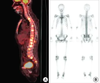
Although fusion whole body positron emission tomogra- phy (PET) showed lesions indicative of multiple bone metas- tases (Fig. 1A), a bone scan showed little osteoblastic reaction (Fig. 1B). Because there was no evidence of malignancy on chest, abdominal and pelvic CT and prostate ultrasonogra- phy, we performed lumbar spine magnetic resonance imag- ing (MRI) (Fig. 2). The MRI findings suggested that the PET lesions were reactive changes of the BM caused by malignant cell infiltration, such as leukemia, lymphoma or multiple myeloma (MM) rather than multiple bone metastases.
A BM biopsy showed an increase in plasma cells (15%), eosinophils and histiocytes, but no malignant cells. Due to suspected MM, we tested the monoclonality of the plasma- cytosis and fluorescence in situ hybridization myeloma panel.
Although b2-microglobulin was 3.6 mcg/mL, there was no evidence of monoclonality in plasmacytosis, IGH/FGFR3 rearrangement, or RB1 or TP53 deletion. The serum concen-
tration of kappa free light chain (FLC) was 81.10 mg/L, the serum concentration of lambda FLC was 139.00 mg/L and the kappa/lambda FLC ratio was 0.58. Serum protein elec- trophoresis and immunoelectrophoresis showed increases in
a-1, 2 and g-globulin fractions, findings consistent with a chronic inflammatory response. CT guided bone biopsies of the lumbar spine and another BM biopsy showed no evidence of malignant cells. The patient chose supportive care, but was readmitted one month later for aggravation of his general con- dition. Abdominal and pelvic CT showed no changes except for apparent bony changes. The patient still wanted support- ive care and died soon afterward.
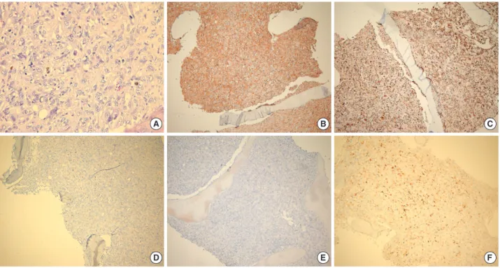
At necropsy, we performed another BM biopsy and a liver biopsy. The liver biopsy showed no evidence of malignancy, except for severe cholestasis with non-specific hepatitis. The BM biopsy, however, revealed a diffuse non-cohesive prolifer- ation of pleomorphic neoplastic cells with large round to oval nuclei, vesicular chromatin and abundant foamy eosinophilic cytoplasm (Fig. 3). Immunohistochemistry showed positive expression of CD68 (macrophage/histiocytic marker), S100 protein, CD31, CD99 and Vimentin. Neoplastic cells were weakly positive for CD21 (mature B-cell and follicular den- dritic cell marker), CD4 (T-cell marker) and lysozyme and negative for CD3 (T-cell marker), CD20/CD79a (B-cell mark- ers), CD56 (NK cell marker), cytokeratin, smooth muscle actin (SMA, a myeloid cell marker), SMMHC and HMB45.
Staining showed Masson-Trichrome positive fine collagen fibers and PAS positivity on megakaryocytes. Together, these morphologic and immunohistochemical features are consis- tent with the BM involvement of HS.
DISCUSSION
HS is a rare and aggressive malignant disorder commonly presenting at extranodal sites and showing a poor response to
314 B.S. Sohn, T. Kim, J.E. Kim, et al.
Fig. 1. Scanning findings for bones. (A) Disseminated hyperme- tabolic lesion (maxSUV=9.0, L2 body) at the whole spine by PET.
A hypermetabolic lesion suggesting a primary malignant lesion was not detected in the lung, intraabdominal and pelvic organs.
(B) No increased bone uptake of hypermetabolic lesions on bone scan, with little osteoblastic effect.
A B
Fig. 2. Diffuse and heterogeneous T1 and T2 showing low SI changes in the lower thoracic and lumbar spine with subtle en- hancement.
therapy, with most patients dying from the disease (1). Some patients show systemic presentation, with features of malig- nant histiocytosis. HS may also be associated with other hema- tological disorders, such as acute leukemia, lymphoma and myelodysplasia (1, 7). Genetic or epigenetic inactivation of PTEN, p16INK4A, and p14ARF has been observed in HS, indicating that these proteins are involved in the development of HS and providing insights into the pathogenesis of these tumors (13).
Differential diagnosis of HS includes Langerhans cell sar- coma (LCS), diffuse large B-cell lymphoma, peripheral T-cell lymphoma, anaplastic large cell lymphoma (ALCL), metastat- ic carcinoma, and melanoma. The diagnosis of HS is based on histological and immunohistochemical evidence of histi- ocytic differentiation and exclusion of other malignancies with epithelial, melanocytic and lymphoid phenotypes (4). Malig- nant HS cells strongly express the hemoglobin scavenger mark- er CD163, which is regarded as a specific marker for HS (4).
Tumor cells in our patient showed expression of histiocyt- ic markers (CD68) and lysozyme with weak expression of CD21. The cells, however, did not express B-cell, T-cell, and myeloid markers, including CD3, CD20, CD56, and CD79a, or SMA and SMMHC. The expression of histiocytic markers was unusual for ALCL, and no rearrangement of the T-cell receptor g-chain gene was identified. Although these cells showed expression of S100 protein, the latter may have been derived from activated normal macrophages (1, 9). Metastat- ic carcinoma and melanoma were ruled out by the absence
of expression of cytokeratins and HMB45 (9).
Morphologically, tumor cells in LCS have nuclei that are grooved, indented, folded, or lobulated, with fine chromatin and a thin nuclear membrane, features that are absent in HS (2, 9). The tumor cells in our patient had abundant clear cyto- plasm, without spindle or fusiform cells, showing a whorled, fascicular or storiform pattern. These morphologic differences could distinguish the disease in our patient from LCS, and from follicular and interdigitating dendritic cell tumors (1).
HS shows a very aggressive clinical course in most patients.
In one study, 6 of 11 patients died of disease within 0.5 to 36 months (10). In a study of 18 patients who underwent chemotherapy, 3 achieved complete remission, 6 had no res- ponse, and 7 died of disease (1). In another study of four adult patients with widespread disease, all failed chemotherapy and died from disease within 0.5 to 14 months after diagnosis (14). In other studies, 6 of 8 adult patients died of disease 5 to 48 months after diagnosis despite aggressive chemother- apy (15). The patient described here died of disease 2 months after his first visit to our hospital.
Due to the rarity of this malignancy, chemotherapy regi- mens have not been standardized, with most studies being retrospective reviews (8). Nonetheless, chemotherapy used to treat large cell lymphomas is a reasonable approach for patients with HS (12), although thalidomide may also ben- efit these patients (3, 11).
In summary, HS is a rare neoplasm difficult to pathologi- cally diagnose. Immunohistochemical features suggesting
A Case of Histiocytic Sarcoma Presenting with Primary Bone Marrow Involvement 315
Fig. 3. Histopathological findings of bone marrow biopsy. (A) Non-cohesive proliferation of large pleomorphic neoplastic cells with large round-to-oval nuclei with vesicular chromatin and abundant foamy cytoplasm (H&E stain, ×400). Immunostaining with antibodies to (B) CD99, (C) CD68, (D) CD56, (E) HMB45 (each, ×100) and (F) S100 (×200).
B C
A
E F
D
HS include the strong expression of histiocytic markers such as CD68, lysozyme and CD163. Proper recognition of HS is important, because the clinical presentation and morpho- logic appearance of this neoplasm may lead to a misdiagno- sis as other hematologic malignancies.
REFERENCES
1. Pileri SA, Grogan TM, Harris NL, Banks P, Campo E, Chan JK, Favera RD, Delsol G, De Wolf-Peeters C, Falini B, Gascoyne RD, Gaulard P, Gatter KC, Isaacson PG, Jaffe ES, Kluin P, Knowles DM, Mason DY, Mori S, Muller-Hermelink HK, Piris MA, Ralfkiaer E, Stein H, Su IJ, Warnke RA, Weiss LM. Tumours of histiocytes and accessory dendritic cells: an immunohistochemical approach to clas- sification from the International Lymphoma Study Group based on 61 cases. Histopathology 2002; 41: 1-29.
2. Weiss LM, Grogan TM, Muller-Hermelink HK, Stein H, Dura WT, Favara B, Pauli M, Feller AC. Histiocytic sarcoma. In: Jaffe ES, Har- ris NL, Stein H, Vardiman JW, eds. WHO Classification of Tumors:
Pathology and Genetics of Tumors of Haematopoietic and Lymphoid Tissues. 2nd ed. Lyon, France: IARC Press 2001: 278-9.
3. Abidi MH, Tove I, Ibrahim RB, Maria D, Peres E. Thalidomide for the treatment of histiocytic sarcoma after hematopoietic stem cell transplant. Am J Hematol 2007; 82: 932-3.
4. Vos JA, Abbondanzo SL, Barekman CL, Andriko JW, Miettinen M, Aguilera NS. Histiocytic sarcoma: a study of five cases including the histiocyte marker CD163. Mod Pathol 2005; 18: 693-704.
5. Buonocore S, Valente AL, Nightingale D, Bogart J, Souid AK. His- tiocytic sarcoma in a 3-year-old male: a case report. Pediatrics 2005;
116: e322-5.
6. Huang SC, Chang CL, Huang CH, Chang CC. Histiocytic sarcoma- a case with evenly distributed multinucleated giant cells. Pathol Res
Pract 2007; 203: 683-9.
7. Feldman AL, Minniti C, Santi M, Downing JR, Raffeld M, Jaffe ES.
Histiocytic sarcoma after acute lymphoblastic leukaemia: a common clonal origin. Lancet Oncol 2004; 5: 248-50.
8. Alexiev BA, Sailey CJ, McClure SA, Ord RA, Zhao XF, Papadim- itriou JC. Primary histiocytic sarcoma arising in the head and neck with predominant spindle cell component. Diagn Pathol 2007; 2: 7.
9. Fukunaga M, Kato H. Histiocytic sarcoma associated with idiopath- ic myelofibrosis. Arch Pathol Lab Med 2004; 128: 1167-70.
10. Kamel OW, Gocke CD, Kell DL, Cleary ML, Warnke RA. True his- tiocytic lymphoma: a study of 12 cases based on current definition.
Leuk Lymphoma 1995; 18: 81-6.
11. Dalle JH, Leblond P, Decouvelaere A, Yakoub-Agha I, Preudhomme C, Nelken B, Mazingue F. Efficacy of thalidomide in a child with histiocytic sarcoma following allogeneic bone marrow transplanta- tion for T-ALL. Leukemia 2003; 17: 2056-7.
12. Hornick JL, Jaffe ES, Fletcher CD. Extranodal histiocytic sarcoma:
clinicopathologic analysis of 14 cases of a rare epithelioid malig- nancy. Am J Surg Pathol 2004; 28: 1133-44.
13. Carrasco DR, Fenton T, Sukhdeo K, Protopopova M, Enos M, You MJ, Di Vizio D, Nogueira C, Stommel J, Pinkus GS, Fletcher C, Hor- nick JL, Cavenee WK, Furnari FB, Depinho RA. The PTEN and INK4A/ARF tumor suppressors maintain myelolymphoid homeosta- sis and cooperate to constrain histiocytic sarcoma development in humans. Cancer Cell 2006; 9: 379-90.
14. Ralfkiaer E, Delsol G, O’Connor NT, Brandtzaeg P, Brousset P, Vejlsgaard GL, Mason DY. Malignant lymphomas of true histiocyt- ic origin. A clinical, histological, immunophenotypic and genotypic study. J Pathol 1990; 160: 9-17.
15. Lauritzen AF, Delsol G, Hansen NE, Horn T, Ersboll J, Hou-Jensen K, Ralfkiaer E. Histiocytic sarcomas and monoblastic leukemias. A clinical, histologic, and immunophenotypical study. Am J Clin Pathol 1994; 102: 45-54.
316 B.S. Sohn, T. Kim, J.E. Kim, et al.