© 2011 The Korean Academy of Medical Sciences.
This is an Open Access article distributed under the terms of the Creative Commons Attribution Non-Commercial License (http://creativecommons.org/licenses/by-nc/3.0) which permits unrestricted non-commercial use, distribution, and reproduction in any medium, provided the original work is properly cited.
pISSN 1011-8934 eISSN 1598-6357
A Case of the Inferior Mesenteric Artery Arising from the Superior Mesenteric Artery in a Korean Woman
Anatomical variations of the inferior mesenteric artery are extremely uncommon, since the inferior mesenteric artery is regularly diverged at the level of the third lumbar vertebra. We found a rare case in which the inferior mesenteric artery arose from the superior mesenteric artery. The findings were made during a routine dissection of the cadaver of an 82-yr-old Korean woman. This is the tenth report on this anomaly, the second female and the first Korean. The superior mesenteric artery normally arising from abdominal aorta sent the inferior mesenteric artery as the second branch. The longitudinal anastomosis vessels between the superior mesenteric artery and inferior mesenteric artery survived to form the common mesenteric artery. This anatomical variation concerning the common mesenteric artery is of clinical importance, performing procedures containing the superior mesenteric artery.
Key Words: Variation; Mesenteric Artery, Inferior; Longitudinal Anastomosis Vessel Seung Jin Yoo, Min Jung Ku, Sa Sun Cho
and Sang Pil Yoon
Department of Anatomy, School of Medicine, Jeju National University, Jeju, Korea Received: 22 June 2011
Accepted: 17 August 2011 Address for Correspondence:
Sang Pil Yoon, MD
Department of Anatomy, School of Medicine, Jeju National University, 66 Jejudaehak-ro, Jeju 690-756, Korea Tel: +82.64-754-3823, Fax: +82.64-725-2593 E-mail: spyoon@jejunu.ac.kr
http://dx.doi.org/10.3346/jkms.2011.26.10.1382 • J Korean Med Sci 2011; 26: 1382-1385
CASE REPORT
Basic Medical Sciences
INTRODUCTION
There are three unpaired visceral branches of the abdominal aorta, the celiac trunk (CT), the superior mesenteric artery (SMA) and the inferior mesenteric artery (IMA), proceeding in a cra- nio-caudal direction. The two upper unpaired visceral branches originate from the aorta in a prefixed site at the level of the first lumbar vertebra, whereas the lower one has more variable points of origin. This is true for all ages and for both genders (1). On the other hand, the IMA is diverged generally at the level of the third lumbar vertebra, considerably below the origin of the SMA and the CT (2). Thus, there have been many reports on the variations between CT and SMA, but the variations of the IMA are found to be extremely rare.
The abdominal vessels, especially the CT and SMA, frequent- ly show diverse anomalies in their origin and course to date (3- 7). Either component of the CT sometimes arises directly from the abdominal aorta or independently from the aorta. In addi- tion, the CT unites with the SMA at their origins to form a com- mon trunk, the celiacomesenteric trunk (CMT) (3). The rare oc- currence of CMT at the level of the first lumbar vertebra is stat- ed to be 1%-2.7%. According to report and review on other ab- dominal arterial anomalies associated with the CMT, a left colic artery arises from the distal portion of the CMT, corresponding to the SMA (4).
The left colic artery arising from the SMA has been reported to occur at a low frequency of 1%. The prevalence of the varia-
tion on the IMA, such as the absence of the IMA or the forma- tion of a common mesenteric artery in which the IMA joins the SMA, is extremely rare (8). We encountered a rare variation of the IMA branching out of the SMA during a routine dissection of an 82-yr-old female cadaver at our university in 2011.
CASE DESCRIPTION
During a routine dissection carried out at Jeju National Univer- sity Medical School in 2011, we found a case in which the IMA arose from the SMA. This variation was observed in an 82-yr-old Korean woman cadaver, whose cause of death was ‘unknown’.
The protocol for the current report did not include any specific issue that needed to be approved by the institutional review board of the Jeju National University and it conformed to the provisions of the Declaration of Helsinki in 1995. Gross dissec- tion was performed in the customary fashion. In order to indi- cate the arteries, the veins were removed. All arterial branches supplying the gastrointestinal tract were examined. The distance between two branches of the abdominal aorta and the external caliber of the main arteries at their origin were also measured.
The typical vascular network (Fig. 1A, B, and B1) of abdominal aorta was absent, and the CT and the SMA were the only un- paired visceral branches out of abdominal aorta (Fig. 1C, and C1). Since the IMA did not arise from the abdominal aorta, the SMA can be also named as the common mesenteric artery or bimesenteric trunk. The CT and the SMA arose at a distance of
Yoo SJ, et al. • The Common Mesenteric Artery in a Korean
http://jkms.org 1383
http://dx.doi.org/10.3346/jkms.2011.26.10.1382
14.3 cm and 12.5 cm respectively from the bifurcation of the ab- dominal aorta to the right and left common iliac arteries, which correspond to the level of the first lumbar vertebra. The SMA had a caliber of 8 mm, and was approximately 18 mm below the CT (10 mm in external caliber at its origin). The SMA gave off its first (inferior pancreatico-duodenal artery) and second branch 27 mm and 35 mm away from its origin, respectively (Fig. 2B). The sec- ond branch (3 mm in external caliber), corresponding in course and distribution to the IMA, gave rise to the classical branches of the IMA, the left colic artery, the sigmoid, and the superior rectal arteries (Fig. 2A). After the second branch, the SMA indi- cated classical branching pattern which proceeded inferiorly and laterally to be attached to the right and middle colon.
DISCUSSION
This is the first report on the common mesenteric artery, which the IMA arises from the SMA, of a Korean. Among a total of ten cases including the present case, the occurrence of common mesenteric artery were observed in cadavers (2, 9-14) except for the case of a common arterial trunk among the CT, SMA and IMA in a radiological description (15). All reports were on male, but the variation observed in the case of Yamasaki et al. (13) had been the only female case until the current case was discovered.
All reported cases were associated with an artery that shared the same characteristics of the ordinary IMA, even though it arose from the SMA instead of the abdominal aorta. In all cases, the IMA always diverged as the first branch of the SMA, except for a Gwyn and Skilton (10). In the present case, the IMA arose as a
A B C
B1 C1
Fig. 1. Schematic diagrams (A-C) and representative photo- graphs (B1 and C1) showing the arrangements of the origin of the celiac trunk (CT), superior mesenteric artery (SMA) and inferior mesenteric artery (IMA) from dorsal aorta (DA). (A) Embryonic dorsal aorta comprises the seven of the ventral splanchnic arteries (VSA) connected by a longitudinal anas- tomosis vessels (LA). (B) and (B1) The typical vascular net- work of abdominal aorta (Ao) formed by the partial disappear- ance of the longitudinal anastomosis vessels. (C) and (C1).
Retention of parts of this primitive arterial channels could give rise to anomalous variation of the SMA and the IMA. G, left gastric artery; H, common hepatic artery; S, splenic artery;
CMA, common mesenteric artery; IVC, inferior vena cava.
Yoo SJ, et al. • The Common Mesenteric Artery in a Korean
1384 http://jkms.org http://dx.doi.org/10.3346/jkms.2011.26.10.1382 second branch 35 mm away from the origin of the SMA, and the
first branch (inferior pancreaticoduodenal artery) was 27 mm distal to the SMA. Besides, Katagiri et al. (4) reported that a left colic artery arose from the CMT and that the sigmoid and the superior rectal arteries branched out of the original IMA.
Although the presence of a CMT is rare (3, 5), the occurrence of the IMA arising from the SMA, rather than the abdominal aor- ta, is even rarer. Benton and Cotter (16) reported a variation of the double IMAs, which arose independently from the abdomi- nal aorta. Other researchers rarely described any other variation of IMA (17, 18), and thus Lippert and Pabst (8) mentioned the frequency of the variation in which the IMA arises from the SMA to be less than 0.1%. Kitamura et al. (12) suggested the embryo- logical explanation for the development of the celiac-mesenteric system (Fig. 1). Namely, the seven primitive splanchnic branches arising from the abdominal aorta in embryo are connected by the ventral longitudinal anastomosis among the roots of the om- phalomesenteric artery, of which some disappear and the clas- sical branches-the left gastric, common hepatic, splenic, of CT, the SMA and the IMA-are formed. The longitudinal anastomo- sis vessels disappear between the SMA and IMA during the pro-
A B
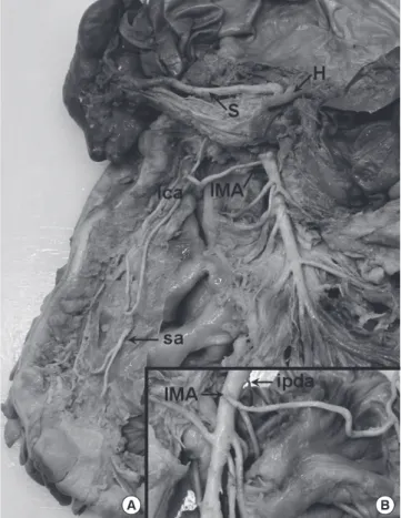
Fig. 2. Photographs of the arterial system in the area of the common mesenteric ar- tery of the reported case (A, posterior aspect; B, anterior aspect). The inferior mesen- teric artery (IMA) arose from the superior mesenteric artery from the left-anterior sur- face as the second branch, while the inferior pancreaticoduodenal artery (ipda) from the right-posterior surface as the first branch. H, common hepatic artery; S, splenic artery; lca, left colic artery; sa, sigmoid artery.
cess of development. The common mesenteric artery can be re- garded as an anomaly of the arterial convergence like in this case (2, 12, 14).
Clinically, the functional results after sigmoid colectomy follow- ing ligation or preservation of the IMA was reported (19). Liga- tion of the IMA caused a higher rate of fecal incontinence; on the other hand, preservation of the IMA during sigmoid colectomy lowered the frequency of postoperative impaired anorectal func- tion. Since both the SMA and IMA supply the whole colon, iden- tification of the IMA is particularly important when performing surgical and radiological procedures. Obstructive diseases such as thromboembolism of the common mesenteric artery (2) and en bloc resection of the head of the pancreas including the supe- rior mesenteric vessels (20) can cause fatal colonic degeneration, associated with the area requiring the blood supply of the IMA.
ACKNOWLEDGMENTS
The authors thank the students (Ji Hoi Kim, Misun Kim, Hyun Sik Park, Sum Kim, Kyusik Choi, Suk Won Chung, Jong Woo Ock, Hyun Jo Shin, and Hyun Joo Oh) of the first year in dissection course at Jeju National University Medical School for their assis- tance.
REFERENCES
1. Yahel J, Arensburg B. The topographic relationships of the unpaired vis- ceral branches of the aorta. Clin Anat 1998; 11: 304-9.
2. Yi SQ, Li J, Terayama H, Naito M, Iimura A, Itoh M. A rare case of inferi- or mesenteric artery arising from the superior mesenteric artery, with a review of the review of the literature. Surg Radiol Anat 2008; 30: 159-65.
3. Cavdar S, Sehirli U, Pekin B. Celiacomesenteric trunk. Clin Anat 1997;
10: 231-4.
4. Katagiri H, Ichimura K, Sakai T. A case of celiacomesenteric trunk with some other arterial anomalies in a Japanese woman. Anat Sci Int 2007;
82: 53-8.
5. Yi SQ, Terayama H, Naito M, Hayashi S, Moriyama H, Tsuchida A, Itoh M. A common celiacomesenteric trunk, and a brief review of the litera- ture. Ann Anat 2007; 189: 482-8.
6. Nayak SR, Prabhu LV, Krishnamurthy A, Ganesh Kumar C, Ramanathan LA, Acharya A, Prasad Sinha A. Additional branches of celiac trunk and its clinical significance. Rom J Morphol Embryol 2008; 49: 247-9.
7. Manoharan B, Aland RC. Atypical coeliomesenteric anastomosis: the pres- ence of an anomalous fourth coelic trunk branch. Clin Anat 2010; 23:
904-6.
8. Lippert H, Pabst R. Arterial variations in man: classification and frequen- cy. Munchen: JF Bergmann Verlag, 1985, p 52-3.
9. Adachi B. Das Fehlen der A. mesenterica inferior bei einem Japaner. Anat Anz 1930; 69: 431-3.
10. Gwyn DG, Skilton JS. A rare variation of the inferior mesenteric artery in man. Anat Rec 1966; 156: 235-7.
11. Mori Y, Ito I, Hatashita S, Yoshikawa K. A rare anomaly of the absence of inferior mesenteric artery. J Osaka Med Coll 1960; 20: 77-9.
Yoo SJ, et al. • The Common Mesenteric Artery in a Korean
http://jkms.org 1385
http://dx.doi.org/10.3346/jkms.2011.26.10.1382
12. Kitamura S, Nishiguchi T, Sakai A, Kumamoto K. Rare case of the inferior mesenteric artery arising from the superior mesenteric artery. Anat Rec 1987; 217: 99-102.
13. Yamasaki M, Nakao T, Ishizawa A, Ogawa R. A rare case of the inferior mesenteric artery and some colic arteries in man. Anat Anz 1990; 171:
343-9.
14. Osawa T, Feng XY, Sasaki N, Nagato S, Matsumoto Y, Onodera M, Nara E, Fujimura A, Nozaka Y. Rare case of the inferior mesenteric artery and the common hepatic artery arising from the superior mesenteric artery. Clin Anat 2004; 17: 518-21.
15. Nonent M, Larroche P, Forlodou P, Senecail B. Celiac-bimesenteric trunk:
anatomic and radiologic description - case report. Radiology 2001; 220:
489-91.
16. Benton RS, Cotter WB. A hitherto undocumented variation of the inferior
mesenteric artery in man. Anat Rec 1963; 145: 171-3.
17. Michels NA, Siddharth P, Kornblith PL, Parke WW. The variant blood supply to the descending colon, rectosigmoid, and rectum based on 400 dissections. Dis Colon Rectum 1965; 8: 251-78.
18. Sierocinski W. Studies on the arteries supplying the descending and sig- moid colon in man. Folia Morphol (Warsz) 1976; 35: 287-306.
19. Dobrowolski S, Hać S, Kobiela J, Sledziński Z. Should we preserve the in- ferior mesenteric artery during sigmoid colectomy? Neurogastroenterol Motil 2009; 21: 1288-e123.
20. Noto M, Miwa K, Kitagawa H, Kayahara M, Takamura H, Shimizu K, Ohta T. Pancreas head carcinoma: frequency of invasion to soft tissue adherent to the superior mesenteric artery. Am J Surg Pathol 2005; 29:
1056-61.