Evaluating the Immunological Adjuvant Activities of Carrageenan and Degraded Carrageenan
Ji-Hun Park1,2 and Tae-Saeng Choi1*
1Department of Microbiology, College of Medicine, Dankook University, Cheonan 330-714, Korea
2Department of Nanobiomedical Science and BK21 PLUS NBM Global Research Center for Regenerative Medicine, Dankook University, Cheonan 330-714, Korea
Received May 16, 2018 /Revised June 6, 2018 /Accepted June 7, 2018
Carrageenan (CGN) has been used as a safe food additive for several decades. CGN has also been widely used to induce inflammation in various animal models. Likewise, degraded CGN (dCGN), which is produced by subjecting CGN to acid hydrolysis, also induces inflammation and does so more effectively than CGN. One of the most important characteristics of an immunological adjuvant is its ability to activate innate immunity. The immune-adjuvant effects of CGN and dCGN have not yet been studied in detail. The purpose of this study was to evaluate the immunological adjuvant activ- ities of both CGN and dCGN, which was done by comparing the levels of an ovalbumin (OVA)-specific antibody after treatment with OVA in the absence or presence of CGN or dCGN in plasma from immunized mice. CGN and dCGN showed similar levels of adjuvant activity, as evi- denced by increased antibody titer. Specifically, both CGN and dCGN significantly increased the lev- els of OVA-specific IgG, IgG1, and IgG2a antibodies in the plasma as compared with OVA alone (the control). However, compared to the positive control (Freund’s adjuvant), both CGN and dCGN caused greater increases in IgG1 than in IgG2a. These results suggest that CGN and dCGN have similar ad- juvant activities and produce more IgG1 antibodies than IgG2a.
Key words : Antibody, carrageenan (CGN), degraded carrageenan (dCGN), immunological adjuvant, TNF-α
*Corresponding author
*Tel : +82-41-550-3872, Fax : +82-41-550-3864
*E-mail : [email protected]
This is an Open-Access article distributed under the terms of the Creative Commons Attribution Non-Commercial License (http://creativecommons.org/licenses/by-nc/3.0) which permits unrestricted non-commercial use, distribution, and reproduction in any medium, provided the original work is properly cited.
Journal of Life Science 2018 Vol. 28. No. 9. 1076~1080 DOI : https://doi.org/10.5352/JLS.2018.28.9.1076
Introduction
Carrageenan (CGN), a type of sulfated polysaccharide from seaweed, has been used as a safe food additive as a stabilizer, emulsifier, or thickener for several decades [6, 14, 18]. On one hand, CGN has been widely used in animal studies to induce inflammation (e.g., models of carra- geenan-induced paw edema, granuloma pouch model, and acute peritonitis) [4, 16]. Degraded CGN (dCGN), which is produced by subjecting CGN to acid hydrolysis at high tem- perature, is believed to be produced from CGN in the acidic environment of the stomach [9, 10]. However, additional re- search has shown that it is difficult to produce dCGN from CGN within the gastrointestinal tract [20]. Additionally, sev- eral studies have shown that dCGN can induce colitis more
effectively than CGN when administered orally [1, 3, 11].
Subsequent studies of dCGN have shown that it induces in- flammation more effectively than CGN in vitro and in vivo.
Moreover, dCGN is recognized as a “possible human carci- nogen” and is not permitted as a food additive, unlike CGN [2, 17].
Immunological adjuvants are substances that are used as an auxiliary to elicit an early, high, and long-lasting immune response. As a result, the use of an immunological adjuvant in vaccination could reduce the quantity of the antigen and the frequency of administration [19]. It is generally accepted that its mechanisms of action are the antigen depot effect at the site of injection and the activation of innate immunity, including the upregulation of cytokines and chemokines, leukocyte recruitment, and the activation of antigen-present- ing cells [12, 13].
As mentioned above, CGN and dCGN induce inflamma- tion. Additionally, CGN solution, which is widely used as a food additive, is highly viscous [21]. This suggests that CGN exhibits an antigen depot effect by preventing antigen dispersion at the injection site. However, until now, the im- munological adjuvant effects of CGN and dCGN have not
been studied in detail. In this study, our results showed that both CGN and dCGN has similar adjuvant effects and pre- dominantly produce the IgG1 antibody relative to IgG2a.
Materials and Methods
Animals
Female BALB/c mice (aged 8-10 weeks) were used for this study. Mice were provided drinking water and a normal diet ad libitum and were maintained under a 12 hr light-dark cycle at 24±1℃ with 50% humidity. All animal studies were conducted in compliance with guidelines set forth by the Care and Use of Research Animals and were approved by the Animal Studies Committee of Dankook University (Approval number: DKU-17-002).
Preparation of CGN and dCGN
λ-CGN (CGN) was purchased from Sigma-Aldrich (St.
Louis, MO, USA). CGN (100 mg) was dissolved in 10 ml Dulbecco’s phosphate-buffered saline (DPBS) by heating to 90℃ for 15 min. For preparation of degraded λ-CGN (dCGN), 100 mg CGN was dissolved in 5 ml of 0.1 M hydro- chloric acid and heated at 60℃ for 4 hr. The reaction was terminated by neutralization (pH 7.0-7.2) with 0.1 M sodium hydroxide. Next, the solution was dialyzed against DPBS (pH 7.4) to eliminate excess salt overnight at 4℃. The final concentrations of both CGN and dCGN were adjusted to 10 mg/ml in DPBS and aliquots were stored at -20℃ before use.
Cell culture and tumor necrosis factor (TNF)-α assay The RAW264.7 mouse macrophage cell line was obtained from the Korean Cell Line Bank (Seoul, Korea) and cultured in Dulbecco’s Modified Eagle’s Medium (DMEM) supple- mented with 10% fetal bovine serum at 37℃ with 5% CO2. Cells were plated onto 96-well plates at a density of 5×10⁵ cells/well and allowed to adhere overnight. To evaluate the effects of CGN and dCGN on TNF-α secretion, cells were treated with 250, 500, or 1,000 μg/ml CGN or dCGN and lipopolysaccharide (LPS; 10 ng/ml) as a positive control for 4 hr. Supernatants from treated cells were collected and TNF-α concentration was determined with a TNF-α enzyme- linked immunosorbent assay (ELISA) kit (BioLegend, San Diego, CA, USA) according to the manufacturer’s in- structions.
Immunization
Chicken egg albumin (OVA; Sigma-Aldrich) was used as an antigen. Mice were randomly divided into six groups containing five mice each. After a 1-week adaptation period, mice from each group were injected subcutaneously twice with OVA (2 μg) alone or OVA plus CGN or dCGN (100 or 500 μg/mouse) at 2-week intervals. As a positive control (CFA/IFA), one group of mice was immunized with OVA containing Complete Freund's adjuvant (CFA) for the first injection followed by injections with Incomplete Freund's adjuvant (IFA) mixed with protein solution at a ratio of 1:1 (vol:vol). Two and four weeks after the first immunization, blood was collected from the tail vein to measure blood plas- ma levels of the OVA-specific antibody.
Measurement of OVA-specific IgG and subclasses Blood was collected from mice before and after immuni- zation (days 0, 14, and 28). For blood plasma preparation, whole blood was obtained by incising the tail vein with a sharp surgical blade and centrifuged at 1,500x g for 15 min at room temperature. The plasma was aliquoted and frozen at -70℃ until use. OVA-specific IgG, IgG1 and IgG2a anti- bodies, were measured by indirect ELISA, which was per- formed in 96-well polystyrene plates. The plate was coated with 100 μl of 20 μg/ml OVA in 0.05 mol/l carbonate–bicar- bonate buffer (pH 9.6). The plates were incubated for 2 hr at 37℃ or overnight at 4℃. Unbound antigen was washed three times with phosphate-buffered saline (PBS) containing Tween-20 (PBS-T; 20 mM PBS, pH 7.4, and 0.05% Tween-20), and the unoccupied sites were blocked with 2% nonfat milk in PBS-T for 1 hr at room temperature. After incubation, the plates were washed three times with PBS-T. The bound anti- bodies were detected using anti-mouse horseradish perox- idase-conjugated IgG, IgG1, or IgG2a antibodies (Santa Cruz Biotechnology, Dallas, TX, USA). The levels of OVA-specific antibodies in plasma were expressed as the mean endpoint titer. The endpoint titers were defined as the highest serum dilution that exceeded 0.1 at an absorbance of 450 nm.
Statistical analyses
Statistical differences among groups were analyzed using one-way analysis of variance. Data are shown as the mean±
standard error of the mean, and significance was defined as p<0.05.
Fig. 1. CGN and dCGN induced TNF-α secretion in RAW264.7 cells. RAW264.7 cells were stimulated with various con- centrations of carrageenan (CGN) or degraded carra- geenan (dCGN) (125-1,000 μg/ml) for 4 hr. Cells were stimulated with 10 ng/ml lipopolysaccharide (LPS; pos- itive control) or medium only (negative control). Super- natants from treated cells were collected and the concen- tration of tumor necrosis factor (TNF)-α was determined with a TNF-α enzyme-linked immunosorbent assay (ELISA) kit. Data are presented as the mean±standard deviation of triplicates. Different superscripts represent significantly different values (p<0.05).
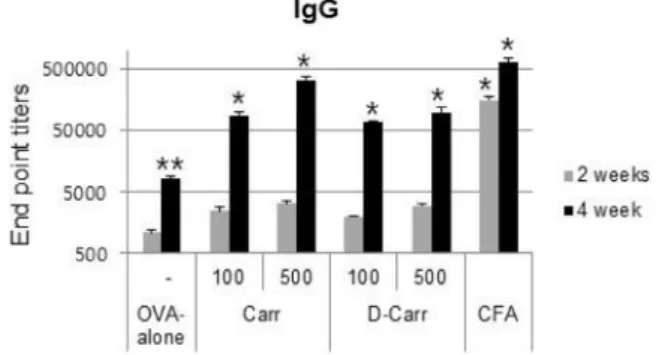
Fig. 2. Adjuvant effects of CGN and dCGN on the induction of OVA-specific total IgG in blood plasma. Plasma was col- lected at 2 and 4 weeks after the first immunization. Mice from each group were injected subcutaneously twice with ovalbumin (OVA; 2 μg) alone or OVA plus CGN or dCGN (100 or 500 μg/mouse) at 2-week intervals. As a positive control, mice were immunized with OVA plus Complete Freund's adjuvant and boosted with Incom- plete Freund’s adjuvant 2 weeks later (CFA/IFA). Total IgG titers were determined by endpoint dilution ELISA.
Different superscripts represent significantly different values (p<0.05).
Results
TNF-α release from RAW264.7 cells following treat- ment with CGN or dCGN
To investigate whether CGN and/or dCGN induced the release of TNF-α in RAW264.7 cells, TNF-α was measured in culture supernatants from CGN- or dCGN-treated cells.
LPS (10 ng/ml) was used as the positive control. Fig. 1 shows that TNF-α secretion increased in a concentration- de- pendent manner following treatment with CGN and dCGN, and dCGN induced TNF-α secretion more efficiently than CGN.
Effects of CGN and dCGN on anti-OVA IgG and subclasses
To evaluate the immunological adjuvant effects of CGN and dCGN, we analyzed OVA-specific antibody levels in blood plasma of mice immunized with OVA or OVA mixed with CGN or dCGN. Immunization was performed twice at 2-week intervals and plasma was collected 2 weeks after the primary immunization and 2 weeks after boosting. Fig.
2 shows that, although total IgG levels in the OVA plus CGN or dCGN group tended to increase slightly at 2 weeks after primary immunization, total IgG levels were significantly higher than in the OVA only group at 2 weeks after boosting. Total IgG in plasma in the high CGN concentration
(500 μg) treatment group was high, similar to the level of the CFA/IFA positive control group at 2 weeks after boosting. We did not detect a significant difference between CGN and dCGN on antibody production, even though a sig- nificant difference in TNF-α release was observed.
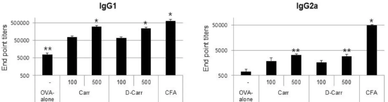
Next, we investigated whether the addition of CGN or dCGN as an adjuvant affected the synthesis of IgG sub- classes (IgG1 and IgG2a). Antibody titers of IgG1 and IgG2a subtypes in mouse plasma collected 2 weeks after boosting were examined. The addition of CGN or dCGN as an ad- juvant efficiently increased the production of both IgG1 and IgG2a compared with OVA alone (Fig. 3). However, the in- duction of IgG2a was relatively low compared with IgG1.
The higher IgG1/IgG2a ratio in the CGN- or dCGN-treated group was evident compared with that of the CFA/IFA pos- itive control group (Fig. 4).
Discussion
According to our current understanding, immunological adjuvants can be broadly divided into two classes based on their mechanisms of action: antigen depot effect and im- mune potentiators (e.g., Toll-like receptor and cytokines).
More specifically, their mechanisms of action include the fol- lowing: the recruitment and activation of immune cells, such as macrophages and dendritic cells, the secretion of cyto-
Fig. 3. Adjuvant effects of CGN and dCGN on the induction of OVA-specific antibodies in blood plasma. Plasma was collected at 4 weeks after the first immunization. Mice from each group were injected twice subcutaneously with OVA (2 μg) alone or OVA plus CGN or dCGN (100 or 500 μg/mouse) at 2-week intervals. Antibody titers of IgG1 (a) and IgG2a (b) were determined by endpoint dilution ELISA. Different superscripts represent significantly different values (p<0.05).
Fig. 4. CGN and dCGN induced Th1-like responses. Each col- umn indicates the ratio of the anti-OVA IgG1/IgG2a an- tibody titer. IgG1 was more predominant than IgG2a in both treatment groups (CGN and dCGN) and specifi- cally in the case of comparison with the positive control (CFA/IFA).
kines and chemokines, and keeping the antigen near the in- jection sites for a longer period of time [5].
Animal studies including the mouse air pouch and the paw edema models have confirmed that CGN induces im- mune cell recruitment and cytokine secretion at injection sites [8, 16]. It is also well known that dCGN exhibits a stron- ger induction effect for an inflammatory response compared with CGN [4, 8, 9, 10]. In addition to these effects, we specu- lated that CGN also exhibits depot effects because of its high viscosity. These identified or estimated effects of CGN and dCGN led us to this study. For these reasons, we examined the adjuvant effects of CGN and d-CGN.
CGN and dCGN induced TNF-α secretion in RAW264.7 cells and dCGN was more effective than CGN, similar to previous studies [7]. The immunization experiments showed that the adjuvant effects of CGN and dCGN on antibody production were significantly higher than without adjuvant treatment. The analysis of IgG subclass concentrations re-
vealed that CGN and dCGN predominantly elicited IgG1 rather than IgG2a. This result suggests that as adjuvants, both CGN and dCGN enhance humoral immunity rather than cellular immunity.
Regarding the initial cellular TNF-α secretion experi- ments, we hypothesized that the adjuvant effect of dCGN would be relatively higher than that of CGN. However, their adjuvant effects were similar. Although we cannot explain this result, this may be attributable to their different vis- cosities.
dCGN, a partially degraded form of CGN formed by acid and heat, has a relatively low viscosity [15]. As mentioned above, we speculated that their viscosities caused a different depot effect of antigen (i.e., CGN has higher viscosity than dCGN and dCGN has higher inflammatory potential than CGN). Taken together, we suggest that the cause of similar adjuvant effects between CGN and dCGN is the combined effects of immune potentiators and depot.
References
1. Anver, M. R. and Cohen, J. 1976. Animal model of human disease. Ulcerative colitis, Animal model: ulcerative colitis induced in guinea pigs with degraded carrageenan. Am. J.
Pathol. 84, 431-434.
2. Ariffin, S. H., Yeen, W. W., Abidin, I. Z., Abdul Wahab, R. M., Ariffin, Z. Z. and Senafi, S. 2014. Cytotoxicity effect of degraded and undegraded Kappa and iota carrageenan in human intestine and liver cell lines. BMC Complement Altern. Med. 14, 508-523.
3. Ashi, K. W., Inagaki, T., Fujimoto, Y. and Fukuda, Y. 1978.
Induction by degraded carrageenan of colorectal tumors in rats. Cancer Lett. 4, 171-176.
4. Barth, C. R., Funchal, G. A., Luft, C., de Oliveira, J. R., Porto, B. N. and Donadio, M. V. 2016. Carrageenan-induced in- flammation promotes ROS generation and neutrophil ex-
초록:Carrageenan과 degraded carrageenan의 면역 보강제로서의 효능 평가
박지훈1,2․최태생1*
(1단국대학교 의과대학 미생물학 교실, 2단국대학교 나노 바이오 의학과 분자 의과학)
Carrageena은 전세계적으로 안전한 식품첨가물로 승인되어 오랜 기간 다양한 식품, 기타 가공품에 사용되어 지고 있다. 다른 한편으로, 이 Carrageenan은 동물 실험에서 염증 유도 물질로 확인되어 염증 유발 실험에 현재까 지도 매우 빈번히 사용 되고 있다. 또한 이 Carrageenan을 고온과 강산에서 처리하여 부분적으로 분해한 de- graded Carrageenan은 염증 유도 능이 Carrageenan 보다 더 강한 것으로 알려져 있다. 면역 보강제의 중요한 특 성 가운데 하나는 선천면역(대표적으로 염증반응)의 활성화 인 것이 잘 알려져 있다. 그러나 현재까지 Carrageenan이나 degraded Carrageenan의 면역 보강제로서의 효과에 관하여 상세한 비교 연구는 수행되어 지지 않았다. 본 연구의 목적은 Carrageenan과 degraded Carrageenan의 면역 보강제로서의 효과를 비교 분석하는데 있다. 실험 동물은 마우스를 사용하였으며, 난 알부민을 항원으로, 피하면역을 수행하여 각각의 면역 보강제 효과 를 항체 형성 정도로 조사하였다. Carrageenan이나 degraded Carrageenan 모두 항원 단독으로 면역한 것과 비교 할 때 유의적으로 높은 IgG 생성 능을 보였다. 추가적으로 항원 특이적 IgG1과 IgG2a를 조사한 결과, 이들 Carrageenan, degraded Carrageenan은 본 실험에서 양성 대조 군으로 사용한 보강제, Complete Freund`s ad- juvant와 비교 할 때 IgG2a 보다는 IgG1 생성 능이 높게 유도되는 것이 확인되었다. 이들 결과를 종합하면 염증 유발 능이 보다 강한 degraded carrageenan의 면역 보강제 효과는 carrageenan과 유사한 정도로 확인되었으며, 이들 모두 IgG2 보다는 IgG1 생성 효과가 강한 것으로 나타났다.
tracellular trap formation in a mouse model of peritonitis.
Eur. J. Immunol. 46, 964-970.
5. Brunner, R., Jensen-Jarolim, E. and Pali-Scholl, I. 2010. The ABC of clinical and experimental adjuvants-a brief over- view. Immunol. Lett. 128, 29-35.
6. Campo, V. L., Kawano, D. F., Silva, D. B. and Carvalho, D. I. 2009. Carrageenans: Biological properties, chemical modifications and structural analysis- A review. Carbohydr.
Polym. 77, 167-180.
7. Chen, H., Wang, F., Mao, H. and Yan, X. 2014. Degraded λ-carrageenan activates NF-kB and AP-1 pathways in mac- rophages and enhances LPS-induced TNF-α secretion through AP-1. Biochim. Biophys. Acta. 1840, 2162-2170.
8. Duarte, D. B., Vasko, M. R. and Fehrenbacher, J. C. 2016.
Models of inflammation: Carrageenan Air Pouch. Curr.
Protoc. Pharmacol. 72, 561-569.
9. Ekstrom, L. G. 1985. Molecular weight distribution and the behavior of kappa-carrageenan on hydrolysis. Carbohydr.
Res. 135. 283-289.
10. Ekstrom, L. G., Kuivinen, J. and Johansson, G. 1983. Molecu- lar weight distribution and hydrolysis behavior of carra- geenans. Carbohydr. Res. 116, 89-94..
11. Fath, R. B Jr., Deschner, E. E., Winawer, S. J. and Dworkin, B. M. 1984. Degraded carrageenan-induced colitis in CF1 mice. Digestion 29, 197-203.
12. Fischetti, L., Zhong, Z., Pinder, C. L., Tregoning, J. S. and Shattock, R. J. 2017. The synergistic effects of combining TLR ligand based adjuvants on the cytokine response are de- pendent upon p38/JNK signaling. Cytokine 99, 287-296.
13. HogenEsch, H. 2002. Mechanisms of stimulation of the im mune response by aluminum adjuvants. Vaccine 20 Suppl
3, S34-39.
14. Jiao, G., Yu, G., Zhang, J. and Ewart, H. S. 2011. Chemical structures and bioactivities of sulfated polysaccharides from marine algae. Mar. Drugs 9, 196-223.
15. Masson, C. R. and Caines, G. W. 1954. Viscosity and molec- ular weight of degraded carrageenan. Can. J. Chem. 32, 51-59.
16. Morris, C. J. 2003. Carrageenan-induced paw edema in the rat and mouse. Methods Mol. Biol. 225, 115-121.
17. Oohashi, Y., Ishioka, T., Wakabayashi, K. and Kuwabara, N. 1981. A study on carcinogenesis induced by degraded carrageenan arising from squamous metaplasia of the rat colorectum. Cancer Lett. 14, 267-272.
18. Prajapati, V. D., Maheriya, P. M., Jani, G. K. and Solanki, H. K. 2014. Carrageenan: a natural seaweed polysaccharide and its applications. Carbohydr. Polym. 105, 97-112.
19. Schijns, V. E. and Tangeras, A. 2005. Vaccine adjuvant tech- nology: from theoretical mechanisms to practical approaches.
Dev. Biol. (Basel) 121, 127-134.
20. Tobacman, J. K. 2001. Review of harmful gastrointestinal ef- fects of carrageenan in animal experiments. Environ. Health Perspect. 109, 983-994.
21. Zia, K. M., Tabasum, S., Nasif, M., Sultan, N., Aslam, N., Noreen, A. and Zuber, M. 2017. A review on synthesis, properties and applications of natural polymer based carra- geenan blends and composites. Int. J. Biol. Macromol. 96, 282- 301.