Ferritin, an Iron Storage Protein, Associates with Kinesin 1 through the Cargo-binding Region of Kinesin Heavy Chains (KHCs)
Won Hee Jang1, Young Joo Jeong1, Won Hee Lee2, Mooseong Kim2, Sang-Jin Kim3, Sang-Hwa Urm4, Il Soo Moon5 and Dae-Hyun Seog1*
1Department of Biochemistry Inje University College of Medicine, Busan 614-735, Korea
2Department of Neurosurgery Inje University College of Medicine, Busan 614-735, Korea
3Department of Neurology Inje University College of Medicine, Busan 614-735, Korea
4Department of Preventive Medicine, Inje University College of Medicine, Busan 614-735, Korea
5Department of Anatomy & Dongguk Medical Institute, College of Medicine, Dongguk University, Gyeongju 780-714, Korea Received February 18, 2016 /Revised March 11, 2016 /Accepted March 11, 2016
The intracellular transport of organelles and protein complexes is mediated by kinesin superfamily proteins (KIFs). The first kinesin, kinesin 1, was identified as a molecular motor protein that moves various organelles and protein complexes along the microtubule rails in cells. Kinesin 1 is a tetramer of two heavy chains (KHCs, also called KIF5s) and two kinesin light chains (KLCs). KIF5s interact with many different proteins through their tail region, but their binding proteins have not yet been fully identified. To identify the interaction proteins for KIF5A, we performed yeast two-hybrid screen- ing and found a specific interaction with ferritin heavy chain (Frt-h), which has a role in iron storage and detoxification. Frt-h bound to the amino acid residues between 800 and 940 of KIF5A and to other KIF5s in the yeast two-hybrid assay. The coiled-coil domain of Frt-h is essential for interaction with KIF5A. In addition, ferritin light chain (Frt-l) interacted with KIF5s in the yeast two-hybrid assay. In addition, these proteins showed specific interactions in the glutathione S-transferase (GST) pull-down assay. An antibody to KHC specifically co-immunoprecipitated Frt-h and Frt-l from mouse brain extracts. These results suggest the kinesin 1 motor protein may transport the ferritin complex in cells.
Key words : Cargo, ferritin, kinesin 1, microtubule motors, protein-protein interaction
*Corresponding author
*Tel : +82-51-890-6974, Fax : +82-51-894-5801
*E-mail : [email protected]
This is an Open-Access article distributed under the terms of the Creative Commons Attribution Non-Commercial License (http://creativecommons.org/licenses/by-nc/3.0) which permits unrestricted non-commercial use, distribution, and reproduction in any medium, provided the original work is properly cited.
Journal of Life Science 2016 Vol. 26. No. 6. 698~704 DOI : http://dx.doi.org/10.5352/JLS.2016.26.6.698
Introduction
The intracellular transport of proteins is crucial for main- tenance and function of a cell. Intracellular transport can be divided into two categories: fast transport and slow transport. Fast transport is responsible for transport of the organelles and vesicles. Slow transport drives the movement of cytoplasmic proteins and protein complexes [8]. Both fast transport and slow transport are powered by molecular mo- tor proteins along microtubules [8]. Microtubules in the cell form tracks on which various cargoes can be transported by motor proteins [9]. Motor proteins fall into two super- families, kinesins and dyneins [8, 13]. Kinesins are main mo- lecular motors that move toward the microtubule plus end
direction, whereas dyneins move toward the microtubule minus end direction [9, 27]. Kinesin 1, a conventional kine- sin, was the first identified and is the most abundant molec- ular motor protein [8, 23]. It is a heterotetramer composed of two kinesin heavy chains (KHCs, also called KIF5s) and two kinesin light chains (KLCs) [8]. KIF5s contain a globular motor domain that binds to microtubules, a neck linker, an α-helical stalk domain, and a tail domain that associates with various cargoes [4, 23]. The cargoes of kinesin 1 fall into six classes: mRNP protein complexes, pathogens, cytoskele- ton subunits, membrane-bound organelles, signaling mole- cules, and protein complexes [2, 5, 7, 10, 14, 18, 22].
In mammals, there are three different KIF5s. KIF5B is ex- pressed in all cell types, whereas KIF5A and KIF5C are only expressed in neurons [15]. To investigate the functions of KIF5s, depletion of KIF5A in mice was performed using con- ditionally targeting by a synapsin-promoted Cre-recombi- nase transgene [28]. The conditional knockout mice of KIF5A showed a reduction in neurofilament axonal transport, para- lysis, and epileptic seizures [19, 28]. Mutations in motor do- main of KIF5A have been identified in dominant forms of
hereditary spastic paraplegia type 10 (SPG10) [21]. Most of these are missense mutations that affect the motor domain function of KIF5A and exhibit a reduction in transport veloc- ity [21]. Also, a mutation of KIF5A has been found in a pa- tient with Charcot-Marie-Tooth disease type 2 (CMT2) [10].
Thus, these findings suggest that KIF5A related with SPG10, epileptic seizures, and CMT2 diseases that affect the intra- cellular transport within cells. In contrast to KIF5A, the de- pletion of KIF5C, the other neuron-specific KIF5, in mice leaded to survival with no abnormality except a reduction of brain size [15].
Although the roles of KIF5A in brain have been reported, not all cargoes have been revealed yet [10]. In addition, a little is known about the binding proteins for KIF5A. To im- prove the understanding of the roles of KIF5A in intra- cellular transport, using the yeast two-hybrid screens, we identified the ferritin heavy chain (Frt-h), a cytosolic iron storage protein as a protein that interacts with KIF5A.
Materials and Methods
Plasmid constructs
A previously described mouse KIF5A cDNA [15] was uti- lized as a template to amplify the region coding for amino acids 800-1027 with the appropriate primers. The amplified fragments were subcloned into pGEM T-easy vector (Prome- ga Corp, Madison, WI, USA). The resulting recombinant plasmid was then cut with EcoRI and XhoI and the insert was subcloned into pLexA (Clontech, Palo Alto, CA, USA).
Complementary DNAs for mouse Frt-h (GeneBank accession number: NM_010239) and ferritin light chain (Frt-l) (Gene- Bank accession number: NM_010240) were amplified by pol- ymerase chain reaction (PCR) from Marathon-ReadyTM cDNA library (Clontech). The amplified cDNA fragments were then inserted into pB42AD (Clontech).
Screening of KIF5A-binding proteins by yeast two- hybrid assay
The Matchmaker LexA two-hybrid system was used for screening according to the manufacturer’s manual (Clon- tech). In brief, the cDNA fragment containing the tail region of mouse KIF5A was fused to the DNA-BD region of the pLexA vector and the plasmid DNA was transformed into yeast strain EGY48 carrying the p8op-lacZ gene. EGY48 yeast cells carrying the KIF5A bait plasmid were trans- formed with the mouse brain cDNA library [12] and grown
on synthetic dextrose (SD) plates supplemented with glucose but with no histidine, tryptophan, or uracil (SD/-His/-Trp/
-Ura). The selection of positive clones was performed on an SD/-His/-Trp/-Ura/-Leu plate containing galactose, raffi- nose, X-gal, and BU salts. Plasmids from positive colonies were isolated and rescued using Escherichia coli strain KC8 strain on ampicillin-resistant plates and inserts were ana- lyzed by restriction digestion. Unique insert DNAs were se- quenced and DNA sequence analysis was performed with the BLAST algorithm at the National Center for Biotechnology Information (NCBI). Sequence-verified clones were tested again for interaction with bait in yeast by retransformation.
β-Galactosidase activity in liquid cultures of yeast The strength of the interactions between Frt-h and KIF5A was assessed by measuring the β-galactosidase activity in liquid cultures. Yeast cells were co-transformed with the ex- pression plasmid from the positive clone and the plasmid expressing KIF5A. The β-galactosidase activity in liquid cul- tures of yeast was assayed as described previously [11]. In brief, mid-log phase transformed yeast cells were collected and permeabilized with 0.1% sodium dodecyl sulphate (SDS) and chloroform. An excess amount of chromogenic substrate o-nitrophenyl-β-D-galactoside was added to the yeast lysate, the mixture was incubated at 30℃, and then the reaction was stopped by increasing pH to 11 by the addi- tion of 1 M Na2CO3. The formation of the reaction product, o-nitrophenol, was determined by measuring absorbance at 420 nm on a spectrophotometer and normalizing for the re- action time and the cell density.
Glutathione S-transferase (GST) pull-down assays Pull-down assays using GST fusion proteins were per- formed as follows. Complementary DNAs encoding the full length of Frt-h and Frt-l were subcloned into pET41a. The recombinant GST-Frt-h and GST-Frt-l fusion proteins were expressed in bacterial strain BL21 GOLD (Stratagene, La Jolla, CA, USA) after induction with0.5 mM isopropyl thio- β-D-galactopyranoside (Fisher Biotech, South Australia, Australia) for 3 hr. The fusion proteins were purifiedusing glutathione-agarose beads (Sigma-Aldrich, St. Louis, MO, USA) according to the manufacturer’s protocol. GST alone or GST fusion proteins were dialyzed for 2 hr in PBS using Slide-A-Lyzer (Pierce, Rockford, IL, USA). Ten μg of each of the GST fusion proteins was then coupled to 50 μl of glu-
A
B
C
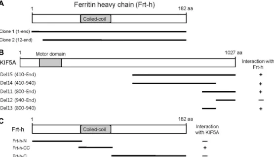
Fig. 1. Identification of the proteins interacting with KIF5A by yeast two-hybrid screening. (A) Schematic diagram of Frt-h. The gray box corresponds to the coiled-coil domain of Frt-h. Clone 1 and 2 isolated from the yeast two-hybrid screen were overlapped at the open reading frame of Frt-h. (B) Minimal Frt-h binding region in KIF5A. KIF5A has motor domain, coiled-coil domain, and tail region. Motor domain is indicated in gray. Several truncated forms of KIF5As were constructed by PCR and tested in the yeast two-hybrid assay for interaction with Frt-h. aa, the amino acid residue number. +, interaction with Frt-h; -, no interaction with Frt-h. (C) Minimal KIF5A binding region in Frt-h. The coiled-coil domains of Frt-h are indicated in gray. Several truncated forms of Frt-h were constructed by PCR and tested in the yeast two-hybrid assay for interaction with KIF5A. aa, the amino acid residue number. +, interaction with KIF5A; -, no interaction with KIF5A.
tathione-agarosebeads by incubating at room temperature for 1 hr, followed by rinsing several times with PBS. Mouse brain lysate was prepared as previously described [11].
Mouse brains were homogenized in ice-cold homogenization buffer (0.32 M sucrose, 4 mM HEPES, pH 7.3) supplemented with protease inhibitors. The homogenate was centrifuged at 12,000× g for 15 min and the resulting supernatant was saved. The supernatant (mouse brain lysate) was incubated overnight at 4℃ with the GST fusion protein-coupled gluta- thione beads. The beads were pelleted by centrifugation, washed three times with the extraction buffer (1% Triton X-100 in PBS containing 10 μg/ml each aprotinin, leupeptin, and pepstatin and 1 μM phenylmethanesulfonyl fluoride), and once withPBS. The bound proteins were eluted from the glutathione beads with 100 μl of Laemmli’s loading buffer. The samples were boiledfor 5 min and then sepa- rated by SDS-PAGE. The proteins were transferred to a ni- trocellulose membrane and subjected to immunoblot analy- sis with anti-KIF5A and anti-KIF5B antibodies [12].
Co-immunoprecipitation
For immunoprecipitation, the mouse brain lysate was di- luted in the same volume of 2X binding buffer (50 mM
HEPES, 240 mM KCl, 2 mg/ml BSA, 0.2% Triton X-100, pH 7.4) and incubated with anti-KHC antibody (Merck Millipore, Darmstadt, Germany) or with control IgG overnight at 4℃, followed by precipitation with protein-A Sepharose (Amer- sham Pharmacia, Piscataway, NJ, USA). The beads were col- lected by brief centrifugation and washed three times with TBS-T (20 mM Tris-HCl, pH 7.5, 0.15 M NaCl, 0.1% Tween 20). The pellets were resuspended with Laemmli’s loading buffer, and the proteins were eluted, denatured by boiling for 2 min, and then processed for SDS-PAGE and immuno- blot analysis with antibodies against KLC1, Frt-h, and Frt-l (Santa Cruz Biotechnology, Santa Cruz, CA, USA).
Results
Identification of KIF5A interacting proteins by yeast two-hybrid screening
Previous studies suggested that the carboxyl (C)-terminal tail region of KIF5s recognize and bind to the various car- goes [10, 23]. Especially, KIF5A has 73 amino acids that have no homology with KIF5B and KIF5C. Using the C-terminal tail region containing fragment (800-1,027 aa) of KIF5A as a bait, 2 positive clones were obtained from 0.7×10⁶ in-
A B C
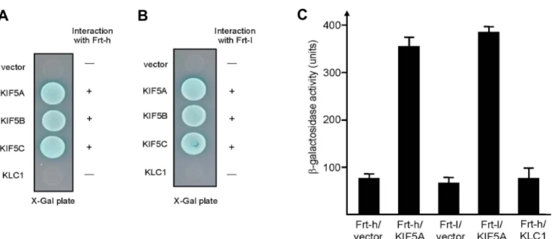
Fig. 2. Interaction between KIF5A and Frt-h. The tail region of each KIF5 and the full length KLC1 were fused to the pLexA DNA binding domain. (A) Frt-h specifically interacted with KIF5s but not with KLC1. +, interaction with Frt-h; -, no interaction with Frt-h. (B) Frt-l specifically interacted with KIF5s but not with KLC1. +, interaction with Frt-l; -, no interaction with Frt-l. (C) The strength of interactions between Frt-h or Frt-l and KIF5A was examined quantitatively using β-galactosidase activity in yeast two-hybrid reporter assay.
dependent mouse brain cDNA library. These two clones were individually isolated, sequenced, and subjected to fur- ther yeast two-hybrid filter assay to confirm the interactions.
Two positive clones turned out to be the cDNA of Frt-h (Fig.
1A). To identify the region of KIF5A required for the inter- action with Frt-h, a series of deletion mutants of KIF5A were constructed and analyzed their interactions with Frt-h using the yeast two-hybrid assay (Fig. 1B). This yeast two-hybrid assay demonstrated that the minimal binding region for Frt-h was located in a small region of KIF5A corresponding to amino acids 800-940, in which a coiled-coil domain exists [15]. Frt-h contains the coiled-coil domain, which seems to interact with various binding proteins [1]. To identify the region of Frt-h required for the interaction with KIF5A, a series of deletion mutants of Frt-h were constructed and ana- lyzed their interactions with KIF5A using the yeast two-hy- brid assay. Only the coiled-coil domain of Frt-h interacted with KIF5A (Fig. 1C). This result indicates that the binding domain for KIF5A was located in the small coiled-coil do- main of Frt-h.
To clarify whether Frt-h interacts with only KIF5A or with other kinesin 1 subunits, the C-terminal tails of KIF5A, KIF5B, and KIF5C, and the full length KLC1 were tested for binding with Frt-h. Frt-h interacted with the tail domains of the KIF5A, KIF5B, and KIF5C in the yeast two-hybrid sys- tem (Fig. 2A). There was no detectable binding between KLC1 and Frt-h (Fig. 2A). This result was not surprising in view of the fact that the KIF5B and KIF5C also have the coiled-coil domain similar to that of KIF5A [15]. Frt-l, other
subunit of ferritin contains the two coiled-coil domain in their primary structure [1]. To clarify whether kinesin 1 sub- units interact with the coiled-coil domain of Frt-l, the C-ter- minal tails of KIF5s and the full length KLC1 were tested for binding with Frt-l. As shown in Fig. 2B, KIF5s interacted with Frt-l but KLC1 did not bind to Frt-l. These data indicate that the coiled-coil domain of Frt-h and Frt-l binds to KIF5s.
To quantify the binding affinity of KIF5A to Frt-h or Frt-l, the KIF5A bait plasmid and the Frt-h or Frt-l expression plas- mids were transformed to yeast and the β-galactosidase ac- tivity was measured in liquid cultures. The interaction of KIF5A with Frt-h or Frt-l yielded approximately 360 units of β-galactosidase activity (Fig. 2C).
Frt-h and Frt-l directly interact with KIF5A at the protein level
As an additional demonstration for the interaction be- tween KIF5s and ferritin complex at the protein level, direct interaction between KIF5s and Frt-h or Frt-l was assayed us- ing a GST pull-down experiment. Recombinant GST-Frt-h and GST-Frt-l fusion proteins were expressed in E. coli. The purified GST fusion proteins are allowed to interact with mouse brain extracts. Immunoblot analyses revealed that both the GST-Frt-h and the GST-Frt-l interacted with KIF5A and KIF5B (Fig. 3A).
To examine whether the interaction between kinesin 1 and ferritin complex takes place in vivo, co-immunoprecipitation analyses were performed. Lysates from mouse brain were incubated with an anti-KHC antibody. Protein G-agarose
A B
Fig. 3. Association of KIF5A with Frt-h in the GST pull-down assay and co-immunoprecipitation. (A) Proteins in the mouse brain lysate were allowed to bind to GST alone, GST-Frt-h fusion protein, or GST-Frt-l fusion protein.
The elution fractions were resolved by SDS-PAGE and immunoblotting was performed using antibodies against KIF5A or KIF5B. (B) Mouse brain lysates were im- munoprecipitated with an anti-KHC antibody or pre- immune serum, and then the precipitates were immuno- blotted with anti-Frt-h, Frt-l, or KLC1 antibodies. Input, 10% of the mouse brain lysates used for each co-im- munoprecipitation assay.
beads selectively precipitated the immuno-complexes, which were subsequently separated by SDS-PAGE and immuno- blotted with anti-Frt-h, Frt-l, and KLC1 antibodies. As shown in Fig. 3B, Frt-h, Frt-l, and KLC1 were co-immunoprecipitated with KHC. These results demonstrate that ferritin complex is a specific binding partner of kinesin 1 in vivo.
Discussion
Mutations in KIF5A are linked to various neurological diseases such as CMT [10], hereditary spastic paraplegia (HSP) [21], and epileptic seizures [19], but the specific func- tion of KIF5A are incompletely understand. In this study, we show that kinesin 1 can interact with ferritin complex.
Using the C-terminal domain of KIF5A as bait, we identified Frt-h in a yeast two-hybrid assay of a mouse brain cDNA library. The coiled-coil domain of Frt-h interacted with KIF5s. Furthermore, using a combination of GST-pull down and co-immunoprecipitation, we confirmed that kinesin 1 interacted with ferritin complex. Taking all of these results together, we hereby propose a model that kinesin 1 trans- ports the ferritin complex in cells.
Several kinesin 1 cargo molecules have been shown to interact with KIF5s [10, 23]. The C-terminal tail region of KIF5s is known as a protein-interacting domain, functioning as a physical linker between kinesin 1 and various proteins
such as glutamate receptor-interacting protein 1 (GRIP1), huntingtin-associated protein 1 (HAP1), and GABAA re- ceptor-associated protein (GABARAP) [18, 19, 24]. GRIP1 and HAP1 have been reported to interact with KIF5s.
However, GABARAP has been identified to interact with on- ly KIF5A. Our data showed that Frt-h and Frt-l interact with all three KIF5s.
Cytosolic ferritin is playing an essential role in iron ho- meostasis of the cell [16]. Ferritin is made up of two compo- nents, Frt-h and Frt-l, which combine to form the 24-subunit protein [17]. Frt-l is required for the long-term storage of iron, while Frt-h has ferroxidase activity in cells [16, 17]. The proportion of Frt-h and Frt-l composing ferritin multimeric complexes is regulated in a tissue-specific manner. Immuno- histochemical study of ferritin in brain showed that neurons express predominantly Frt-h, microglia express Frt-l, and both Frt-h and Frt-l are found in oligodendrocytes [3]. In this study, although we did not show the interaction of kine- sin 1 with Frt-h and Frt-l in a tissue-specific manner, our observations suggest a mechanism that kinesin 1 is linked directly to ferritin complex and could transport to ferritin complex in the cell.
Ferritin has typically been reported as a cytoplasmic pro- tein [6]. However, several reports have found the presence of ferritin in nucleus [6, 20, 26]. Ferritin that is present in the nucleus is comprised of the same ferritin found in the cytoplasm [26]. A previous study reported that the phos- phorylation of Frt-h is important for the specific nuclear translocation of ferritin [25]. These finding suggest that iron may be necessary for the activity of nuclear enzymes for DNA repair or RNA transcription [26]. However, the trans- port mechanism of ferritin from cytoplasm to nucleus is still unclear. To address this issue, it would be worth to identify the specific transport motor protein for ferritin from cyto- plasm to nucleus. In this study, it is proposed that ferritin is a new cargo of kinesin 1. The direct interaction between Frt-h and KIF5s sheds new light on the mechanisms of ferri- tin complex transport to nucleus from cytoplasm, giving at the same time one more example of kinesin 1 cargo.
Acknowledgment
This research was supported by Basic Science Research Program though the National Research Foundation of Korea (NRF) funded by the Ministry of Education, Science and Technology (2010-0021296).
References
1. Beaumont, C., Dugast, I., Renaudie, F., Souroujon, M. and Grandchamp, B. 1989. Transcriptional regulation of ferritin H and L subunits in adult erythroid and liver cells from the mouse. J. Biol. Chem. 264, 7498-7504.
2. Brendza, R. P., Serbus, L. R., Duffy, J. B. and Saxton, W.
M. 2000. A function for kinesin I in the posterior transport of oskar mRNA and Staufen protein. Science 289, 2120-2122.
3. Connor, J. R., Boeshore, K. L., Benkovic, S. A. and Menzies, S. L. 1994. Isoforms of ferritin have a specific cellular dis- tribution in the brain. J. Neurosci. Res. 37, 461-465.
4. Diefenbach, R. J., Mackay, J. P., Armati, P. J. and Cunning- ham, A. L. 1998. The C-terminal region of the stalk domain of ubiquitous human kinesin heavy chain contains the bind- ing site for kinesin light chain. Biochemistry 37, 16663-16670.
5. Diefenbach, R. J., Miranda-Saksena, M., Diefenbach, E., Holland, D. J., Boadle, R. A., Armati, P. J. and Cunningham, A. L. 2002. Herpes simplex virus tegument protein US11 interacts with conventional kinesin heavy chain. J. Virol. 76, 3282-3291.
6. Friedman, A., Arosio, P., Finazzi, D., Koziorowski, D. and Galazka-Friedman, J. 2011. Ferritin as an important player in neurodegeneration. Parkinsonism Relat. Disord. 17, 423- 430.
7. Gorska-Andrzejak, J., Stowers, R. S., Borycz, J., Kostyleva, R., Schwarz, T. L. and Meinertzhagen, I. A. 2003. Mitochon- dria are redistributed in Drosophila photoreceptors lacking milton, a kinesin-associated protein. J. Comp. Neurol. 463, 372-388.
8. Hirokawa, N. 1998. Kinesin and dynein superfamily pro- teins and the mechanism of organelle transport. Science 279, 519-526.
9. Hirokawa, N. and Takemura, R. 2005. Molecular motors and mechanisms of directional transport in neurons. Nat. Rev.
Neurosci. 6, 201-214.
10. Hirokawa, N., Niwa, S. and Tanaka, Y. 2010. Molecular mo- tors in neurons: transport mechanisms and roles in brain function, development, and disease. Neuron 68, 610-638.
11. Jang, W. H. and Seog, D. H. 2013. Kinesin superfamily-asso- ciated protein 3 (KAP3) mediates the interaction between Kinesin-II motor subunits and HS-1-associated protein X-1 (HAX-1) through direct binding. J. Life Sci. 23, 978-983.
12. Jang, W. H., Kim, S. J., Jeong, Y. J., Jun, H. J., Moon, I. S.
and Seog, D. H. 2012. APP tail 1 (PAT1) interacts with Kinesin light chains (KLCs) through the tetratricopeptide re- peat (TPR) domain. J. Life Sci. 22, 1608-1613.
13. Kamal, A. and Goldstein, L. S. 2000. Connecting vesicle transport to the cytoskeleton. Curr. Opin. Cell Biol. 12, 503- 508.
14. Kanai, Y., Dohmae, N. and Hirokawa, N. 2004. Kinesin transports RNA: isolation and characterization of an RNA- transporting granule. Neuron 43, 513-525.
15. Kanai, Y., Okada, Y., Tanaka, Y., Harada, A., Terada, S. and
Hirokawa, N. 2000. KIF5C, a novel neuronal kinesin en- riched in motor neurons. J. Neurosci. 20, 6374-6384.
16. Lawson, D. L., Treffry, A., Artymiuk, P. J., Harrison, P. M., Yewdall, S. J., Luzzago, A., Cesareni, G., Levi, S. and Arosio, P. 1989. Identification of the ferroxidase centre in ferritin.
FEBS Lett. 254, 207-210.
17. Levi, S., Santambrogio, P., Cozzi, A., Rovida, E., Corsi, B., Tamborini, E., Spada, S., Albertini, A. and Arosio, P. 1994.
The role of the L-chain in ferritin iron incorporation: studies of homo and heteropolymers, J. Mol. Biol. 238, 649-654.
18. Li, X. J., Li, S. H., Sharp, A. H., Nucifora Jr, F. C., Schilling, G., Lanahan, A., Worley, P., Snyder, S. H. and Ross, C. A.
1995. A huntingtin-associated protein enriched in brain with implications for pathology. Nature 378, 398-402.
19. Nakajima, K., Yin, X., Takei, Y., Seog, D. H., Homma, N.
and Hirokawa, N. 2012. Molecular motor KIF5A is essential- for GABA(A) receptor transport, and KIF5A deletion causes epilepsy. Neuron 76, 945-961.
20. Pountney, D., Trugnan, G., Bourgeois, M. and Beaumont, C. 1999. The identification of ferritin in the nucleus of K562 cells, and investigation of a possible role in the transcrip- tional regulation of adult beta globin gene expression. J. Cell Sci. 112, 825-831.
21. Reid, E., Kloos, M., Ashley-Koch, A., Hughes, L., Bevan, S., Svenson, I. K., Graham, F. L., Gaskell, P. C., Dearlove, A., Pericak-Vance, M. A., Rubinsztein, D. C. and Marchuk, D.
A. 2002. A kinesin heavy chain (KIF5A) mutation in heredi- tary spastic paraplegia (SPG10). Am. J. Hum. Genet. 71, 1189-1194.
22. Rietdorf, J., Ploubidou, A., Reckmann, I., Holmström, A., Frischknecht, F., Zettl, M., Zimmermann, T. and Way, M.
2001. Kinesin-dependent movement on microtubules pre- cedes actin-based motility of vaccinia virus. Nat. Cell Biol.
3, 992-1000.
23. Seog, D. H., Lee, D. H. and Lee, S. K. 2004. Molecular Motor Proteins of the Kinesin superfamily proteins (KIFs): Struc- ture, Cargo and Disease. J. Kor. Med. Sci. 19, 1-7.
24. Setou, M., Seog, D. H., Tanaka, Y., Kanai, Y., Takei, Y., Kawagishi, M. and Hirokawa, N. 2002. Glutamate-receptor- interacting protein GRIP1 directly steers kinesin to dendrites.
Nature 417, 83-87.
25. Surguladze, N., Patton, S., Cozzi, A., Fried, M. G. and Connor, J. R. 2005. Characterization of nuclear ferritin and mechanisms of translocation. Biochem. J. 388, 731-740.
26. Thompson, K. J., Fried, M. G., Ye, Z., Boyer, P. and Connor, J. R. 2002. Regulation, mechanisms and proposed function of ferritin translocation to cell nuclei. J. Cell Sci. 115, 2165- 2177.
27. Welte, M. A. 2004. Bidirectional transport along micro- tubules. Curr. Biol. 14, 525-537.
28. Xia, C. H., Roberts, E. A., Her, L. S., Liu, X., Williams, D.
S., Cleveland, D. W. and Goldstein, L. S. 2003. Abnormal neurofilament transport caused by targeted disruption of neuronal kinesin heavy chain KIF5A. J. Cell Biol. 161, 55-66.
초록:철 저장 단백질 ferritin과 kinesin 1 결합 규명
장원희1․정영주1․이원희2․김무성2․김상진3․엄상화4․문일수5․석대현1*
(1인제대학교 의과대학 생화학교실, 2인제대학교 의과대학 신경외과학교실, 3인제대학교 의과대학 신경과학교실,
4인제대학교 의과대학 예방의학교실, 5동국대학교 의과대학 해부학교실)
세포내소기관과 단백질 복합체의 운반은 kinesin superfamily proteins (KIFs)에 의해 매개된다. 처음으로 밝혀진 ki- nesin인 kinesin 1은 motor단백질로서 세포 내에서 미세소관을 따라 이동하며, 다양한 세포내 소기관이나 단백질복합체 를 운반한다. Kinesin 1은 장쇄(KHC, 또한 KIF5s로도 통칭) 2분자와 단쇄(KLCs) 2분자로 구성된 4합체(tetramer) 구조를 가진다. KIF5s의 운반체 결합영역을 포함하는 말단영역은 다수의 운반체와 결합하지만, 결합운반체에 관하여 아직 충분 히 밝혀지지 않았다. 본 연구에서 KIF5A의 결합 단백질을 동정하기 위하여 효모 two-hybrid screening을 수행하였고 철 저장 및 해독 기능을 하는 단백질인 ferritin heavy chain (Frt-h)을 찾아내었다. Frt-h은 KIF5A의 아미노산 800번과 940번 사이의 부위와 결합하며, 다른 KIF5s와도 결합함을 효모 two-hybrid assay로 확인하였다. 또한 Frt-h의 coiled-coil 도메인이 KIF5A와의 결합에 필수영역임을 밝혔다. 한편, ferritin light chain (Frt-l) 또한 KIF5s와 결합함을 효모 two-hy- brid assay로 확인하였다. 이러한 단백질간의 결합을 glutathione S-transferase (GST) pull-down assay를 통하여 검증하 였다. 추가적으로 생쥐의 뇌 파쇄액을 항 KHC 항체로 면역침강을 행한 결과, KLC1뿐만 아니라 Frt-h와 Frt-l도 같이 침강하였다. 이러한 결과들은 세포 내에서 kinesin 1이 ferritin 복합체를 운반함을 시사한다.