• 검색 결과가 없습니다.

Presence of Leukemia-maintaining Cells in Differentiation-resistant Fraction of K562 Chronic Myelogenous Leukemia

N/A
N/A
Protected

Academic year: 2021

Share "Presence of Leukemia-maintaining Cells in Differentiation-resistant Fraction of K562 Chronic Myelogenous Leukemia"

Copied!
10
0
0

로드 중.... (전체 텍스트 보기)

전체 글

(1)

Presence of Leukemia-maintaining Cells in Differentiation-resistant Fraction of K562 Chronic Myelogenous Leukemia

Hong-Rae Lee 1† , Mi-Ju Kim 1 , Gahee Ha 1 , So-Jung Kim 2 , Sun-Hee Kim 1 * and Chi-Dug Kang 1 *

1 Department of Biochemistry, Pusan National University School of Medicine, Yangsan 626-870, Korea

2 MD-PhD program, Pusan National University School of Medicine, Yangsan 626-870, Korea Received January 18, 2013 /Revised February 13, 2013 /Accepted February 14, 2013

The present study investigated whether leukemia-maintaining cells reside in a differentiation-resistant fraction using a megakaryocytic differentiation model of K562 cells. Treatment with phorbol-12-myr- istate-13-acetate (PMA) significantly inhibited the colony-forming efficiency of the K562 cells. At a PMA concentration of 1 nM or higher, colony was not formed, but approximately 40% of K562 cells still survived in soft agar. Approximately 70% of colony-forming cells that were isolated following the removal of PMA after exposure to the agent were differentiated after treatment with 10 nM PMA for 3 days. The differentiation rate of the colony-forming cells was gradually increased and reached about 90% 6 weeks after colony isolation, which was comparable to the level of a PMA-treated K562 control.

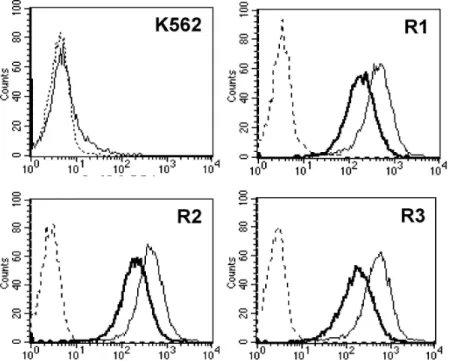
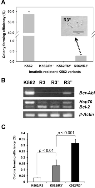
Meanwhile, imatinib-resistant variants from the K562 cells, including K562/R1, K562/R2, and K562/R3 cells, did not show any colony-forming activity, and most imatinib-resistant variants were CD44 positive. After 4 months of culture in drug-free medium, the surface level of CD44 was de- creased in comparison with primary imatinib-resistant variants, and a few colonies were formed from K562/R3 cells. In these cells, Bcr-Abl, which was lost in the imatinib-resistant variants, was re-ex- pressed, and the original phenotypes of the K562 cells were partially recovered. These results suggest that leukemia-maintaining cells might reside in a differentiation-resistant population. Differentiation therapy to eliminate leukemia-maintaining cells could be a successful treatment for leukemia if the leukemia-maintaining cells were exposed to a differentiation inducer for a long time and at a high dose.

Key words : Chronic myelogenous leukemia, differentiation-resistance, imatinib, leukemia-maintaining cells, phorbol-12-myristate-13-acetate

*Corresponding authors

*Tel:+82-51-510-8082, Fax:+82-51-510-8086

*E-mail : [email protected] (C. D. Kang)

*Tel:+82-51-510-8081, Fax:+82-51-510-8086

*E-mail : [email protected] (S. H. Kim)

Present address : Research Center, Dongnam Institute of Radiological & Medical Science, Busan, Korea

This is an Open-Access article distributed under the terms of the Creative Commons Attribution Non-Commercial License (http://creativecommons.org/licenses/by-nc/3.0) which permits unrestricted non-commercial use, distribution, and reproduction in any medium, provided the original work is properly cited.

ISSN (Online) 2287-3406 Journal of Life Science 2013 Vol. 23. No. 2. 197~206 DOI : http://dx.doi.org/10.5352/JLS.2013.23.2.197

Introduction

Although modern tumor therapy has achieved consid- erable progress, cancer is still one of the leading causes of death. Current failure with cancer treatment is not usually due to a lack of primary clinical responses including com- plete remission, but to recurrence or metastasis after initial therapies. The concept that cancer is driven by cancer-initiat- ing or maintaining cells (popularly known as cancer stem cells, CSC) has recently attracted a great deal of attention

[37]. While conventional cancer therapies have targeted well the bulk tumor cells, there is now compelling evidence that cancer-initiating cells may be responsible for recurrence or metastasis of cancers after successful initial induction of re- mission due to their resistance to traditional cancer treat- ments such as surgery, radiotherapy and chemotherapy [46].

It has been demonstrated that the cancer stem cells share several important characteristics of normal stem cells, in- cluding the capacity for self-renewal, the ability to differ- entiate, migrate and metastasize, a relative quiescence, acti- vation of telomerase and antiapoptotic pathways, the in- creased expression of multidrug-resistance proteins, and ro- bust DNA repair activity [25, 44], and consequently show high resistance to anticancer drugs [11] and radiotherapy [13].

Current therapies have been developed largely against the

bulk tumor cells, since they have been usually developed

by their ability to shrink tumors. Therefore, even therapies

that cause complete regression of tumors might spare

enough cancer stem cells to allow regrowth of the tumors.

(2)

A number of therapeutic strategies directed at CSCs are be- ing studied experimentally. The approaches include ablation using antitumor agents that target prospective markers of CSCs (e.g., monoclonal antibodies and activated immune cells), reversal of chemo- or radioresistance mechanisms op- erative in CSC, CSC pathway interference, differentiation therapy, disruption of protumorigenic CSC-microenviron- ment interactions, antiangiogenic or antivasculogenic ther- apy, and disruption of immunoevasion pathways [18, 19].

These strategies can be potentially combined with current anticancer treatments to enhance responsiveness and reduce the possibility of recurrence and dissemination.

Among these strategies, differentiation therapy may be attractable at least in leukemia, since acute promyelocytic leukemia (APL) can be successfully treated with all-trans reti- noic acid (ATRA), a natural derivative of vitamin A, which unlike other chemotherapies, does not directly kill the malig- nant cells. ATRA induces the terminal differentiation of the leukemic cells, after which these differentiated leukemic cells undergo spontaneous apoptosis [1, 2, 22]. In addition, it has been demonstrated in experimental models that the quies- cent CSCs could be differentiated into more mature tumor cells in various solid tumors, such as human glioblastoma by activation of bone morphogenic protein-signaling path- ways [35], medulloblastoma by inhibition of Notch pathway [16], breast cancer by expression of the let-7 miRNA [45]

and treatment with salinomycin [21], and epigenetic differ- entiation therapy [29].

However, it has been reported that 2 or 3 years after ATRA plus chemotherapy-based regimens more than 7 % of patients had relapsed [17, 36]. The relapse might be due to the dormant leukemic stem cells, which may be resistant to differentiation induction. Since CSCs have self-renewal ac- tivity, they should be resistant to differentiation induction.

Or not, CSC population could not be maintained and tumor would be regressed by differentiation therapy. In the present study, it was studied if leukemia-maintaining cells reside in differentiation-resistant fraction, using a megakaryocytic dif- ferentiation model of K562 cells, which was characterized as a multipotential leukemia stem cell line [30].

Materials and Methods Cell and culture

K562 cell line was obtained from the American Type Culture Collection (Manassas, VA, USA). The imatinib-re-

sistant K562 variants, including K562/R1, K562/R2, and K562/R3 cells, were isolated from K562 cells by culturing in the presence of gradually increasing concentrations of im- atinib [28]. The cells were grown in suspension in RPMI 1640 medium (GIBCO Invitrogen cell culture, Grand Island, NY, USA) supplemented with 10% (v/v) heat-inactivated FBS (GIBCO Invitrogen cell culture), 100 units/ml penicillin (Sigma-Aldrich Corp. St. Louis, MO, USA) and 100 mg/ml streptomycin (Sigma-Aldrich Corp.). Cells were maintained at 37°C in a humidified atmosphere containing 5% CO 2 in 95% air and fed with fresh medium every 2 or 3 days. Viable cell counts were performed by trypan blue dye exclusion.

Soft agar colony assay

The soft agar colony assay was performed as described below. Each well of a 96-well culture plate was coated with 50 μl bottom agar mixture in RPMI 1640 medium containing 10% FBS, 0.5% agar (Sigma-Aldrich Corp.). The bottom layer was overlaid with 50 μl top agar mixture in RPMI 1640 me- dium containing 10% FBS, 0.35% agar containing 100 cells for K562 cells or 1×10 4 cells for K562/R3 cells. After in- cubation at 37˚C, 5% CO₂for 14 days in a humidified atmos- phere, colonies larger than 100 μm in diameter were scored by counting under an inverted microscope (Olympus CKX 41, Tokyo, Japan) equipped with a camera (Olympus DP72, Tokyo, Japan) and image analyzer (Olympus DP2-BSW, Tokyo, Japan) to determine colony size. Sometimes, colonies were stained with 1 mg/ml MTT solution (Sigma-Aldrich Corp.).

Cell proliferation assay

Cell proliferation was analyzed with Cell Counting Kit-8 (Sigma-Aldrich Corp.), according to manufacturer’s manual.

Briefly, cells (2×10 3 cells/200 μl/well) were seeded in a 96-well plate. After incubation, 10 μl of the CCK-8 solution was added to each well of the plate. After incubation for 1 hour in the 37 o C incubator, the absorbance was measured at 450 nm using a PowerWave X340 Microplate Reader (Bio-Tek Instruments, Winooski, VT, USA). A calibration curve was prepared using the data obtained from the wells that contain known numbers of viable cells. All experiments were repeated with at least two experiments in triplicate.

Flow cytometric analysis

The flow cytometric analysis was performed on a

FACSCanto II Flow Cytometer (BD Biosciences, San Jose,

(3)

0 10 20 30 40 50 60 70 80

0 0.25 0.5 1 2 4

C ol ony f or m ing e ff ic ie nc y ( % )

PMA concentration (nM)

A)

0 10 20 30 40 50

1 2 4

A li v e s ingl e c e ll ( % )

PMA concentration ( nM )

B)

C)

0.25 nM

0 nM 0.5 nM

1 nM 2 nM 4 nM

A) B)

C)

0.25 nM

0 nM 0.5 nM

1 nM 2 nM 4 nM

A B

C

Fig. 1. Inhibition of soft agar colony forming efficiency of K562 cells by treatment with PMA. A) The colony forming efficiency of K562 cells was determined after 2 weeks culture in soft agar in the presence or absence of PMA at various concentrations.

The colonies larger than 100 μm in diameter were counted. B) The number of single or double cells, which survived in soft agar in the presence of PMA at high concentrations. The soft agar containing cells was stained with MTT after 2 weeks culture and the stained cells were counted. C) The photographs of MTT-stained colonies in soft agar were taken after 2 weeks culture on inverted microscope at magnification of x40. The length of scale bar is 200 μm. Each bar represents the mean value±SE for 3 independent experiments

CA, USA). Cell suspensions were analyzed after staining with mouse anti-human CD44-FITC (BD Biosciences). At least 10,000 events were acquired and analyzed using FACSDiva software (BD Biosciences).

RT-PCR

Total RNA was isolated using an RNeasy Mini Kit (QIAGEN, Valencia, CA, USA) according to the manu- facturer’s protocol from K562 and its imatinib-resistant var- iants and one microgram of extracted total RNA was used to synthesize cDNA. RT-PCR was performed with PTC-100 Peltier Thermal Cycler-100 (MJ Research, INC., Waltham, MA, USA) using with the following primers [28]. Bcr-Abl (forward), 5’-GACATGCCATAGGTAGCAATTTCCC-3’, and (reverse), 5’-ACATCACGC CAGTCAACAGTCTGG-3’;

Hsp70 (forward), 5’-TCATCTCTGCATGTAGA AACCGGA- 3’, and (reverse), 5’-CGAGGCCGACAAGAAG AAGGTG-3’;

Bcl-2 (forward), 5’-CCGCTACCGCCGCGACTTC-3’, and (reverse), 5’-AAACAGAGGCCGCATGCTG-3’; ACTB (forward),

5’-TCCATCCTGGCCT CGCTGTC-3’, and (reverse), 5’

-GCATTTGCGGTGGAC GATGG-3’.

Statistical analysis

For comparison of groups, the unpaired Student t-test was performed. A P value below 0.05 was considered statistically significant in all experiments.

Results

Inhibition of soft agar colony forming efficiency of K562 Cells by PMA-induced differentiation

To determine if leukemia-maintaining cells can form colo-

nies in differentiation-inducing condition, K562 cells were

treated with phorbol-12-myristate-13-acetate (PMA, Sigma-

Aldrich Corp.), which is known to induce megakaryocytic

differentiation of K562 cells through activation of the protein

kinase C/extracellular signal-regulated kinase/90-kDa ribo-

somal S6 kinase/nuclear factor-κB pathway [26, 27]. When

(4)

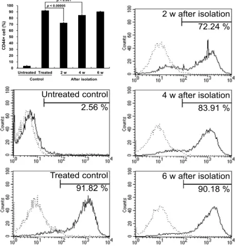
2.56 % Untreated control

91.82 % Treated control

72.24 % 2 w after isolation

90.18 % 6 w after isolation 83.91 % 4 w after isolation

0 10 20 30 40 50 60 70 80 90 100

Untreated Treated 2 w 4 w 6 w

CD44+ cell (%)

Control After isolation p < 0.00005

p < 0.001

Fig. 3. Resistance of colony forming cells in the presence of PMA to PMA-induced differentiation. The colonies formed by removal of PMA after exposure to 4 nM PMA for 3 days were isolated and expanded for the indicated times. After that, the cells were treated with 10 nM PMA for 3 days and determined with flow cytometer for the expression of CD44 surface marker of PMA-induced differentiation. Each bar represents the mean value±SD for three independent experiments. A representative flow cytometric analysis of three independent experiments was shown. Dot line indicated the isotype control.

K562 cells were treated with PMA at various concentrations for 2 weeks in soft agar, colony forming efficiency and col- ony size of K562 cells were significantly inhibited and at 1 nM or more concentration of PMA, colony was not formed (Fig. 1 A and C). However, at this high concentration of PMA approximately 40% of K562 cells still survived as single or double cells in soft agar (Fig. 1 B and C). These results demonstrate that induction of differentiation can inhibit col- ony-forming activity in soft agar, which is highly correlated with tumorigenicity [3, 6].

Recovery of colony forming activity of PMA-treated K562 cells after removal of PMA

Since there were single or double cells surviving in dor- mant state after exposure to PMA, it was examined if the PMA-treated dormant K562 cells would be able to regrow and form colony after removal of PMA. After exposure of K562 cells to various concentrations of PMA for various peri-

0 2 4 6 8 10 12 14 16 18

1 day 2 day 3 day 4 day 5 day

C o lo n y f o rm in g e ff ic ie n c y ( % )

1 nM 2 nM 4 nM

Fig. 2. Recovery of the colony forming activity of PMA-treated

K562 cells after removal of PMA. The K562 cells were

cultured in the presence of high concentrations of PMA

(1 nM, 2 nM, and 4 nM) in soft agar and after the in-

dicated periods PMA was washed out with complete

medium 12 times every one hour. After washing out of

PMA the cells were cultured further for additional 2

weeks for colony observation. The colonies larger than

100 μm in diameter were counted. Each bar represents

the mean value±SE for 3 independent experiments.

(5)

Fig. 4. Change in expression of CD44 surface marker in imatinib-resistant K562 variants during long-term drug free culture. After 4 month culture in drug-free medium, the surface expression level of CD44 was compared between the primary imatinib-re- sistant variants, including K562/R1, K562/R2, and K562/R3 cells (thin lines) and the secondary imatinib-resistant variants cultured in drug free condition, which were renamed as K562/R1’, K562/R2’, and K562/R3’ cells, respectively (thick lines).

Dot line indicated the isotype control.

ods, PMA was washed-out and colony formation was ob- served with MTT staining after 2 weeks (Fig. 2). When K562 cells were treated with 1 nM PMA, the colony forming activ- ity could be recovered by removal of PMA after 5 days. At concentrations of 2 and 4 nM PMA, the colony forming activ- ity could not be recovered by removal of PMA after treat- ment with PMA for 5 and 4 days, respectively. These results suggest that the single or double cells survived after ex- posure to PMA may be leukemia-maintaining cells, and the colony forming activity of leukemia-maintaining cells could be suppressed by induction of differentiation for long time with high concentration of PMA.

Resistance of colony forming cells in the presence of PMA to PMA-induced differentiation

If the cells, which can form colonies after exposure to PMA, are leukemia-maintaining cells, these cells should maintain the resistance to differentiation induction.

Therefore, colonies formed by removal of PMA after ex- posure to 4 nM PMA for 3 days were isolated and it was determined if the colony forming cells were resistant to PMA-induced differentiation (Fig. 3). When PMA was not treated, the colony forming cells showed an undifferentiated

phenotype, as untreated K562 control (data not shown).

When K562 cells were treated with 10 nM PMA for 3 days, more than 90% cells were CD44-positive, which is a PMA-in- duced megakaryocytic differentiation marker [26, 32, 40].

However, approximately 70% of the colony forming cells was positive for CD44 after treatment with 10 nM PMA for 3 days at 2 weeks after colony isolation. This differentiation rate was gradually increased and reached about 90% com- parable to the level of the PMA-treated K562 control cells at 6 weeks after colony isolation. This result indicated that K562 cells have leukemia-maintaining cells, which are re- sistant to PMA-induced differentiation and can recover the original phenotype.

Association of colony forming activity and Bcr-Abl expression in imatinib-resistant K562 variants

Since it was difficult to isolate and expand the leuke-

mia-maintaining cells from K562 cells after treatment with

PMA, we tried to identify the leukemia-maintaining cells

from the imatinib-resistant K562 variants including

K562/R1, K562/R2, and K562/R3 cells, which were isolated

from K562 cells in the presence of imatinib [28]. Since it is

well known that leukemic stem cells in CML are insensitive

(6)

202 생명과학회지 2013, Vol. 23. No. 2

to imatinib treatment [5, 20], and CML stem cells are quies- cent and undifferentiated, resulting in resistance to chemo- therapy [33], the imatinib-resistant K562 variants may have more leukemia-maintaining cells and consequently can form more colonies, compared with their parental K562 cells.

Unexpectedly, the imatinib-resistant K562 variants did not show any colony forming activity in soft agar (data not shown), and were CD44-positive (Fig. 4. thin line), explain- ing why the imatinib-resistant variants did not have a colony forming activity. After 4 month-culture in drug-free me- dium, the surface expression level of CD44 was decreased in comparison with the primary imatinib-resistant variants, including K562/R1, K562/R2, and K562/R3 cells (Fig. 4.

thick line), although almost all cells of imatinib-resistant var- iants remained still as CD44-positive. The secondary im- atinib-resistant variants cultured in drug-free condition were renamed as K562/R1’, K562/R2’, and K562/R3’ cells, respectively.

Since the expression level of CD44 was decreased in the secondary imatinib-resistant variants, it was determined if these cells could form colonies in soft agar. A few tiny colo- nies were observed in K562/R3’ cells (Fig. 5A). Since it has been shown that Bcr-Abl and Hsp70 were decreased and Bcl-2 was increased remarkably in the primary imatinib-re- sistant variants [28], there was a possibility that recovery of expression of the molecules might be associated with this reversion of clonogenicity, albeit very low. Therefore, the ti- ny colonies were picked up and cultured in drug-free me- dium (named as K562/R3’’ cells), and RT-PCR was per- formed to determine the level of Bcr-Abl, Hsp70 and Bcl-2.

Bcr-Abl was barely detected in K562/R3 and K562/R3’ cells, but reappeared in K562/R3’’ cells (Fig. 5B). In addition, the decreased Hsp70 and the increased Bcl-2 in K562/R3 cells were being increased and decreased in K562/R3’’ cells, respectively. These results were followed by an increased clonogenicity of K562/R3’’ cells, compared with K562/R3’

and K562/R3 cells (Fig. 5C).

Discussion

Currently cancer research is being focused on the molecular and cellular analysis of the cancer stem cells, and subsequent development of new therapeutic modalities targeting CSCs. It has been demonstrated that several properties of CSCs make them difficult to eradicate, for example, the activity of multiple drug resistance transporters

B) K562 R3 R3’ R3’’

b -Actin Hsp70 Bcl-2 Bcr-Abl

C)

p < 0.001

p < 0.01

Imatinib-resistant K562 variants K562 K562/R1' K562/R2' K562/R3'

C o lo n y f o rm in g e ff ic ie n c y ( % )

0.0 0.5 1.0 40.0 50.0 60.0

R3’’

A)

B) K562 R3 R3’ R3’’

b-Actin Hsp70 Bcl-2 Bcr-Abl

C)

p < 0.001

p < 0.01

R3’’

A)

B) K562 R3 R3’ R3’’

b-Actin Hsp70 Bcl-2 Bcr-Abl

C)

0 0.05 0.1 0.15 0.2 0.25 0.3 0.35 0.4

K562/R3 K562/R3' K562/R3''

C o lo n y f o rm in g e ff ic ie n c y ( % ) p < 0.001 p < 0.01

R3’’

A

B

C

Fig. 5. Slow recovery of colony forming efficiency and molec- ular changes in the imatinib-resistant K562 variants after long-term drug free culture. A) Clonogenicity of the sec- ondary imatinib-resistant variants cultured in drug-free condition, renamed as K562/R1’, K562/R2’, and K562/R3’ cells, respectively, was determined in soft agar as described in the ‘Materials and Methods’ section.

Each bar represents the means of triplicates±SD. For K562/R1’, K562/R2’, and K562/R3’ cells, colonies larger than 50 μm in diameter were counted. (Inset: a photo- graph of a small colony formed from K562/R3’ cells, from which K562/R3’’ cells were expanded.) B) The mRNA levels of Bcr-Abl, Hsp70 and Bcl-2, which were modulated in the imatinib-resistant K562/R3 variants, were analyzed with RT-PCR in K562/R3’ and K562/R3’’

cells. C) Clonogenicity of K562/R3, K562/R3’, and K562/R3’’ cells was determined in soft agar as described in the ‘Materials and Methods’ section. Each bar repre- sents the mean value±SE for 3 independent experiments.

The colonies larger than 50 μm in diameter were

counted.

(7)

[14], overexpression of antiapoptotic proteins [42], great abilities for DNA repair [4, 15, 39], and cell cycle restriction [43]. Since elimination of CSCs is not easy due to these properties, differentiation induction of cancer stem cells will be considered as one way to avoid these CSC problems. In this context, differentiation-inducing drugs such as BMPs (bone morphogenetic proteins), and histone deacetylase inhibitors as well as ATRA, have drawn growing attention [7, 31, 35].

It had been known that K562 cells have the capacity to express characteristics of erythrocytic, monocytic, and mega- karyocytic differentiation when exposed to various agents, and can be induced to differentiate into cells with monocytic and/or megakaryoblastic characteristics by PMA [41]. This differentiation process is characterized by changes in cell morphology, adhesiveness, and expression of mega- karyocytic markers as well as cell growth arrest [24, 38]. In the present study, when K562 cells were treated with PMA at high concentration for long time, the colony forming activ- ity of K562 cells could be completely suppressed, indicating that the clonogenicity, which is highly correlated with tu- morigenicity [3, 6], would be suppressed by inducing differ- entiation in chronic myelogenous leukemic cells.

It was demonstrated that BMP4 could reduce the tu- mor-initiating cell pool of glioblastoma multiformes (GBM) by triggering the Smad signalling cascade followed by a re- duction in proliferation, and increased expression of markers of neural differentiation, with no effect on cell viability, and the concomitant reduction in clonogenic ability, in the size of the CD133+ population and in the growth kinetics of GBM cells [35]. Although it has been shown that retinoids also induce astrocytic differentiation with down regulation of te- lomerase activity and enhance sensitivity to taxol for apopto- sis in glioblastoma cells [10], the effectiveness of ATRA has been studied in stem-like glioma cells (SLGCs) for the first time, demonstrating that ATRA-based differentiation can target the AC133/CD133-positive SLGCs population, induc- ing long-term antiangiogenic, antimigratory, antitumorigenic, proapoptotic, and therapy-sensitizing effects [8]. Therefore, these results highlighted the potential of differentiation ther- apy to target the stem-like cell population in cancers includ- ing leukemias as well as glioblastoma.

However, when PMA was removed after inducing mega- karyocytic differentiation of K562 cells, some clonogenic cells were reappeared, more easily with lower doses and shorter duration of PMA treatment. ATRA is able to induce com-

plete remission in about 90% of newly diagnosed patients with acute promyelocytic leukemia through in vivo differ- entiation of APL blasts. However, it cannot eliminate com- pletely the leukemic clone and to be more effective must be used in combination with anthracycline-based chemo- therapy [12, 34]. The reappearance of clonogenic cells may be probably due to resistance of leukemic stem cells to differentiation. To reduce the incidence of relapse of APL, maintenance treatment with ATRA, and possibly in combi- nation with low-dose chemotherapy is required [12]. Since it was demonstrated in the present study that the colony forming activity of K562 cells could be completely sup- pressed by treatment for long time with high concentration of PMA, long-term maintenance therapy with high dose of differentiation inducer would be required to prevent re- currence of cancers.

Previously, we reported that three imatinib-resistant K562/R1, R2 and R3 variants showed a gradual loss of Bcr-Abl at mRNA and protein levels, consequently un- responsiveness to imatinib [28]. Since it has been demon- strated that the primitive, quiescent, Philadelphia-positive stem cells from patients with chronic myeloid leukemia are insensitive to STI571 in vitro [20] and in vivo [5], and that human CML stem cells do not depend on BCR-ABL activity for survival and are thus not eliminated by imatinib therapy [9], it was expected that leukemic stem cells with self-renew- al capacity might be enriched in imatinib-resistant K562/R1, R2 and R3 variants and consequently colony forming effi- ciency would be higher, compared with their parental K562 cells. However, they did not have any colony forming activ- ity in soft agar and were CD44-positive, explaining the ab- sence of colony forming activity in the imatinib-resistant variants. We could isolate some tiny colonies from K562/R3’

cells after culture in drug-free medium for more than 4 months. This result suggests that the imatinib-resistant K562 variants contain the dormant leukemia-maintaining cells, al- though rare.

In response to therapy, cancer cells may reduce their de-

pendence on a particular hallmark capability, becoming

more dependent on another and consequently obtain a quite

different form of acquired drug resistance [23]. The im-

atinib-resistant variants of K562 showed several changes in

levels of apoptosis-modulating molecules such as an increase

in antiapoptotic Bcl-2 and Ku70 levels, and a decrease in

proapoptotic Bax and Hsp70, which might be involved in

the compensation for Bcr-Abl loss during the obtaining of

(8)

imatinib resistance [28]. After isolation of clonogenic cells from K562/R3 variant, the level of CD44 was being de- creased and Bcr-Abl was reappeared in K562/R3’’ cells, com- pared with primary imatinib-resistant variants. In addition, the decreased Hsp70 and the increased Bcl-2 also began to return slowly to the original level of K562 cells. Therefore, these results suggest that megakaryocytic differentiation and loss of Bcr-Abl of K562 cells induced by long-term culture in the presence of imatinib could be responsible for loss of clonogenicity of imatinib-resistant K562 variants, and the re- appearance of original phenotypes of K562 cells during cul- ture in imatinib-free medium may confer the clonogenicity to K562/R3’’ cells, again.

Whereas a number of differentiating agents have been studied over the past decades, only ATRA is being used to treat APL by causing immature blood cells to differentiate, and the capability of drugs to induce differentiation in CSCs has not yet been seriously considered [37]. From the present study including two models of differentiation of K562 cells, it could be suggested that differentiation therapy may be effective to eliminate cancer maintaining cells, when differ- entiating agents would be able to be used to get rid of cancer maintaining cells at high dose and for long time without side effects, albeit differentiation-resistant population might be cancer maintaining/initiating/stemcells.

Acknowledgement

This work was supported by a 2-Year Research Grant of Pusan National University.

References

1. Ades, L., Chevret, S., De Botton, S., Thomas, X., Dombret, H., Beve, B., Sanz, M., Guerci, A., Miguel, J. S., Dela Serna, J., Garo, C., Stoppa, A. M., Reman, O., Stamatoulas, A., Fey, M., Cahn, J. Y., Sotto, J. J., Bourhis, J. H., Parry, A., Chomienne, C., Degos, L. and Fenaux, P. 2005. Outcome of acute promyelocytic leukemia treated with all trans reti- noic acid and chemotherapy in elderly patients: the European group experience. Leukemia 19, 230-233.

2. Al Bahar, S., Pandita, R., Bavishi, K. and Kreze, O. 2004.

All-transretinoic acid and chemotherapy in the treatment of acute promyelocytic leukemia. Indian J Cancer 41, 125-128.

3. Alison, M. R., Islam, S. and Wright, N. A. 2010. Stem cells in cancer: instigators and propagators? J Cell Sci 123, 2357-2368.

4. Bao, S., Wu, Q., McLendon, R. E., Hao, Y., Shi, Q., Hjelmeland, A. B., Dewhirst, M. W., Bigner, D. D. and Rich,

J. N. 2006. Glioma stem cells promote radioresistance by preferential activation of the DNA damage response. Nature 444, 756-760.

5. Bhatia, R., Holtz, M., Niu, N., Gray, R., Snyder, D. S., Sawyers, C. L., Arber, D. A., Slovak, M. L. and Forman, S. J. 2003. Persistence of malignant hematopoietic progeni- tors in chronic myelogenous leukemia patients in complete cytogenetic remission following imatinib mesylate treatment. Blood 101, 4701-4707.

6. Bigner, S. H., Bullard, D. E., Pegram, C. N., Wikstrand, C.

J. and Bigner, D. D. 1981. Relationship of in vitro morpho- logic and growth characteristics of established human glio- ma-derived cell lines to their tumorigenicity in athymic nude mice. J Neuropathol Exp Neurol 40, 390-409.

7. Bots, M. and Johnstone, R. W. 2009. Rational combinations using HDAC inhibitors. Clin Cancer Res 15, 3970-3977.

8. Campos, B., Wan, F., Farhadi, M., Ernst, A., Zeppernick, F., Tagscherer, K. E., Ahmadi, R., Lohr, J., Dictus, C., Gdynia, G., Combs, S. E., Goidts, V., Helmke, B. M., Eckstein, V., Roth, W., Beckhove, P., Lichter, P., Unterberg, A., Radlwimmer, B. and Herold-Mende, C. 2010. Differentiation therapy exerts antitumor effects on stem-like glioma cells.

Clin Cancer Res 16, 2715-2728.

9. Corbin, A. S., Agarwal, A., Loriaux, M., Cortes, J., Deininger, M. W. and Druker, B. J. 2011. Human chronic myeloid leu- kemia stem cells are insensitive to imatinib despite in- hibition of BCR-ABL activity. J Clin Invest 121, 396-409.

10. Das, A., Banik, N. L. and Ray, S. K. 2008. Retinoids induced astrocytic differentiation with down regulation of telomer- ase activity and enhanced sensitivity to taxol for apoptosis in human glioblastoma T98G and U87MG cells. J Neurooncol 87, 9-22.

11. Dean, M., Fojo, T. and Bates, S. 2005. Tumour stem cells and drug resistance. Nat Rev Cancer 5, 275-284.

12. Degos, L. and Wang, Z. Y. 2001. All trans retinoic acid in acute promyelocytic leukemia. Oncogene 20, 7140-7145.

13. Diehn, M. and Clarke, M. F. 2006. Cancer stem cells and radiotherapy: new insights into tumor radioresistance. J Natl Cancer Inst 98, 1755-1757.

14. Elliott, A., Adams, J. and Al-Hajj, M. 2010. The ABCs of cancer stem cell drug resistance. IDrugs 13, 632-635.

15. Facchino, S., Abdouh, M., Chatoo, W. and Bernier, G. 2010.

BMI1 confers radioresistance to normal and cancerous neu- ral stem cells through recruitment of the DNA damage re- sponse machinery. J Neurosci 30, 10096-10111.

16. Fan, X., Matsui, W., Khaki, L., Stearns, D., Chun, J., Li, Y.

M. and Eberhart, C. G. 2006. Notch pathway inhibition de- pletes stem-like cells and blocks engraftment in embryonal brain tumors. Cancer Res 66, 7445-7452.

17. Fenaux, P., Chastang, C., Chevret, S., Sanz, M., Dombret,

H., Archimbaud, E., Fey, M., Rayon, C., Huguet, F., Sotto,

J. J., Gardin, C., Makhoul, P. C., Travade, P., Solary, E.,

Fegueux, N., Bordessoule, D., Miguel, J. S., Link, H.,

Desablens, B., Stamatoullas, A., Deconinck, E., Maloisel, F.,

Castaigne, S., Preudhomme, C. and Degos, L. 1999. A

randomized comparison of all transretinoic acid (ATRA) fol-

(9)

lowed by chemotherapy and ATRA plus chemotherapy and the role of maintenance therapy in newly diagnosed acute promyelocytic leukemia. The European APL Group. Blood 94, 1192-1200.

18. Frank, N. Y., Schatton, T. and Frank, M. H. 2010. The ther- apeutic promise of the cancer stem cell concept. J Clin Invest 120, 41-50.

19. Garvalov, B. K. and Acker, T. 2011. Cancer stem cells: a new framework for the design of tumor therapies. J Mol Med (Berl) 89, 95-107.

20. Graham, S. M., Jorgensen, H. G., Allan, E., Pearson, C., Alcorn, M. J., Richmond, L. and Holyoake, T. L. 2002.

Primitive, quiescent, Philadelphia-positive stem cells from patients with chronic myeloid leukemia are insensitive to STI571 in vitro. Blood 99, 319-325.

21. Gupta, P. B., Onder, T. T., Jiang, G., Tao, K., Kuperwasser, C., Weinberg, R. A. and Lander, E. S. 2009. Identification of selective inhibitors of cancer stem cells by high-through- put screening. Cell 138, 645-659.

22. Gupta, V., Yi, Q. L., Brandwein, J., Lipton, J. H., Messner, H. A., Schuh, A. C., Wells, R. A. and Minden, M. D. 2005.

Role of all-trans-retinoic acid (ATRA) in the consolidation therapy of acute promyelocytic leukaemia (APL). Leuk Res 29, 113-114.

23. Hanahan, D. and Weinberg, R. A. 2011. Hallmarks of cancer:

the next generation. Cell 144, 646-674.

24. Hocevar, B. A., Morrow, D. M., Tykocinski, M. L. and Fields, A. P. 1992. Protein kinase C isotypes in human eryth- roleukemia cell proliferation and differentiation. J Cell Sci 101 ( Pt 3), 671-679.

25. Iovino, F., Meraviglia, S., Spina, M., Orlando, V., Saladino, V., Dieli, F., Stassi, G. and Todaro, M. 2010. Immunotherapy targeting colon cancer stem cells. Immunotherapy 3, 97-106.

26. Jacquel, A., Herrant, M., Defamie, V., Belhacene, N., Colosetti, P., Marchetti, S., Legros, L., Deckert, M., Mari, B., Cassuto, J. P., Hofman, P. and Auberger, P. 2006. A survey of the signaling pathways involved in megakaryocytic dif- ferentiation of the human K562 leukemia cell line by molec- ular and c-DNA array analysis. Oncogene 25, 781-794.

27. Kim, K. W., Kim, S. H., Lee, E. Y., Kim, N. D., Kang, H.

S., Kim, H. D., Chung, B. S. and Kang, C. D. 2001.

Extracellular signal-regulated kinase/90-KDA ribosomal S6 kinase/nuclear factor-kappa B pathway mediates phorbol 12-myristate 13-acetate-induced megakaryocytic differ- entiation of K562 cells. J Biol Chem 276, 13186-13191.

28. Lee, S. M., Bae, J. H., Kim, M. J., Lee, H. S., Lee, M. K., Chung, B. S., Kim, D. W., Kang, C. D. and Kim, S. H. 2007.

Bcr-Abl-independent imatinib-resistant K562 cells show aberrant protein acetylation and increased sensitivity to his- tone deacetylase inhibitors. J Pharmacol Exp Ther 322, 1084-1092.

29. Lotem, J. and Sachs, L. 2006. Epigenetics and the plasticity of differentiation in normal and cancer stem cells. Oncogene 25, 7663-7672.

30. Lozzio, B. B., Lozzio, C. B., Bamberger, E. G. and Feliu, A.

S. 1981. A multipotential leukemia cell line (K-562) of hu-

man origin. Proc Soc Exp Biol Med 166, 546-550.

31. Massard, C., Deutsch, E. and Soria, J. C. 2006. Tumour stem cell-targeted treatment: elimination or differentiation. Ann Oncol 17, 1620-1624.

32. Moll, J., Khaldoyanidi, S., Sleeman, J. P., Achtnich, M., Preuss, I., Ponta, H. and Herrlich, P. 1998. Two different functions for CD44 proteins in human myelopoiesis. J Clin Invest 102, 1024-1034.

33. Naka, K., Hoshii, T. and Hirao, A. 2010. Novel therapeutic approach to eradicate tyrosine kinase inhibitor resistant chronic myeloid leukemia stem cells. Cancer Sci 101, 1577-1581.

34. Ohno, R., Asou, N. and Ohnishi, K. 2003. Treatment of acute promyelocytic leukemia: strategy toward further increase of cure rate. Leukemia 17, 1454-1463.

35. Piccirillo, S. G., Reynolds, B. A., Zanetti, N., Lamorte, G., Binda, E., Broggi, G., Brem, H., Olivi, A., Dimeco, F. and Vescovi, A. L. 2006. Bone morphogenetic proteins inhibit the tumorigenic potential of human brain tumour-initiating cells. Nature 444, 761-765.

36. Sanz, M. A., Martin, G., Gonzalez, M., Leon, A., Rayon, C., Rivas, C., Colomer, D., Amutio, E., Capote, F. J., Milone, G. A., De La Serna, J., Roman, J., Barragan, E., Bergua, J., Escoda, L., Parody, R., Negri, S., Calasanz, M. J. and Bolufer, P. 2004. Risk-adapted treatment of acute promyelocytic leu- kemia with all-trans-retinoic acid and anthracycline mono- chemotherapy: a multicenter study by the PETHEMA group. Blood 103, 1237-1243.

37. Scatena, R., Bottoni, P., Pontoglio, A. and Giardina, B. 2011.

Cancer stem cells: the development of new cancer therapeutics. Expert Opin Biol Ther 11, 875-892.

38. Shelly, C., Petruzzelli, L. and Herrera, R. 2000. K562 cells resistant to phorbol 12-myristate 13-acetate-induced growth arrest: dissociation of mitogen-activated protein kinase acti- vation and Egr-1 expression from megakaryocyte differentiation. Cell Growth Differ 11, 501-506.

39. Tamura, K., Aoyagi, M., Wakimoto, H., Ando, N., Nariai, T., Yamamoto, M. and Ohno, K. 2010. Accumulation of CD133-positive glioma cells after high-dose irradiation by Gamma Knife surgery plus external beam radiation. J Neurosurg 113, 310-318.

40. Tringali, C., Lupo, B., Cirillo, F., Papini, N., Anastasia, L., Lamorte, G., Colombi, P., Bresciani, R., Monti, E., Tettamanti, G. and Venerando, B. 2009. Silencing of mem- brane-associated sialidase Neu3 diminishes apoptosis resist- ance and triggers megakaryocytic differentiation of chronic myeloid leukemic cells K562 through the increase of gan- glioside GM3. Cell Death Differ 16, 164-174.

41. Tsiftsoglou, A. S., Pappas, I. S. and Vizirianakis, I. S. 2003.

Mechanisms involved in the induced differentiation of leu- kemia cells. Pharmacol Ther 100, 257-290.

42. van Stijn, A., van der Pol, M. A., Kok, A., Bontje, P. M.,

Roemen, G. M., Beelen, R. H., Ossenkoppele, G. J. and

Schuurhuis, G. J. 2003. Differences between the CD34+ and

CD34- blast compartments in apoptosis resistance in acute

myeloid leukemia. Haematologica 88, 497-508.

(10)

초록:만성 골수성 백혈병 K562세포의 분화 내성 분획에서 백혈병 유지 세포의 동정 이홍래 1 ․김미주 1 ․하가희 1 ․김소중 2 ․김선희 1 *․강치덕 1 *

( 1 부산대학교 의학전문대학원 생화학교실, 2 부산대학교 의학전문대학원 의사과학자 복합학위과정)

본 연구에서는 K562 만성 골수성 백혈병 세포를 이용하여, 분화 유도에 의해 암 유지/개시 세포의 자기 재생 능력이 소실되는 지를 조사하였다. K562 세포의 집락(colony) 형성 능력은 PMA 처리에 의하여 현저히 억제되었 고, 1 nM 이상의 PMA 처리시에는 집락이 형성되지 않았으나, 약 40%의 세포는 여전히 연한천(soft agar)에서 살아 있었다. PMA 4 nM을 3일간 처리하고 제거한 후 분리한 집락 형성 세포에 다시 10 nM PMA를 3일간 처리 하였을 때, 약 70% 정도의 세포가 분화되었고, 6주 후에 PMA를 처리하였을 때는 분화율이 약 90%로 K562 모세 포에 PMA를 처리한 수준에 도달하였다. 한편, imatinib-내성 K562 변종 세포들은 연한천에서 집락을 형성하지 않았으며, 대부분의 세포가 CD44 양성이었다. Imatinib 무첨가 배지에서 4개월 배양 후, 이 세포들의 표면 CD44 발현량은 감소하였고, K562/R3 imatinib-내성 변종 세포에서는 연한천에서 작은 집락이 형성되었다. 이 세포에서 는 imatinib-내성 변종 세포에서 소실되었던 Bcr-Abl이 다시 발현되기 시작하였고, 다른 표현형들도 부분적으로 회복되었다. 이러한 결과는 백혈병 유지 세포가 분화에 내성을 나타내는 세포이며, 분화 유도제를 오랜 기간 동안 고농도로 처리할 수 있다면 백혈병 줄기 세포를 제거하기 위한 분화 요법이 백혈병 치료에 적용될 수 있음을 시사하였다.

43. Viale, A., De Franco, F., Orleth, A., Cambiaghi, V., Giuliani, V., Bossi, D., Ronchini, C., Ronzoni, S., Muradore, I., Monestiroli, S., Gobbi, A., Alcalay, M., Minucci, S. and Pelicci, P. G. 2009. Cell-cycle restriction limits DNA damage and maintains self-renewal of leukaemia stem cells. Nature 457, 51-56.

44. Wicha, M. S., Liu, S. and Dontu, G. 2006. Cancer stem cells:

an old idea--a paradigm shift. Cancer Res 66, 1883-1890; dis- cussion 1895-1886.

45. Yu, F., Yao, H., Zhu, P., Zhang, X., Pan, Q., Gong, C., Huang, Y., Hu, X., Su, F., Lieberman, J. and Song, E. 2007. let-7 regu- lates self renewal and tumorigenicity of breast cancer cells.

Cell 131, 1109-1123.

46. Zhou, B. B., Zhang, H., Damelin, M., Geles, K. G., Grindley,

J. C. and Dirks, P. B. 2009. Tumour-initiating cells: chal-

lenges and opportunities for anticancer drug discovery. Nat

Rev Drug Discov 8, 806-823.

수치

Fig. 1. Inhibition of soft agar colony forming efficiency of K562 cells by treatment with PMA
Fig. 2. Recovery of the colony forming activity of PMA-treated K562 cells after removal of PMA
Fig. 4. Change in expression of CD44 surface marker in imatinib-resistant K562 variants during long-term drug free culture
Fig. 5. Slow recovery of colony forming efficiency and molec- molec-ular changes in the imatinib-resistant K562 variants after long-term drug free culture

참조

관련 문서