Inhibition of Neurogenesis of Subventricular Zone Neural Stem Cells by 5-ethynyl-2’-deoxyuridine (EdU)
Ki-Youb Park1*, Hyun-Chang Oh1, Ji-Yong Lee1 and Man Su Kim2
1Korea Science Academy of KAIST 105-47 Baegyanggwanmun-ro, Busanjin-Gu, Busan 614-100, Korea
2College of Pharmacy, Inje University, Gimhae 50834, Korea
Received January 24, 2017 /Revised February 27, 2017 /Accepted February 27, 2017
In the subventricular zone (SVZ) and the subgranular zone of the brain, neurogenesis occurs through- out one’s lifespan. Neural stem cells (NSCs) in these regions divide to maintain their stem cell pools as well as differentiate into neurons and glial cells. To monitor cell division, a thymidine analogue such as 5-ethynyl-2’-deoxyuridine (EdU) has been used. In some cases, EdU was applied to label new- ly born neurons. Here, we report about the effects of EdU on the proliferation and differentiation of NSCs cultured from mouse SVZ. First, when NSCs were cultured in a proliferation medium contain- ing EdU for 24 hr, they did not generate any neurons under the following differentiation conditions.
When EdU was applied to the proliferating NSCs for 1 hr prior to differentiation, neurogenesis was still substantially reduced. Second, EdU decreased cell proliferation of NSCs in dose- and time-de- pendent manners. Finally, EdU inhibited differentiation into oligodendrocyte lineage, while the num- ber of glial fibrillary acidic protein (GFAP)-positive astrocytes increased. To our knowledge, these findings are the first to show the effects of EdU on the differentiation of SVZ NSCs and suggest that cell division is necessary for differentiation into neurons and oligodendrocytes.
Key words : Cell proliferation, 5-ethynyl-2’-deoxyuridine (EdU), neural stem cells (NSCs), neuronal differentiation, subventricular zone (SVZ)
*Corresponding author
*Tel : +82-51-606-2225, Fax : +82-51-606-2226
*E-mail : [email protected]
This is an Open-Access article distributed under the terms of the Creative Commons Attribution Non-Commercial License (http://creativecommons.org/licenses/by-nc/3.0) which permits unrestricted non-commercial use, distribution, and reproduction in any medium, provided the original work is properly cited.
Journal of Life Science 2017 Vol. 27. No. 6. 623~631 DOI : https://doi.org/10.5352/JLS.2017.27.6.623
Introduction
Cell proliferation can be detected in many ways, among which detecting incorporation of nucleoside analogues into the DNA has been frequently used to examine cell division.
Among nucleoside analogues, 5-bromo-2’-deoxyuridine (BrdU) has been mostly used as a thymidine analogue.
However, detection of incorporated BrdU requires denatu- ration of the DNA in the cells so that antibodies against BrdU can get an access to the DNA [2]. A newly developed thymidine analogue, EdU, contains an alkyne group that can react with a fluorescent-tagged azide through so called
‘click chemistry’ [21]. This method does not require denatu- ration process, which preserves cell structures during EdU detection. Also, the intensity of fluorescence from the click chemistry is more linearly correlated with the level of EdU incorporated than antibody-based detection of BrdU.
Thanks to these advantages, EdU has been used to label S-phase of the cell cycle in various biological experiments since its emergence [2]. Yet, cytotoxicity of EdU has been reported in many previous studies. Inhibition of cell pro- liferation by EdU has been shown in glioblastoma cells, hu- man breast cancer cell lines and other cancer cell line [7, 13, 20, 24]. Especially, EdU incorporation induced cell cycle arrest in mouse embryonic stem cells, while BrdU did not [13]. EdU inhibited thymidylate synthetase by competing against 2’-deoxyuridylate [6]. Even though EdU has been used to label proliferating cells in SVZ and subgranular zone in hippocampus, [12, 17, 23], direct effects of EdU on NSCs have not been examined.
SVZ in the brain is the region where life-long neuro- genesis occurs. It contains multiple types of cells such as ependymal cells, type B cells, transit-amplifying progenitor (TAP) type C cells and type A cells (neuroblasts) [8]. Type B cells can exist as both GFAP+EGFR (Epidermal Grwoth Factor Receptor)-Nestin- quiescent stem cells and GFAP+ EGFR+Nestin+ activated stem cells [4]. Activated type B cells give rise to type C cells and type C cells undergo multiple cell divisions and finally commit to neuronal lineage by be- coming type A neuroblasts [9]. Live imaging revealed slow- dividing NSCs and fast-dividing NSCs when they were cul-
tured directly from SVZ and maintained in serum-and growth factor-free medium [5]. Half of the fast-dividing NSCs divided once before becoming post-mitotic neuro- blasts. The other half of them divided 2 to 5 times before becoming neuroblasts. Consistently, antimitotic drug cyto- sine-beta-D-arabinofuranoside (Ara-C) infusion to the ani- mals killed off proliferating type A and type C cells of SVZ [9, 11, 19]. However, whether or not cell division is necessary and essential for neuronal differentiation has not been di- rectly tested in vitro.
Initially, we intended to map the fate of SVZ NSCs using EdU in our in vitro culture system. Instead, to our great sur- prise, 24-hr treatment of EdU during cell proliferation com- pletely eliminated neurogenesis during the following differ- entiation period. This was accompanied by reduction of total cell numbers by EdU treatment. Short-term treatment such as 1-hr treatment of EdU also greatly inhibited neurogenesis during the following differentiation. We think these might not be due to nonspecific and general cytotoxic effects, be- cause incubation of proliferating NSCs in EdU-containing medium did not kill off cells, but rather seemed to slow down overall cell proliferation. When numbers of cells were counted at different time points, EdU did not affect cell pro- liferation at 6 and 24 hr, but reduced the number of cells after 48 hr. Finally, no Olig2-positive cells were found after differentiation in EdU-treated samples, while the number of GFAP-positive astrocytes was increased. Our report showed that EdU inhibited both neurogenesis and oligodendro- genesis. All these results suggest that cell proliferation is in- deed required for differentiation of NSCs to neurons and oligodendrocytes.
Materials and Methods
Mouse SVZ NSC culture
To obtain mouse SVZ NSCs, postnatal mice (5 to 7 day- old CD1 (ICR) mice from Orient Bio, Sungnam, Korea) were euthanized using carbon dioxide. Then, brains were taken out from the mice to cut out tissues of SVZ. Preparation of cells from the SVZ tissues was done in the same way as we reported previously [18]. This animal work was ap- proved by Inje University Animal Care and Use Committee (approval ID number: 2016-011) and all the procedures were performed under the national guidelines for animal care and use of laboratory animals.
Cultured cells were grown in a proliferation medium, N5
medium, which contains DMEM/F12-GlutaMAXTM supple- ment (Gibco, ThermoFisher, Waltham, MA, USA), 5% fetal bovine serum (GenDEPOT, Texas, USA), N2 supplement (Gibco, ThermoFisher, Waltham, MA, USA), 35 μg/ml bo- vine pituitary extract (Gibco, ThermoFisher, Waltham, MA, USA), 20 ng/ml epidermal growth factor (EGF, Invitrogen, ThermoFisher, Waltham, MA, USA), 20 ng/ml basic fibro- blast growth factor (bFGF, Gibco, ThermoFisher, Waltham, MA, USA), and antibiotic/antimycotic (Gibco, ThermoFisher, Waltham, MA, USA). NSCs were maintained in N5 medium at 37°C with 5% carbon dioxide. Cells were passaged every 2 or 3 days depending on confluency. All the SVZ NSCs used in this study were passaged 5 to 7 times before being used for actual experiments.
Neuronal differentiation of cultured SVZ NSCs Proliferating NSCs were plated on a laminin-coated 8-well Lab-Tek CC2 chamber slide (Nunc, ThermoFisher, Waltham, MA, USA) at 1 day prior to differentiation. For laminin coat- ing, 5 µg/ml laminin (Invitrogen, ThermoFisher, Waltham, MA, USA) dissolved in phosphate-buffered saline (PBS) was added to cover the surface of the chamber slide and in- cubated for 4 hr to overnight at 37°C. Before plating, the chamber slide was rinsed with PBS. To start differentiation, cells were briefly rinsed with N6 medium that is the same medium as N5, but lacking EGF, bFGF, and fetal bovine serum. Then, cells were incubated in fresh N6 medium for 5 days before fixation.
EdU incorporation and detection
For EdU incorporation and detection, all the procedures followed manufacture’s instructions by using Click-iT Plus EdU Imaging Kit (Invitrogen, ThermoFisher, Waltham, MA, USA). Proliferating SVZ NSCs were incubated with EdU dis- solved in N5 medium for 1 day and then, with fresh N6 medium to initiate differentiation. For the 1-hr pulse label- ing, EdU-containing N5 medium was added to the pro- liferating NSCs at 1 hr prior to medium switch to N6. Cells were fixed in 4% paraform aldehyde (Sigma, St. Louis, MO, USA) for 15 min, rinsed with 3% bovine serum albumin (BSA) in PBS twice, incubated in blocking solution with 10%
goat serum (Cell Signaling, Danvers, MA, USA) and 0.1~0.3%
triton X-100 (Sigma, St. Louis, MO, USA) in PBS. Finally, EdU detection procedure was done following manu- facturer’s instructions. Alexa Fluor® 594 picolyl azide was added to make a reaction cocktail solution that was added
A
B C D
E
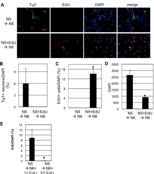
Fig. 1. Neurogenesis of SVZ NSCs was blocked by EdU. A. NSCs were maintained in proliferating medium (N5) in the absence of EdU (upper lane) or in the presence of 5 μM EdU (lower lane) for 1 day before differentiation. To initiate neurogenesis, NSCs were incubated in the differentiation medium (N6) for 5 days. Immunocytochemistry was done to detect neuronal specific marker (Tuj1) and DAPI for nucleus. Also, EdU incorporation into the cells was detected. Scale bar=20 μm. B.
Tuj1-positive neurons were counted and divided by total number of DAPI. C. EdU-positive cells were counted and divided by total number of DAPI. D. Total number of DAPI counted is shown. E. Five μM EdU was added for 24 hr at the 1st day of differentiation (N5àN6+1st EdU) or 2nd day of differentiation (N5àN6+2nd EdU). B-E. In each condition, average and standard deviation from three different wells of cells are shown (*p<0.01, Student’s T-test).
to the cells for 30 min in dark. For all the rinsing, 3% BSA in PBS was used.
Immunocytochemistry and imaging
To stain cells using antibodies, the same method as pre- viously reported [18] was performed. Primary and secon- dary antibodies and their dilutions were as followings:
mouse anti-Tuj1 (Novex, ThermoFisher, Waltham, MA, USA) at 1:500 dilution, mouse anti-GFAP (Millipore, Billerica, MA, USA) at 1:500 dilution, rabbit anti-Olig2 (Millipore, Billerica, MA, USA) at 1:500 dilution (all primary antibodies were diluted in blocking solution), Alexa-488–conjugated
anti-mouse, and Alexa-594–conjugated anti-rabbit secon- dary antibodies (1:500 in PBS; both from Jackson Immuno Research, West Grove, PA, USA). For nuclear staining, cells were incubated with DAPI (4',6-diamidino-2-phenylindole, Sigma, St. Louis, MO, USA) at 1:1,000 dilution. In the case of EdU treatment, EdU detection preceded the incubation of cells with primary antibodies.
Images were taken using a fluorescence microscope (Olympus, Tokyo, Japan). To count cells, 4 to 5 non-over- lapping images were taken per well. The number of DAPI was counted using the cell count macro in iSolution software (Olympus, Tokyo, Japan). Others such as Tuj1, GFAP, Olig2,
A B
Fig. 2. The longer treatment of EdU during proliferation of SVZ NSCs resulted in the less neurogenesis. A. NSCs were incubated in N5 medium and 5 μM EdU was added to the N5 at 1 or 24 hr before starting differentiation using N6 medium. Negative control without EdU is 0 hr time-point condition. At 5 days of differentiation, cells were fixed for immunocytochemistry and EdU detection. Tuj1-positive neurons were counted and divided by total number of DAPI. B. Total number of DAPI counted for each condition is shown. A, B. Data are average and standard deviation from three different wells of cells for each condition (*p<0.01, Student’s T-test).
or EdU-positive cells were manually counted from 3 wells for each condition.
Cell proliferation assay
The same numbers of NSCs were plated onto dish in N5 medium containing different concentrations of EdU, 0, 5, 15, or 45 μM. At 1 day of incubation in EdU, cells were trypsi- nized and harvested for cell counting manually using hematocytometer. For the time-point experiment, cells were kept in N5 containing 0, 5, or 15 μM of EdU. Then, at 6, 24, and 48 hr of EdU treatment, cells were harvested and the live cells were counted using EVE automatic cell counter (NanoEnTek, Seoul, Korea).
Results
Inhibition of neurogenesis by EdU
In general, proliferation and differentiation of stem cells have been regarded as separate processes. If cells are under- going proliferation, they are not differentiating and vice versa. However, cells divide during neuronal differentiation of SVZ NSCs. In SVZ, NSCs produce transit amplifying cells which divide multiple rounds and later become migratory neuroblasts [15]. To examine relationship between pro- liferation and differentiation more closely, we used EdU, a thymindine analogue, which incorporates into DNA during S-phase. Cultured SVZ NSCs were maintained in N5 me- dium in the absence or presence of 5 μM EdU for 1 day and then, medium was switched to N6 medium to initiate
neuronal differentiation. After 5 days of differentiation, cells were fixed for EdU detection and immunocytochemistry. To our great surprise, NSCs treated with EdU failed to generate any neurons, while about 6 % of cells were Tuj1+ neurons in NSCs without EdU treatement (Fig. 1A, Fig. 1B).
Approximately 14 % of total cells were EdU+ in NSCs treat- ed with EdU (Fig. 1A, Fig. 1C). Also, EdU treatment de- creased total number of cells by 65%(Fig. 1D). Upon in- corporation into the DNA, EdU probably blocked further process of cell division by holding cell cycle progression.
This interruption might have been involved in complete loss of neurogenesis (Fig. 1A - Fig. 1D). When EdU was added as a pulse for the first or second day of N6 medium, approx- imately 9% or 0% of total cells were EdU-positive, re- spectively (Fig. 1E). This indicates that cells still divide dur- ing the first 24 hr of differentiation and then, stop dividing afterwards.
To further study about effect of EdU on neuronal differ- entiation, shorter incubation with EdU was performed. SVZ NSCs were incubated in N5 medium containing 5 μM EdU for 1 or 24 hr prior to differentiation. Consistently to the previous result, 24-hr EdU treatment abolished neuro- genesis, while 53% of the total cells were Tuj1+ neurons in NSCs without EdU treatment (Fig. 2A). When NSCs were treated with EdU for 1 hr, 19% of the total cells were Tuj1+
neurons. These trends correlated with the total number of cells counted by DAPI staining (Fig. 2B). All these data sug- gest that uninterrupted cell division process upon the cue of differentiation might be required to produce neurons.
A B
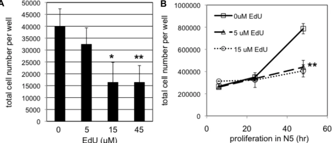
Fig. 3. Cell proliferation was inhibited by EdU in dose- and time-dependent manners. A. Same number of SVZ NSCs were plated onto culture dish. Then, they were incubated in N5 medium containing different concentration of EdU (0, 5, 15, or 45 μM).
At 24 hr, number of cells was counted. Data are average and standard deviation from four different wells of cells. B. NSCs were incubated in N5 medium containing 5 or 15 μM EdU. As a negative control, NSCs were grown in N5 medium in the absence of EdU (0 μM EdU). At 6, 24, 48 hr post EdU treatment, number of cells were counted. Data are average and standard deviation from three different wells of cells. A, B. Statistical significance compared with the negative control (0 μM) is shown (*p<0.05, **p<0.01, Student’s T-test).
Inhibition of cell proliferation by EdU
According to DAPI counting in figures 1 and 2, the num- ber of cells decreased by EdU treatment. To examine the effect of EdU on cell proliferation, we applied different doses of EdU for various time periods. The same numbers of pro- liferating SVZ NSCs were treated with 0, 5, 15, and 45 μM of EdU for 1 day and numbers of live cells were counted.
While 5 μM of EdU did not decrease the number of cell significantly, 15 or 45 μM of EdU decreased numbers of cells almost by half (Fig. 3A). Separate groups of proliferating NSCs were incubated in 0, 5, or 15 μM EdU-containing N5 medium and total numbers of cells were counted at 6, 24, and 48 hr post EdU treatment. After 6- or 24-hr treatment, numbers of cells were not significantly different (p>0.05) in all conditions (Fig. 3B). After 48-hr treatment, NSCs treated with 5 or 15 μM EdU were significantly less than control cells (**p<0.01). Since EdU did not kill off NSCs right after treatment, but rather it took time to reduce cell numbers, we think that EdU had held cell cycle progression in NSCs as previously reported in other cell types instead of having non-specific cytotoxic effects [13].
Effects of EdU on gliogenesis
Next, we investigated whether EdU has any effects on differentiation of glial cells such as astrocytes and oligoden- drocytes. As in Fig. 1, SVZ NSCs were incubated in EdU-containing N5 medium for 1 day before the medium
switch to N6 for differentiation. After 5 days of differ- entiation, cells were fixed for immunocytochemisty to stain GFAP, a marker for astrocytes, and Olig2, a maker for im- mature oligodendrocytes (Fig. 4A). GFAP+ cells were 6% of the total cells in no EdU condition, while GFAP+ cells were 17% in EdU-treated NSCs (Fig. 4B). More strikingly, Olig2+
cells were very few (0.2% of the total cells) in EdU-treated NSCs, while Olig2+ cells were 5% in no EdU condition (Fig.
4C). Total numbers of cells were significantly less in EdU-treated condition as expected (Fig. 4D). Therefore, per- turbed cell cycle by EdU seemed to inhibit generation of oligodendrotye lineage cells from SVZ NSCs, but increase differentiation to astrocytes.
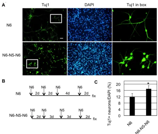
We think at least some of GFAP-positive cells in figure 4 might be quiescent NSCs, because (1) quiescent NSCs are GFAP-positive and resistant to antimitotic drug, EdU in our case [4, 10] and (2) addition of serum and growth factors in the form of N5 medium during differentiation increased neurogenesis (Fig. 5). As a control, cultured mouse SVZ NSCs were treated with differentiation medium (N6) for 10 days and Tuj1-positive neurons were detected in ~12% of the total cells (Fig. 5A, Fig. 5B, upper panels). As an ex- perimental group, cells were treated with N6 medium for 5 days, then, incubated in N5 proliferation medium for 3 days before the final incubation in N6 for 2 days (Fig. 5A, Fig. 5B, lower panels). This medium switch with N5 in the middle of differentiation resulted in increased neurogenesis
A
B C D
Fig. 4. Effects of EdU on glial differentiation of SVZ NSCs. A. Cultured NSCs were incubated in N5 without (upper pannel) or with 5 μM EdU (lower pannel) for 1 day. Then, medium was switched to N6 to start differentiation. After 5 days of differ- entiation, cells were fixed for immunocytochemistry to detect astrocytes (GFAP) and oligodendrocytes (Olig2). DAPI was used for nuclear staining. Scale bar=20 μm. B-D. GFAP-positive cells (B) and Olig2-positive cells (C) were counted and divided by total number of DAPI. Total number of DAPI is shown in D. Data are average and standard deviation from three different wells of cells (*p<0.01, Student’s T-test).
by 44% (Fig. 5C). These results are consistent with the pre- vious report that showed an increased level of neurogenic transcription factor Dlx2 transcript by similar medium switch paradigm [17]. Since many neurons in this ex- perimental group had shorter neuronal processes than in the control group, they might be immature neurons possibly dif- ferentiated from the final medium switch from N5 to N6 (Fig. 5A). Therefore, it is tempting to think that serum and growth factors activated quiescent cells to undergo neuro- genesis, which is supported by previous report that showed re-entering of cell cycle of quiescent NSCs by EGF/FGF2 treatment [5].
Discussion
Since its development, EdU has been widely used to mon- itor cell proliferation as a final readout of the experiments.
Also, EdU has been used to trace cell fate of NSCs in vivo [12, 17]. According to our results and others’, EdU seems to inhibit cell proliferation possibly by arresting cell cycle at G2/M phase [7, 13]. Interestingly, another thymidine ana- logue and more widely used BrdU was also reported to in- duce cell cycle arrest in embryonic NSCs [22]. Therefore,
careful analysis of lineage tracing and cell fate mapping us- ing EdU and other nucleoside analogue is needed. Due to this inhibition of cell proliferation, the possibility of using EdU as an anti-cancer drug has been raised [16, 20, 24].
However, still other side effects including loss of neuro- genesis as shown in this present study and disrupted DNA function and stability [1, 6] call for careful therapeutic appli- cations of EdU.
In this present study, we showed that pre-treatment of EdU for 1 day prior to differentiation completely inhibited neurogenesis and oligodendrogenesis of SVZ NSCs. These suggest that cell division is necessary in differentiation to both neurons and oligodendrocytes, which have been specu- lated in the previous review publication [14]. According to the review, type B cells generate type C cells and the type C cells become neurogenic intermediate progenitor cells (nIPCs) or oligodendrocytic intermediate progenitor cells (oIPCs). The review focused more on neurogenesis, indicat- ing cell division of nIPCs without mentioning of cell division of oIPCs. However, our present results suggest that oIPCs might undergo cell division as well. Still, it is not known why cell divisions are needed before fully committing to the specific cell fate. The most possible reason might be to in-
A
B C
Fig. 5. Neurogenesis of SVZ NSCs was increased by proliferation medium during differentiation. A. In the case of N6 (upper pannel), NSCs were incubated in N6 medium for 10 days and medium was changed with fresh N6 medium every 2 or 4 days during the differentiation, as indicated in B (upper pannel). In the case of N6-N5-N6 (lower pannel), medium was changed as shown in the schematic in B (lower pannel). Scale bar=50 μm. The two images on the far right are zoomed ones of the boxed area. B. Experimental scheme is shown. At the end of the medium switch, cells were fixed for immunocytochemistry as in A. C. Tuj1+ neurons were counted and divided by total number of cells stained in DAPI. Average and standard deviation from three different wells of cells are shown (*p<0.05, Student’s T-test).
crease the number of finally differentiated cells. We spec- ulate that an unknown process occurring during cell divi- sions might direct cell differentiation, because this hypoth- esis explains why the lack of cell division impairs cellular differentiation.
Type B NSCs exist in two flavors, activated (aNSCs) and quiescent (qNSCs) states [3]. Both aNSCs and qNSCs express astroctye marker, GFAP, but only aNSCs express EGFR that can transmit extrinsic mitogenic signals. In our study, EdU seemed to eliminate aNSCs leading to a loss of neurogenesis, but qNSCs might have survived EdU treatment. That might be the reason why we found more GFAP+ cells in EdU-treat- ed samples in Fig. 4, even though it is still possible that more astrocytes (GFAP+) were generated at the cost of neu- rogenesis and oligodendrogenesis. The former speculation is supported by the increase of neurogenesis upon medium switch with N5 in Fig. 5. After 5 days in differentiation me- dium, most of aNSCs might have become type C cells and
only qNSCs have remained in Fig. 5. These qNSCs might have been reactivated by growth factors in N5 medium and become type C cells, which are consistent to the previous report showing activation of qNSCs by growth factors [5].
Yet, it is puzzling that how EGFR-negative qNSCs can re- spond to EGF and FGF. One possibility is that growth factor lacking condition like N6 medium in our case might some- how stimulate qNSCs to escape their quiescence [3]. Finally, even though our speculation turns out to be right, still we have to investigate why qNSCs were not activated at the first place during proliferation period in the initial N5 me- dium, but activated by medium switch with N5 medium af- ter 5 days of differentiation.
For the last several decades, there have been many pub- lications about neural stem cells in vivo and in vitro.
However, cellular behaviors and molecular components in- volved in maintenance and differentiation of neural stem cells still wait for much investigation. Here, we report that
cell division is necessary for neuronal differentiation of SVZ NSCs by using EdU. This opens a door to further study why and how cell division contributes cell fate decision. Also, our report implies that therapeutic approaches using EdU should be taken with much caution.
Acknowledgment
This work was supported by the Korea Science Academy of KAIST with funds from the Ministry of Science, ICT and Future Planning and by Basic Science Research Program through the National Research Foundation of Korea (NRF) funded by the Ministry of Science, ICT and Future Planning (2015R1C1A1A02037078).
References
1. Cassiman, J. J., de Clercq, E. and van den Berghe, H. 1983.
Induction of sister-chromatid exchange by 5-substituted 2'-deoxyuridines. Mutat. Res. 117, 317-327.
2. Cavanagh, B. L., Walker, T., Norazit, A. and Meedeniya, A. C. Thymidine analogues for tracking DNA synthesis.
Molecules 16, 7980-7993.
3. Chaker, Z., Codega, P. and Doetsch, F. 2016. A mosaic world: Puzzles revealed by adult neural stem cell hetero- geneity. Wiley Interdiscip. Rev. Dev. Biol. 5, 640-658.
4. Codega, P., Silva-Vargas, V., Paul, A., Maldonado-Soto, A.
R., Deleo, A. M., Pastrana, E. and Doetsch, F. 2014.
Prospective identification and purification of quiescent adult neural stem cells from their in vivo niche. Neuron 82, 545-559.
5. Costa, M. R., Ortega, F., Brill, M. S., Beckervordersandforth, R., Petrone, C., Schroeder, T., Gotz, M. and Berninger, B.
2011. Continuous live imaging of adult neural stem cell divi- sion and lineage progression in vitro. Development 138, 1057-1068.
6. Danenberg, P. V., Bhatt, R. S., Kundu, N. G., Danenberg, K. and Heidelberger, C. 1981. Interaction of 5-ethynyl-2'-de- oxyuridylate with thymidylate synthetase. J. Med. Chem. 24, 1537-1540.
7. Diermeier-Daucher, S., Clarke, S. T., Hill, D., Vollmann- Zwerenz, A., Bradford, J. A. and Brockhoff, G. 2009. Cell type specific applicability of 5-ethynyl-2'-deoxyuridine (edu) for dynamic proliferation assessment in flow cytometry.
Cytometry A 75, 535-546.
8. Doetsch, F., Garcia-Verdugo, J. M. and Alvarez-Buylla, A.
1997. Cellular composition and three-dimensional organ- ization of the subventricular germinal zone in the adult mammalian brain. J. Neurosci. 17, 5046-5061.
9. Doetsch, F., Caille, I., Lim, D. A., Garcia-Verdugo, J. M. and Alarez-Buylla, A. 1999. Subventricular zone astrocytes are neural stem cells in the adult mammalian brain. Cell 97, 703-716.
10. Doetsch, F., Garcia-Verdugo, J. M. and Alvarez-Buylla, A.
1999. Regeneration of a germinal layer in the adult mamma- lian brain. Proc. Natl. Acad. Sci. USA 96, 11619-11624.
11. Ghanbari, A., Esmaeilpour, T., Bahmanpour, S., Golmoham- madi, M. G., Sharififar, S. and Azari, H. 2015. Depletion of neural stem cells from the subventricular zone of adult mouse brain using cytosine b-arabinofuranoside. Brain Behav.
5, e00404.
12. Hwang, W. W., Salinas, R. D., Siu, J. J., Kelley, K. W., Delgado, R. N., Paredes, M. F., Alvarez-Buylla, A., Oldham, M. C. and Lim, D. A. 2014. Distinct and separable roles for ezh2 in neurogenic astroglia. Elife 3, e02439.
13. Kohlmeier, F., Maya-Mendoza, A. and Jackson, D. A. Edu induces DNA damage response and cell death in mesc in culture. Chromosome Res. 21, 87-100.
14. Kriegstein, A. and Alvarez-Buylla, A. 2009. The glial nature of embryonic and adult neural stem cells. Annu. Rev.
Neurosci. 32, 149-184.
15. Lim, D. A. and Alvarez-Buylla, A. The adult ventricular- subventricular zone (v-svz) and olfactory bulb (ob) neuro- genesis. Cold Spring Harb. Perspect. Biol. 8, doi:
10.1101/cshperspect.a018820.
16. Meneni, S., Ott, I., Sergeant, C. D., Sniady, A., Gust, R. and Dembinski, R. 2007. 5-alkynyl-2'-deoxyuridines: Chroma- tography-free synthesis and cytotoxicity evaluation against human breast cancer cells. Bioorg. Med. Chem. 15, 3082-3088.
17. Park, D. H., Hong, S. J., Salinas, R. D., Liu, S. J., Sun, S.
W., Sgualdino, J., Testa, G., Matzuk, M. M., Iwamori, N.
and Lim, D. A. 2014. Activation of neuronal gene expression by the jmjd3 demethylase is required for postnatal and adult brain neurogenesis. Cell Rep. 8, 1290-1299.
18. Park, K. Y., Na, Y. and Kim, M. S. 2016. Role of nox4 in neuronal differentiation of mouse subventricular zone neu- ral stem cells. J. Life Sci. 26, 8-16.
19. Pastrana, E., Cheng, L. C. and Doetsch, F. 2009. Simultaneous prospective purification of adult subventricular zone neural stem cells and their progeny. Proc. Natl. Acad. Sci. USA 106, 6387-6392.
20. Ross, H. H., Rahman, M., Levkoff, L. H., Millette, S., Martin- Carreras, T., Dunbar, E. M., Reynolds, B. A. and Laywell, E. D. 2011. Ethynyldeoxyuridine (edu) suppresses in vitro population expansion and in vivo tumor progression of hu- man glioblastoma cells. J. Neurooncol. 105, 485-498.
21. Salic, A. and Mitchison, T. J. 2008. A chemical method for fast and sensitive detection of DNA synthesis in vivo. Proc.
Natl. Acad. Sci. USA 105, 2415-2420.
22. Schneider, L. and d'Adda di Fagagna, F. 2012. Neural stem cells exposed to brdu lose their global DNA methylation and undergo astrocytic differentiation. Nucleic Acids Res. 40, 5332-5342.
23. Yeung, S. T., Myczek, K., Kang, A. P., Chabrier, M. A., Baglietto-Vargas, D. and Laferla, F. M. 2014. Impact of hip- pocampal neuronal ablation on neurogenesis and cognition in the aged brain. Neuroscience 259, 214-222.
24. Zhao, H., Halicka, H. D., Li, J., Biela, E., Berniak, K., Dobrucki, J. and Darzynkiewicz, Z. DNA damage signaling,
초록:5-ethynyl-2'-deoxyuridine (EdU)에 의한 뇌실하 영역 신경줄기세포의 신경 세포로의 분화 억제
박기엽1*․오현창1․이지용1․김만수2
(1KAIST 부설 한국과학영재학교, 2인제대학교 약학대학)
뇌실하 영역과 subgranular zone은 뇌에서 평생 새로운 신경 세포를 만들어 내는 곳이다. 이 부위에 있는 신경 줄기세포는 세포 분열을 통해서 줄기 세포군을 계속 유지할 뿐만 아니라, 신경 세포와 신경 교세포로 분화한다.
세포 분열을 측정하기 위해 thymidine 유사체인 5-ethynyl-2'-deoxyuridine (EdU)가 사용되어 왔다. 몇몇의 경우 에서는 새롭게 만들어지는 신경 세포를 표지하려는 목적으로 사용되었다. 이번 연구에서는, EdU가 쥐의 뇌실 하 영역에서 분리해낸 신경줄기세포의 분열과 분화에 어떠한 영향을 미치는 지를 보여주었다. 첫째, 신경줄기세포가 EdU를 포함하는 세포 증식 배양액에서 24시간 동안 배양되었을 때, 추후에 분화를 유도하여도 신경세포로 분화 가 전혀 일어나지 않았다. EdU를 1시간 동안 처리했을 때도 신경세포로의 분화가 상당부분 저해되었다. 둘째, EdU는 농도가 높을수록, 처리시간이 많을수록 신경줄기세포의 증식을 더욱 많이 저해하였다. 끝으로, EdU는 신 경 교세포 중에서 oligodendrocyte으로의 분화는 억제하였지만, astrocyte로의 분화는 오히려 증가시켰다. 본 연구 결과는 뇌실하 영역 신경줄기세포의 분화에 EdU가 어떠한 영향을 미치는 지를 처음으로 보여주었고, 이러한 결 과들은 신경 세포와 oligodendrocyte로의 분화에 세포 분열이 반드시 필요하다는 것을 제안하고 있다.
impairment of cell cycle progression, and apoptosis trig- gered by 5-ethynyl-2'-deoxyuridine incorporated into DNA.
Cytometry A 83, 979-988.