Evaluation of the Healing Process of Autogenous Tooth Bone Graft Material Nine Months after Sinus Bone Graft: Micromorphometric and Histological Evaluation
전체 글
수치


관련 문서
Clinical and histomorph ometric evaluation of extraction sockets treated with an autologous bone marro w graft... Barone A, Aldini NN, Fini M, Giardino R, Calvo Guirado
The OSFE (osteotome sinus floor elevation) technique has been used for maxillary sinus augmentation.. The implants were clinically and radiographically followed
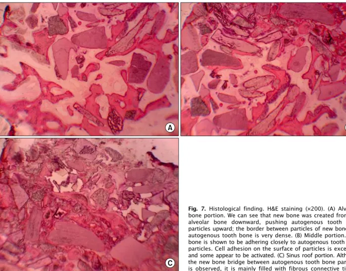
5) After GBR, the membrane was removed i n i ni ti al ti me, the usage of nonabsorbabl e membrane and autogenous bone resul ted i n the mostfavorabl e bone formati
success rates of dental implants placed at the time of or after alveolar ridge augmentation with an autogenous mandibular bone graft and titanium mesh: a 3-to
In 4-week group, the group filled with bone graft with decortication revealed larger new bone formation area than shown in the group that had a defect area
Histologic evaluation of early human bone response to different implant surfaces2. Histologic evaluation of human bone integration on machined and
The result of using PLGA nanofiber membrane with bone graft material showed that the PLGA nanofiber membrane in the experimental group of 2 weeks were ten times more new
of mineralized cancellous bone allograft(Puros) and anorganic bovine bone matrix(Bio-oss) for sinus augmentation: Histomorphometry at 26 to 32 weeks after
