• 검색 결과가 없습니다.

Changes in Compound Muscle Action Potential Depending on Pressure Level of Blood Flow During KAATSU Training

N/A
N/A
Protected

Academic year: 2021

Share "Changes in Compound Muscle Action Potential Depending on Pressure Level of Blood Flow During KAATSU Training"

Copied!
9
0
0

로드 중.... (전체 텍스트 보기)

전체 글

(1)

| Abstract |

Purpose: In recent years, there has been increasing interest in using blood flow-restricted exercise (BFRE) or KAATSU training.

The KAATSU training method, which partially restricts arterial inflow and fully restricts venous outflow in the working musculature during exercise at reduced exercise intensities, has been proven to result in substantial increases in both muscle hypertrophy and strength. The purpose of this study was to investigate the proper level of pressure for KAATSU training using compound muscle action potential (CMAP) analysis.

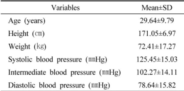
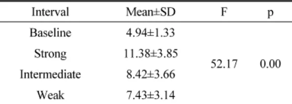
Methods: Twenty-two healthy adults voluntarily participated in this study. CMAP was conducted by measuring the terminal latency and amplitude using a motor nerve conduction velocity test. For reference-line, supramaximal electrical stimulation was applied to the median nerves of the participants to obtain CMAP for the abductor pollicis brevis. For baseline, the intensity of the electrical stimulation was decreased to a level at which the CMAP amplitude was about a third of the CMAP amplitude obtained by the supramaximal electrical stimulation. The pressure levels for the KAATSU were set as a systolic blood pressure (strong pressure), the median values of systolic and diastolic blood pressure (intermediate pressure), and diastolic blood pressure (weak pressure). In the KAATSU condition, CMAP was performed under the same conditions as baseline after low-intensity thumb abduction exercises were performed at the subjects’ own pace for one minute.

Results: As the pressure increased, the CMAP amplitude was significantly increased, signifying that more muscle fibers were recruited.

Conclusion: This study found that KAATSU training recruited more muscle fibers than low-intensity exercise without the restriction of blood flow.

Key Words: Blood flow-restricted exercise (BFRE), KAATSU training, Compound muscle action potential (CMAP)

†Corresponding Author : Jong-Soon Kim ([email protected])

Original Article Open Access

가압훈련의 혈류 압박 정도에 따른 복합근 활동전위의 변화

김종순

1)

부산가톨릭대학교 보건과학대학 물리치료학과

Changes in Compound Muscle Action Potential Depending on Pressure Level of Blood Flow During KAATSU Training

Jong-Soon Kim, P.T., Ph.D.

Department of Physical Therapy, College of Health Sciences, Catholic University of Pusan Received: August 13, 2020 / Revised: September 5, 2020 / Accepted: September 9, 2020

ⓒ 2020 Journal of Korea Proprioceptive Neuromuscular Facilitation Association

This is an Open Access article distributed under the terms of the Creative Commons Attribution Non-Commercial License

(http://creativecommons.org/licenses/by-nc/3.0) which permits unrestricted non-commercial use, distribution, and reproduction

in any medium, provided the original work is properly cited.

(2)

Ⅰ. 서 론

KAATSU는 압력을 부가한다는 의미인 가압(加壓) 의 일본식 발음이다. 따라서 KAATSU 훈련은 가압훈 련이라고 할 수 있다. 가압훈련은 Yoshiaki Sato 박사에 의해 개발된 훈련 방법으로 영문 표현은 혈류 제한 훈련(blood flow restriction training, BFRT)이라고 하는 데 운동을 하는 표적 근육으로의 혈류 공급을 일시적, 부분적으로 제한을 하여 저강도의 저항 운동을 실시 하는 경우 고강도 운동의 효과를 거둘 수 있다고 보고 된 운동 방법이다(Sato, 2005; Wernbom et al., 2008).

즉, 운동을 하는 동안 표적이 되는 근육으로의 동맥혈 유입을 부분적으로 차단하고 정맥혈 유출을 완전히 차단하는 운동 방법이다(Scott et al., 2015). 이러한 혈 류의 차단은 젖산(lactic acid)과 같은 대사산물을 근육 의 사이질(interstitium)에 축적시켜 근력의 증가와 근 육의 성장을 유도하는데(Spranger et al., 2015) 가압훈 련에 의한 혈류의 차단은 무산소 대사 과정을 유발하 고 무산소 대사로 만들어지는 젖산은 포도당신생합성 (gluconeogenesis) 물질을 제공 할뿐 아니라 젖산 탈수 소효소(lactate dehydrogenase)를 통해 피루브산 (pyruvic acid)으로 전환되어 에너지원으로 재사용될 수 있다(Brooks, 2002; Febbraio & Pedersen 2005). 근 수축 시 골격근에서 발현되는 활성 인자들을 마이오카 인(myokine)이라 하는데(Pedersen et al., 2003) 수축하는 골격근에서 분비되는 마이오카인 가운데 myostatin, IL-4(interleukin-4), IL-6, IL-10, 그리고 IL-15 등은 근육 의 비대(hypertrophy)와 근육신생합성(myogenesis)의 조절에 관여한다(Henningsen et al., 2010). 마이오카인 은 운동의 강도가 증가함에 따라 혈장에서 급격히 증 가하는데 가압훈련은 무산소 대사를 유발하고 이는 고강도 운동과 유사한 운동 환경을 조성하여 마이오 카인의 발현을 증가시킬 수 있다(Kim et al., 2016). 전 통적인 관점에서 근육의 강화와 근육의 비대를 도모 하기 위해서는 6RM (repetition maximum) 이하나 1RM 의 60∼80%의 강도로 저항 훈련을 실시하여야 한다 (Garber et al., 2011). 그러나 장애인이나 노인, 급성

손상 환자, 수술 후 환자, 심호흡계 문제를 가진 환자나 만성 질환자 등에서는 이러한 고강도의 운동을 수행 하는 것이 매우 어려운 실정이다. 따라서 가압훈련은 물리치료를 필요로 하는 많은 환자군들에게 대안적인 운동 방법이 될 수 있다.

가압훈련의 효과를 보고한 선행 연구들을 살펴보 면 신체 기능의 향상(Buford et al., 2015), 골격근 비대 와 근력 증가(Abe et al., 2010; Credeur et al., 2010;

Lowery et al., 2014; Moore et al., 2004), 미세혈관 여과 능력의 향상(Evans et al., 2010), 복합근 활동전위의 증가(Kim, 2009), 지구력 개선(Loeppky et al., 2005;

Sumide et al., 2009), 성장호르몬의 분비 촉진(Manini et al., 2012) 등에서 고강도 운동과 유사한 효과를 보인 다고 보고되고 있다. 그러나 가압훈련의 많은 효과 보고에도 불구하고 혈류를 차단한 상태에서 실시하는 가압훈련에 대한 우려가 동시에 공존하고 있다.

가압훈련의 안전성 면에서 제기되는 우려는 먼저, 혈전의 생성이다. Noto 등(2017)은 가압훈련의 영향으 로 팔의 깊은 정맥 혈전증(deep vein thrombosis)인 Paget-Schroetter 증후군이 발생한 환자를 보고하였다.

그러나 Clark 등(2011)과 Madarame 등(2013)의 연구를 살펴보면 가압훈련으로 인한 혈관 내 혈전 생성이나 트롬빈(thrombin) 발생 증거는 없다고 보고하고 있다.

또 다른 위험 요인으로 제기되는 문제는 근육의 손상

이다. Clark과 Manini (2017)는 가압훈련을 위해 가해

지는 압력에 의한 가로무늬근융해증(rhabdomyolysis)

과 심각한 지연발생 근육통(delayed onset muscle

soreness)이 발생하였다고 보고하였다. 그러나 Nielsen

등(2017)은 가압훈련으로 근세포 포식세포 침윤 및

열충격 단백질(heat shock protein)의 상향 조정이 나타

나지만 근육 손상의 명백한 증거는 없었다고 하여 다

른 결과를 보고하고 있다. 신경의 문제로는 가압훈련

으로 인해 신경의 저산소증과 퇴행이 발생할 수 있는

데(Shen et al., 2020) Ozawa 등(2015)은 가압훈련으로

심박수와 성장호르몬 수치의 변화로 인해 망막중심정

맥 폐색(central retinal vein occlusion)이 발생하여 가압

훈련을 받은 환자가 시력을 상실하였다고 보고하였다.

(3)

이와 같이 가압훈련의 많은 효과와 부작용에 대한 보고가 이어져 오고 있으나 안전한 가압훈련에 대한 지침은 매우 부족한 실정이다. 특히 가압훈련의 특징 인 혈관에 가해지는 안전하고 효과적인 압박 정도에 대한 연구는 전무한 실정이다. 이에 본 연구는 가압훈 련의 안전하고 효과적인 압박정도를 신경의 흥분성 유도 관점에서 분석 연구하고자 하였다.

Ⅱ. 연구 방법 1. 연구설계 및 대상

본 연구의 연구 대상자는 본 연구의 목적과 방법, 그리고 연구과정에서 수반되는 가압에 의한 부작용과 위험성 정도에 대하여 충분한 설명을 듣고 자발적으 로 본 연구에 참여하기로 동의한 신경학적 이상 및 정형외과적 손상이나 신경외과적 손상의 과거력이 없 는 건강한 성인 22명을 대상으로 실시하였다.

2. 측정도구 및 방법 1) 측정도구

가압훈련의 압력 정도에 의한 신경의 흥분성 변화 정도를 관찰하기 위하여 근전도 기기(Keypoint, Danteck, Denmark)를 이용하였으며, 검사실 내의 온도 는 22°C 이상, 검사 부위 표피 온도는 30°C∼34°C를 유지하였다(Preston & Shapiro, 1998). 신경의 흥분성 변 화 정도를 분석하기 위한 복합근 활동전위(compound muscle action potential)의 획득은 정중운동신경 전도속 도(median motor nerve conduction velocity) 검사를 통해 실시하였다. 정중운동신경 전도속도 검사는 정행 측 정방법을 통해 분석하였다. 정중운동신경 전도속도 검사를 위해 전극의 부착은 힘살-힘줄 방법(belly- tendon method)에 따라 기록전극(recording electrode)은 짧은엄지벌림근(abductor pollicis brevis)의 힘살(muscle

belly)에 부착하였고 관련전극(reference electrode)은 짧은엄지벌림근의 힘줄에 부착하였으며 접지전극 (ground electrode)은 아래팔에 부착하였다. 전기자극 은 막대전극(bar electrode)을 이용하여 손목 주름에서 정중신경을 자극하였다. 자극은 1Hz의 자극 주파수를 정방향으로 하여 자극하였으며 저역통과 필터링(low- pass filtering) 10㎑, 고역통과 필터링(high-pass filtering) 10㎐, 민감도 5㎷/D, 그리고 소인 속도(sweep speed)는 5㎳/D로 설정하였다. 정중운동신경전도속도 검사의 잠복시는 복합근 활동전위가 기초선에서 최초 음의 위상으로 변위되는 지점을 측정하였으며 진폭은 양극 정점(positive peak)에서 음극정점(negative peak)까지 측정하였다.

2) 측정방법

먼저, 혈압계(UA-772C, A&D Company Ltd, Japan) 를 이용하여 연구대상자의 수축기 혈압과 이완기 혈 압을 측정하여 강한 압력(수축기 혈압), 중간 압력(수 축기 혈압과 이완기 혈압의 중간 값), 약한 압력(이완 기 혈압)을 설정하였다. 그 다음, 압박을 가하지 않은 상태에서 초최대 자극(supramaximal stimulation) 후 연 구대상자의 정중운동신경 잠복시와 진폭을 기록하였 다(참고선). 참고선(reference line)을 획득한 다음 기초 선(baseline)을 설정하기 위하여 압박을 가하지 않은 상태에서 초최대 자극으로 획득된 진폭의 1/3 정도로 복합근 활동전위의 진폭이 획득되도록 자극 강도를 낮추어 자극한 다음 잠복시와 진폭을 측정하였다.

가압훈련을 위한 일정한 압력 적용을 위해 혈압계

(HM-1101, HICO, Japan)를 사용하였다. 강한 압력에서

의 가압훈련 후 신경의 흥분성 변화를 분석하기 위해

자극조건을 기초선과 동일하게 둔 다음 팔꿉관절 바

로 아래에 혈압계를 대고 수축기 혈압으로 압박을 가

한 다음 연구대상자에게 엄지손가락의 벌림 운동을

자유 속도로 1분간 실시하도록 지시하였다. 연구대상

자가 1분간의 가압운동을 실시한 다음 즉시 압박을

제거한 후, 기초선과 동일한 조건으로 자극하여 잠복

(4)

시와 진폭을 구하였다.

중간 압력과 약한 압력에서의 신경의 흥분성 변화 정도의 분석은 강한 압력에서의 가압 훈련 후 측정과 동일한 방법으로 실시하였다. 각 압력별 측정 후 연구 대상자들에게 5분간의 휴식을 취하도록 한 다음, 다른 압력 단계의 가압 훈련을 수행하였으며 각 조건별 자 극에 의한 영향을 배제하기 위하여 가압 훈련의 압력 정도는 무작위로 적용하였다.

3. 자료 분석

연구 과정에서 수집된 자료는 부호화한 후 자료 처리는 유의 수준 α를 0.05로 하여 통계 패키지 SPSS for Windows (ver. 25.0)를 이용하여 분석하였다. 먼저 연구대상자들의 일반적 특성은 평균 및 표준 편차를 산출하였으며 안정시 초최대 자극의 1/3 자극강도(기 초선)에서 획득된 정중운동신경 전도속도 검사의 잠 복시와 진폭 그리고 강한 압력, 중간 압력, 약한 압력을 가한 상태에서 획득된 정중운동신경 전도속도 검사의 잠복시와 진폭을 바탕으로 신경의 흥분성 정도 차이 를 비교하기 위해 반복측정 분산분석(repeated measure ANOVA)을 실시하였다. 압력의 차이에 따른 조건별 차이 분석은 대비 검정(contrast test)을 실시하였다.

Ⅲ. 연구 결과 1. 연구 대상자의 일반적인 특성

본 연구의 대상자들은 총 22명으로 남성이 14명 (73.60%), 여성이 8명(36.40%)이였다. 연구 대상자들 의 평균 연령은 29.64세, 평균 신장은 171.05㎝, 평균 몸무게는 72.41㎏, 수축기 혈압은 125.45㎜Hg, 중간 혈압은 102.27㎜Hg, 이완기 혈압은 78.64㎜Hg이였다 (Table 1).

2. 가압훈련 압력 정도에 따른 잠복시의 변화 가압훈련의 압력 적용 전 기초선 자극으로 획득된 잠복시는 3.07㎳였다. 강한 압력을 적용한 상태에서 시행한 가압훈련 후 획득된 잠복시는 3.01㎳, 중간 압 력을 적용한 상태에서 시행한 가압훈련 후 잠복시는 3.06㎳, 약한 압력을 적용한 상태에서 시행한 가압훈 련 후 잠복시는 3.03㎳으로 기초선 자극과 비교하여 가압훈련 후 잠복시의 유의한 차이는 없었다(p>0.05) (Table 2).

3. 가압훈련 압력 정도에 따른 잠복시 변화 비교 가압훈련의 압력 정도에 따른 잠복시를 비교한 결

Variables Mean±SD

Age (years) 29.64±9.79

Height (㎝) 171.05±6.97

Weight (㎏) 72.41±17.27

Systolic blood pressure (㎜Hg) 125.45±15.03 Intermediate blood pressure (㎜Hg) 102.27±14.11 Diastolic blood pressure (㎜Hg) 78.64±15.82 Table 1. General characteristics of subjects (n=22)

Interval Mean±SD F p

Baseline 3.07±0.37

1.28 0.29

Strong 3.01±0.38

Intermediate 3.06±0.37

Weak 3.03±0.36

Table 2. Mean of terminal latency under each condition

Contrast time F p

Baseline vs. Strong 2.06 0.17 Baseline vs. Intermediate 0.15 0.71

Baseline vs. Weak 3.21 0.09

Strong vs. Intermediate 1.59 0.22

Strong vs. Weak 0.39 0.54

Intermediate vs. Weak 0.94 0.34 Table 3. Contrast test of terminal latency among the

four different conditions

(5)

과 압력 정도의 차이에 의한 유의한 변화는 없었다 (p>0.05)(Table 3).

4. 가압훈련 압력 정도에 따른 진폭의 변화

가압훈련의 압력 적용 전 기초선 자극으로 획득된 진폭은 4.94㎷였다. 강한 압력을 적용한 상태에서 시 행한 가압훈련 후 획득된 진폭은 11.38㎷, 중간 압력을 적용한 상태에서 시행한 가압훈련 후 진폭은 8.42㎷, 약한 압력을 적용한 상태에서 시행한 가압훈련 후 진 폭은 7.43㎷로 기초선 자극과 비교하여 가압훈련을 위한 압력 적용 전⋅후, 유의한 차이를 보였다 (p<0.05)(Table 4).

Interval Mean±SD F p

Baseline 4.94±1.33

52.17 0.00 Strong 11.38±3.85

Intermediate 8.42±3.66

Weak 7.43±3.14

Table 4. Mean CMAP amplitude for each condition

5. 가압훈련 압력 정도에 따른 진폭의 변화 비교

가압훈련의 압력 정도에 따른 진폭을 비교한 결과 압력을 적용하지 않은 기초선에 비해 압력을 적용한 모든 조건에서 진폭의 유의한 변화가 있었다(p<0.05).

압력 적용 정도에 따른 차이를 비교한 결과 강한 압력 을 적용한 경우와 중간 압력, 약한 압력을 적용한 경우

를 비교한 결과 유의한 진폭의 차이가 있었다(p<0.05).

중간 강도의 압력을 적용한 경우와 약한 압력 적용한 경우를 비교한 결과에서는 진폭의 유의한 차이는 없 었다(p>0.05)(Table 5).

Ⅳ. 고 찰

가압훈련은 전통적인 고강도 운동의 단점을 보완 할 수 있고 운동의 수월성으로 인해 Sato (2005)에 의해 문헌으로 보고된 이래 지난 15년간 수많은 연구가 진 행되었고 임상과 스포츠 현장에서 다양한 환자 및 스 포츠 선수들에게 많이 적용되고 있다. 그러나 많은 효과 보고(Abe et al., 2010; Credeur et al., 2010; Evans et al., 2010; Kim, 2009; Loeppky et al., 2005; Lowery et al., 2014; Manini et al., 2012; Moore et al., 2004;

Sumide et al., 2009)와 부작용에 대한 보고(Clark &

Manini, 2017; Noto et al., 2017; Ozawa et al., 2015; Shen et al., 2020)가 이어져 오고 있어 안전하고 효과적인 가압훈련 방법에 대한 연구가 필요한 실정이다. 이에 본 연구는 신경의 흥분성 변화 정도를 분석하여 가압 훈련에 적용할 수 있는 안전하고 효과적인 압력 정도 를 알아보고자 하였다.

가압훈련에서 적용하는 압력 정도에 대한 선행 연 구들을 살펴보면 인간의 정상 혈압인 수축기 혈압 120

㎜Hg, 이완기 혈압 80㎜Hg를 기준으로 이완기 혈압 이하인 50㎜Hg의 약한 압력을 적용한 연구(Kubota et al., 2011), 수축기 혈압 정도인 120㎜Hg의 압력을 적용 한 연구(Kim, 2009; Kim, 2015), 수축기 혈압의 30%를 상회하는 정도를 적용한 연구(Cook et al., 2010), 125∼

215㎜Hg의 압력을 적용한 연구(Buford et al., 2010), 가압훈련을 시작하는 2주 동안은 160㎜Hg를 적용하 고 가압훈련을 진행함에 따라 매 2주마다 20㎜Hg씩 압력을 증가시켜 나가는 방법(Madarame et al., 2008;

Takarada et al., 2002), 200㎜Hg를 적용한 연구(Iida et al., 2007; Kubota et al., 2008), 160∼300㎜Hg를 적용한 연구(Cook et al., 2007) 등 연구자의 주관에 의해 다양

Contrast time F p

Baseline vs. Strong 100.73 0.00 Baseline vs. Intermediate 35.34 0.00 Baseline vs. Weak 23.72 0.00 Strong vs. Intermediate 47.47 0.00

Strong vs. Weak 72.18 0.00

Intermediate vs. Weak 4.59 0.44 Table 5. Contrast test of CMAP amplitude among the

four different conditions

(6)

한 범위의 압력들이 가압훈련에 적용되고 있으나 이 에 대한 명확한 기준은 마련되어 있지 않다. 이에 본 연구에서는 안전한 연구를 수행하기 위해, 선행 연구 들을 참고하여 수축기 혈압, 수축기 혈압과 이완기 혈압의 중간 값, 이완기 혈압을 각각 강한 압력, 중간 압력, 약한 압력으로 정의하고 연구를 수행하였다. 또 한, 본 연구에서는 신경의 흥분성 변화 정도를 복합근 활동전위의 잠복시 및 진폭 분석을 통해 알아보고자 하였다. 잠복시는 얼마나 빠르게 신경이 흥분을 하였 는지를 의미하고 진폭은 얼마나 많은 신경 섬유들이 흥분하였는지를 의미한다. 그러므로 신경의 흥분성 변화는 신경전도속도 검사의 잠복시와 진폭의 비교 분석을 통해 알아 볼 수 있다(Aminoff, 1998; Kimura, 2013; Lee et al., 2018).

본 연구의 결과 안정시 초최대 자극의 1/3 자극강도 (기초선)에서 획득된 복합근 활동전위의 잠복시와 수 축기 혈압과 수축기 혈압과 이완기 혈압의 중간 혈압, 그리고 이완기 혈압 정도의 압력을 적용한 상태에서 실시한 가압훈련 후, 획득된 잠복시를 비교한 결과 유의한 차이는 나타나지 않았다(p=0.29). 기초선에서 획득된 잠복시는 평균 3.07㎳로 우리나라 정상 성인의 정중운동신경 잠복시 2.80㎳(Kim, 2009)에 비해 약간 지연된 경향을 보였다. 이는 약한 전기자극의 영향의 의한 잠복시 지연으로 사료되는데 본 연구에서 기초 선 설정을 위해 시행했던 초최대 자극(참고선 검사)에 서 획득된 잠복시는 2.86㎳로 Kim (2009)에 의해 보고 된 정상 잠복시와 차이가 없었다. 또한 수축기 혈압 정도의 강한 압박과 함께 시행한 가압훈련 후 획득된 잠복시는 3.01㎳로 참고선 자극에서 획득된 잠복시나 정상 성인의 잠복시로 보고된 결과 값에 비해 다소 지연된 소견을 보였으나 기초선 검사에서 획득된 잠 복시에 비해서는 0.06㎳ 빠르게 자극을 전달하는 것으 로 나타났다.

신경자극에 의해 활성화된 근육 섬유의 합을 의미 하는 복합근 활동전위 진폭의 경우 본 연구의 결과 기초선에서 획득된 진폭과 수축기 혈압과 수축기 혈 압과 이완기 혈압의 중간 혈압, 그리고 이완기 혈압

정도의 압력을 적용한 상태에서 실시한 가압훈련 후, 획득된 진폭을 비교한 결과 유의한 차이를 보였다 (p=0.00). 이를 각 조건별로 비교 검증한 결과 기초선에 서 획득된 진폭에 비해 압력을 가한 상태에서 시행한 가압훈련 후 획득된 복합근 활동전위의 진폭이 유의 하게 증가된 양상을 보였다. 특히, 압력이 증가함에 따라 복합근 활동전위의 진폭은 평균 50.40%∼

130.36% 증가하여 압력 증가에 따라 신경의 흥분성이 증가함을 알 수 있었다. Kubota 등(2011)은 건강한 피 검자를 대상으로 2주간 고정을 실시하여 근육의 인위 적인 위축과 근력 약화를 유도 하는 동안 반복적으로 50㎜Hg의 약한 가압을 적용한 결과 대조군에 비해 근력 감소가 적었다고 보고하여 약한 압박의 효과를 보고하였다. 본 연구에서도 이완기 혈압 정도의 약한 압력을 적용한 상태에서 시행한 가압훈련 후 복합근 활동전위의 진폭은 기초선에서 획득된 진폭에 비해 평균 2.49㎷ 높게 나타나 약한 압력을 적용한 가압훈련 에서 의미 있는 효과가 있음을 확인할 수 있었다.

강한 압력을 적용한 상태에서 시행한 가압훈련의 경우를 살펴보면 2주간 고정을 하고 있는 동안 가압훈 련을 위해 200㎜Hg의 압력을 적용한 결과 무용성 근 약화를 예방할 수 있었다는 보고가 있었으며(Kubota et al., 2008), Cook 등(2010)은 안정 상태의 수축기 혈압 보다 30% 정도 더 압력을 가하여 가압훈련을 적용한 경우 근력, 지구력, 그리고 근육의 크기가 유의하게 개선되었다고 보고하고 있다. Segal 등(2015)은 가압훈 련 시작 처음 1분간은 30∼40㎜Hg의 압력을 가한 다음 1분 후에는 100∼180㎜Hg 압력을 증가시켜 최종적으 로 160∼200㎜Hg 압력을 가하여 골관절염 환자에게 가압훈련을 적용한 결과 무릎관절 폄근의 근력이 증 가되었다고 보고하였다. 본 연구에서도 수축기 혈압 정도의 강한 압력을 적용한 상태에서 수행한 가압훈 련 후 획득된 복합근 활동전위의 진폭은 기초선에서 획득된 진폭에 비해 130% 이상의 증가 소견을 보였다.

이는 강한 압력에 의해 신경의 흥분성이 증가되어 많

은 근육 섬유를 동원한 결과로 사료된다.

(7)

Ⅴ. 결 론

본 연구의 결과 가압훈련에 적용되는 압력이 증가 함에 따라 신경의 흥분성을 증가시켜 보다 많은 근육 섬유를 동원시킴을 알 수 있었다. 이완기 혈압 정도의 압력에 의해서도 신경의 흥분성 증가로 인한 근육 섬 유 동원 증가가 관찰되었다. 따라서 최소 이완기 혈압 이상의 압력을 적용하여 점진적으로 압력 정도를 증 가시켜 가압훈련을 실시하고 최대 수축기 혈압을 초 과하지 않는 정도의 압력을 적용하는 것이 효과적이 고 안전한 가압훈련 방법으로 사료된다. 그러나 본 연구의 결과는 1분간의 가압훈련을 통해 획득된 결과 로서 이를 일반화 하기에는 무리가 있는 것으로 판단 된다. 따라서 추후 연구에서는 일반적인 훈련 시간 정도의 가압을 적용한 상태에서 압박 정도와 복합근 활동전위 변화의 상관성에 대한 연구가 이루어져야 할 것으로 사료된다.

Acknowledgements

이 논문은 2018년도 부산가톨릭학교 교내연구비에 의하여 연구되었음

References

Abe T, Fujita S, Nakajima T, et al. Effects of low-intensity cycle training with restricted leg blood flow on thigh muscle volume and VO2max in young men. Journal of Sports Science and Medicine. 2010;9(3):452-458.

Aminoff MJ. Electromyography in clinical practice: clinical and electrodiagnostic aspects of neuromuscular disease, 3rd ed. New York. Churchill Livingstone.

1998.

Brooks GA. Lactate shuttles in nature. Biochemical Society Transactions. 2002;30(2):258-264.

Buford TW, Fillingim RB, Manini TM, et al. Kaatsu training to enhance physical function of older adults with knee osteoarthritis: design of a randomized controlled trial. Contemporary Clinical Trials. 2015;43:217-222.

Clark BC, Manini TM, Hoffman RL, et al. Relative safety of 4 weeks of blood flow-restricted resistance exercise in young, healthy adults. Scandinavian Journal of Medicine & Science in Sports. 2011;21(5):653–662.

Clark BC, Manini TM. Can KAATSU exercise cause rhabdomyolysis? Clinical Journal of Sport Medicine.

2017;27(1):e1-e2.

Cook SB, Clark BC, Ploutz-Snyder LL. Effects of exercise load and blood-flow restriction on skeletal muscle function. Medicine & Science in Sports & Exercise.

2007;39(10):1708-1713.

Cook SB, Brown KA, Deruisseau K, et al. Skeletal muscle adaptations following blood flow-restricted training during 30 days of muscular unloading. Journal of Applied Physiology. 2010;109(2):341-349.

Credeur DP, Hollis BC, Welsch MA. Effects of handgrip training with venous restriction on brachial artery vasodilation.

Medicine & Science in Sports & Exercise. 2010;42(7):

1296-1302.

Evans C, Vance S, Brown M. Short-term resistance training with blood flow restriction enhances microvascular filtration capacity of human calf muscles. Journal of Sports Science. 2010;28(9):999-1007.

Febbraio MA, Pedersen BK. Contraction-induced myokine production and release: is skeletal muscle an endocrine organ? Exercise and Sport Sciences Reviews. 2005;

33(3):114-119.

Garber CE, Blissmer B, Deschenes MR, et al. American college

of sports medicine position stand. Quantity and quality

of exercise for developing and maintaining

cardiorespiratory, musculoskeletal, and neuromotor

fitness in apparently healthy adults: guidance for

prescribing exercise. Medicine & Science in Sports

(8)

& Exercise. 2011;43(7):1334-1359.

Henningsen J, Rigbolt KT, Blagoev B, et al. Dynamics of the skeletal muscle secretome during myoblast differentiation. Molecular & Cellular Proteomics.

2010;9(11):2482-2496.

Iida H, Kurano M, Takano H, et al. Hemodynamic and neurohumoral responses to the restriction of femoral blood flow by KAATSU in healthy subjects. European Journal of Applied Physiology. 2007;100(3):275-285.

Kim JS. Changes of α-motor neuron excitability after low-intensity exercise with transient restriction of blood flow. Journal of Korean Society of Physical Medicine. 2015;10(1): 63-69.

Kim LJ. Changes of compound muscle action potential after low-intensity exercise with transient restriction of blood flow: a randomized, placebo-controlled trial.

Journal of Physical Therapy Science. 2009;21(4):

361-366.

Kim SJ, Kim HS, Moon SH, et al. Effects of lactic acid on the expression of myokines in C2C12 myotubes.

Exercise Science. 2016;25(3):174-182.

Kimura J. Electrodiagnosis in diseases of nerve and muscle:

principle and practice, 4th ed. New York. Oxford University Press. 2013.

Kubota A, Sakuraba K, Sawaki K, et al. Prevention of disuse muscular weakness by restriction of blood flow.

Medicine & Science in Sports & Exercise. 2008;40(3):

529-534.

Kubota A, Sakuraba K, Koh S, et al. Blood flow restriction by low compressive force prevents disuse muscular weakness. Journal of Science and Medicine in Sport.

2011;14(2):95-99.

Lee DR, Rhee MH, Eom JR, et al. Changes in nerve excitability during neural stretching. PNF and Movement.

2018;16(2):287-294.

Loeppky JA, Gurney B, Kobayashi Y, et al. Effects of ischemic training on leg exercise endurance. Journal of

Rehabilitation Research & Development. 2005;42(4):

511-522.

Lowery RP, Joy JM, Loenneke JP, et al. Practical blood flow restriction training increases muscle hypertrophy during a periodized resistance training programme.

Clinical Physiology and Functional Imaging.

2014;34(4):317-321.

Madarame H, Neya M, Ochi E, et al. Cross-transfer effects of resistance training with blood flow restriction.

Medicine & Science in Sports & Exercise. 2008;

40(2):258-263.

Madarame H, Kurano M, Fukumura K, et al. Haemostatic and inflammatory responses to blood flow-restricted exercise in patients with ischaemic heart disease: a pilot study. Clinical Physiology Functional Imaging.

2013;33(1):11-17.

Manini Todd, Yarrow J, Buford T. Growth hormone responses to acute resistance exercise with vascular restriction in young and old men. Growth Hormone & IGF Research. 2012;22(5):167-72

Moore D, Burgomaster K, Schofield L, et al. Neuromuscular adaptations in human muscle following low intensity resistance training with vascular occlusion. European Journal of Applied Physiology. 2004;92(4-5):399- 406.

Noto T, Hashimoto G, Takagi T, et al. Paget-Schroetter syndrome resulting from thoracic outlet syndrome and KAATSU training. Internal Medicine. 2017;56(19):2595-2601.

Nielsen JL, Aagaard P, Prokhorova TA, et al. Blood flow restricted training leads to myocellular macrophage infiltration and upregulation of heat shock proteins, but no apparent muscle damage. The Journal of Physiology. 2017;595(14):4857-4873.

Ozawa Y, Koto T, Shinoda H, et al. Vision loss by central retinal vein occlusion after kaatsu training: a case report. Medicine. 2015;94(36):e1515.

Pedersen BK, Steensberg A, Fischer C, et al. Searching for

(9)

the exercise factor: is IL-6 a candidate? Journal of Muscle Research and Cell Motility. 2003;24(2-3):

113-119.

Preston D, Shapiro B. Electromyography and neuromuscular disorder, Massachusetts. Butterworth-Heinemann.

1998.

Sato Y. The history and future of KAATSU training.

International Journal of Kaatsu Training Research.

2005;1:1-5.

Scott BR, Loenneke JP, Slattery KM, et al. Exercise with blood flow restriction: an updated evidence-based approach for enhanced muscular development. Sports Medicine. 2015;45(3):313-325.

Segal NA, Williams GN, Davis MC, et al. Efficacy of blood flow-restricted, low-load resistance training in women with risk factors for symptomatic knee osteoarthritis.

Physical Medicine & Rehabilitation. 2015; 7(4):

376-384.

Shen L, Li J, Chen Y, et al. L-carnitine’s role in KAATSU

training-induced neuromuscular fatigue. Biomedicine

& Pharmacotherapy. 2020;125:109899.

Spranger M, Krishnan A, Levy P, et al. Blood flow restriction training and the exercise pressor reflex: a call for concern. American Journal of Physiology-Heart and Circulatory Physiology. 2015;309(9):1440-1452.

Sumide T, Sakuraba K, Sawaki K, et al. Effect of resistance exercise training combined with relatively low vascular occlusion. Journal of Science and Medicine in Sport. 2009;12(1):107-112.

Takarada Y, Takazawa H, Sato Y, et al. Effects of resistance exercise combined with vascular occlusion on muscle function in athletes. European Journal of Applied Physiology. 2002;86(4):308-314.

Wernbom, M, Augustsson, J, Raastad, T. Ischemic strength training: a low-load alternative to heavy resistance exercise? Scandinavian Journal of Medicine &

Science in Sports. 2008;18(4):401-416.

수치

Table 2. Mean of terminal latency under each condition
Table  4.  Mean  CMAP  amplitude  for  each  condition

참조

관련 문서

• Generation of different specialized kinds of cells from zygote (fertilized egg) or other precursor cells.. – Generate blood cells, muscle

Hele-Shaw flow (potential flow) Van Dyke, Album of Fluid Motion (1982).. 2017

With these results, we can suggest the hypothesis that increased cardiac output in the patients who administered ketamine increased muscle blood flow, and this is the

Physical fitness changes on college students who played ultimate frisbee showed increases in muscular strength, muscle endurance(, cardiopulmonary endurance,

In the health fitness category of the adult women who underwent instrument pilates exercise, they were statistically significant in muscle strength,

1) Zone of viscous action (boundary layer): frictional effects cannot be neglected. 2) Flow in the reservoir and central core of the tube: primary forces are pressure

In conclusion, it was confirmed that the winter training program is an effective exercise program by inducing positive changes in the physical fitness and

&#34;Two-phase pressure drop of refrigerants during flow boiling in small channels : an experimental investigation and correlation development&#34;,