INTRODUCTION
Dental implantation has been benefited greatly from research into the biological characteristics of titanium.
According to the existing protocol, to obtain predictable osseointegration, loading should be avoided while the subperiosteum heals during two-stage surgery.
However, the discomfort and anxiety that occur during the healing period are issues that must be resolved for both the physician and patient. Therefore, attempts have been made to apply immediate loading after surgery with the extensive support of clinicians
1).
We are faced with two conflicting approaches to dental implantation: the traditional method requiring a long
healing period and the ultrafast method of applying ear- ly loading. Although only a few years ago, immediate or early loading was considered possible only in areas with good bone quality, such as the anterior portion of the mandible, early loading is possible with a single implant even in areas with poor bone quality, such as the posteri- or portion of the maxilla. We therefore need to reevalu- ate the Branemark protocol
1).
A healing period without loading is still considered a prerequisite for implant integration
2), which leads to extended treatment periods, often with delayed func- tional improvement for the patient
3,4). The elimination or reduction of the postsurgical interval between implant placement and implant loading is a challenge for dental implantology. Early loading has been found to induce micromotion at the bone-implant interface, which may lead to fibrous encapsulation instead of direct bone
* Corresponding author Su-Gwan Kim
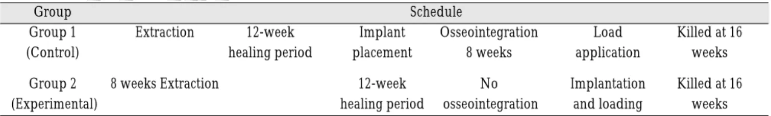
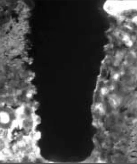
Histomorphometric analysis of an immediate non-functional loaded implant in dogs
Jeong-Wan Ha
1, Su-Gwan Kim
1, Hak-Hyun Kim
1, Seong-Yong Moon
1, Sung-Chul Lim
21
Department of Oral & Maxillofacial Surgery, College of Dentistry, Chosun University, Gwangju, Korea
2