한림대학교 성심병원 호흡기 알레르기내과, 병리학교실, 진단방사선학교실, 흉부외과학교실 최원섭1, 조재현1, 황용일1, 장승훈1, 김동규1, 전선영2, 민광선2, 이인재3, 이재웅4, 정기석1
A Case of Bronchus-Associated Lymphoid Tissue(BALT) Lymphoma Treated with Lobectomy
Won Sub Choi, M.D.1, Jae Hyun Cho, M.D.1, Young Il Hwang M.D.1, Seung Hun Jang, M.D.1, Dong-Gyu Kim, M.D.1, Sun-Young Jun, M.D.2, Kwangseon Min, M.D.2, In Jae Lee, M.D.3, Jae Woong Lee, M.D.4, Ki-Suck Jung M.D.,Ph.D.1
1Division of Pulmonary, Allergy and Critical Care Medicine of Hallym University Medical Center, 2Department of Pathology,
3Department of Radiology, 4Department of Thorasic and Cardiovascular Surgery Hallym University Sacred Heart Hospital, Hallym University College of Medicine, Anyang, Korea
The bronchus-asociated lymphoid tissue(BALT) lymphoma is a low-grade primary malignant lymphoma that originates from bronchus associated lymphoid tissue. A 67-year-old woman was admitted for evaluation of cough, sputum, rhinorrhea which had persisted for one month. Physical examination showed decreased breathing sound on the left upper lung field. High resolution chest computed tomography demonstrated consolidation which showed air-bronchogram and surrounding ground glass opacity in left upper lobe. These findings implicated inactive tuberculosis, organizing pneumonia, or bronchiolo-alveolar carcinoma. The histologic findings from percutaneous needle aspiration biopsy revealed aggregated atypical small lymphoid cells with lymphoepithelial lesions. With immunohistochemical staining, the atypical lymphoid cells reacted positively with CD 20 antibody and negatively with CD 3 antibody. Thus, we could diagnosed her as a patient with BALT lymphoma. After left upper lobectomy, she has been well without recurrence of the disease for 14 months. In this country of Republic of Korea, it was the 1st case of BALT lymphoma surgically treated when histological diagnosis had been done. Based on this case, we wanted to demonstrate the importance of early histological diagnosis and treatment of BALT lymphoma. (Tuberc Respir Dis 2007; 62: 427-431)
Key Words: BALT, Lung, Mucossa-associated lymphoid tissue (MALT) lymphoma.
Address for Correspondence: Ki-Suck Jung, M.D.
Division of Pulmonary, Allergy and Critical Care Medicine of Hallym University Medicine, Hallym University College of Medicine 896 Pyeongchon-dong, Dongan-gu, Anyang, Gyeonggi-do, 431-070, Korea Phone: 82-31-380-3717, FAX: 82-31-380-3973 E-mail: [email protected]
Received: Dec. 28. 2006 Accepted: Apr. 24. 2007
서 론
원발성 기관지 연관 림프조직(bronchus-associated lymphoid tissue, BALT) 림프종은 기관지 점막 연관 림프조직 (mucossa-associated lymphoid tissue, MALT)에서 기원하는 것으로 알려진 드문 질환으로 저등위 B 세포 림프종의 하나이다. 발생 기전은 정확 하게 밝혀져 있지 않으나, 호흡기에 대한 만성적 자 극, 감염 그리고 면역체계 이상이 본 질환과 관계가 있을 것으로 추정되고 있다. 저자들은 비특이적인 호 흡기 증상만을 나타낸 환자를 경피 미세침흡인생검술
을 통해 BALT 림프종으로 진단하고, 수술로 치료한 증례를 국내에서 최초로 경험하였기에 문헌 고찰과 함께 보고하는 바이다.
증 례
환 자: 여자, 66세
주 소: 누우면 심해지는 기침
현병력: 평소 건강하였으나, 내원 20일 전부터 하얀 색의 가래를 동반한 기침, 콧물이 시작되었으며 발열 및 체중 감소는 없었다. 항히스타민제와 점액용해제 등을 복용하였으나 효과 없어 외래를 통해 입원하였 다.
과거력: 3년 전 고혈압 진단 받고 티아지드 계통의 항고혈압약을 복용중임.
가족력: 특이 사항 없음.
개인력: 음주와 흡연력 없음.
신체검사 소견: 환자는 뚜렷한 병색을 보이지는 않
Figure 1B. HRCT demonstrated consolidation which showed air-bronchogram and surrounding ground glass opacity in left upper lobe and the lesion was mainly adjacent to pleura and fissure.
Fiure 2. In gross appearance after operation, lesion demonstrated an irregular diffuse consolidation with a solid tan-colored cut surface abutting visceral pleura.
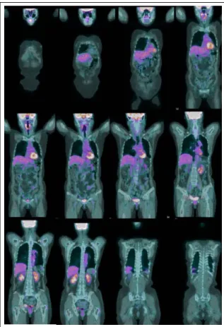
Figure 3. On 12 months After lobectomy, whole body PET CT did not demonstrate any evidence of local recurrence or distant metastasis of BALT lymphoma.
Figure 1A. Consolidation combined with linear opacities was noted on left upper lobe.
았고, 활력 징후는 혈압 140/60 mmHg, 맥박 수 78 회 /분, 호흡수 25 회/분, 체온 36.5℃이었으며, 의식은 명 료하였다. 두경부와 안면부 진찰상 종괴나 안구 돌출, 경정맥 확장, 인후부 발적, 편도비대 등은 보이지 않 았으며, 경부 강직도 없었고, 림프절도 만져지지 않았 다. 흉부 청진에서 좌상엽의 호흡음이 감소되었으나 촉진 시 특이한 점은 없었다. 복부 진찰 상 복부 팽만
은 없었고, 장음은 정상이었으며, 간, 비종대나 만져지 는 종괴는 없었다. 척추 늑골각 압통은 없었고, 피부
A B
C D
Figure 4. (A) Atypical lymphocytes with interstitial multinodular infiltration pattern and multiple foci of perivascular lymphoid infiltration ("lymphotic trackings", arrows) were seen. (H&E, x100) (B) The tumor consisted of small lymphocytes which showed clear abundant cytoplasm. They grew in monotonous sheets and infiltrated to respiratory epithelial cells("lymphoepithelial lesion", arrows). (H&E, x200) (C) Atypical lymphocytes reacted with CD 20 antibody positively (B cell maker). (immunohistochemistry, x200) (D) Epithelial cells in the lymphoepithelial lesions reacted with cytokeratin positively . (immunohistochemistry, x200)
에 특이한 병변은 관찰되지 않았다.
검사실 소견: 말초혈액검사에서 백혈구 6,900/mm3 (호중구 54.1%, 림프구 33.7%, 단핵구 8.8%), 혈색소 12.0 g/dL, 혈소판 410,000/mm3, 혈액화학검사에서 총 단백 6.8 g/dL, 알부민 3.6 g/dL, 총 빌리루빈 0.3 mg/dL, AST 61 IU/L, ALT 17 IU/L, BUN/Cr 11.0/
0.5 mg/dL, glucose 89 mg/dL, alkaline phosphatase 137 IU/L, Na/K 138/4.0 mmol/L, ESR 18 mm/hr, C 반응 단백은 2.98 mg/L이었다. PT INR 1.11, aPTT 31.5초였고, 소변 검사는 정상이었다.
방사선 소견: 단순 흉부 방사선 사진에서 선상 혼탁 을 동반한 말단 경화 소견이 좌상엽에서 관찰되었으 며(Figure 1A), 흉부 전산화 단층 촬영에서 종격동 림 프절 비대나 흉수는 없었고, 좌상엽에서 엽간과 흉막
에 연결된 경화가 간유리 혼탁에 둘러싸인 공기 기관 지조영과 함께 나타나는 등 세기관지 폐포암종, 기질 화 폐렴, 비활동성 결핵 등이 의심되는 소견을 보였다 (Figure 1B).
치료 및 경과: 내원 2일째 기관지경을 실시한 결과 기관지내 특이 병변은 없었으며, 기관지 폐포 세척액 에서 호중구 0%, 림프구 17%, 단핵구 83%의 소견을 보였다. 가래에서 특이한 미생물이 동정되지 않았으 며, 결핵균 다중연쇄반응, 항산염색과 배양, 가래 세포 검사에서 모두 음성 반응이 나타났다. 환자의 기침과 콧물은 코데인과 점액용해제 사용 후 완화되었다. 내 원 3일째, 종괴에 대해 경피 미세침흡입생검술을 시행 하였다. 시술 후 얻은 조직에서 BALT 림프종이 발견 되어, 좌상엽 절제술을 시행하여 병변을 제거하였다.
수술로 얻은 조직은 육안 소견에서(Figure 2) 단면은 황갈색을 띠었고, 확장된 기관지들이 보였으며, 장경 4 cm 정도의 불규칙한 경화 병변이 허파 쪽 가슴막에 인접해 있었다. 수술 시행 후 14개월 동안 환자는 특 이한 증상이나 질병의 재발 없이 외래에서 추적 관찰 중이다. 수술 후 약 12개월 되는 시점에서 실시한 골 수생검과 전신 양전자단층촬영술에서 국소재발이나 원격 전이의 소견이 보이지 않았다(Figure 3).
조직 소견: 현미경 소견에서 투명한 세포질을 갖는, 작은 크기의 비전형 림프구들로 구성되어 있었고, 이 비전형 림프구들은 기관지상피세포로 침윤하여 림프 상피성 병변을 형성하고 있었다(Figure 4A). 면역조 직화학염색소견에서 비전형 림프구들이 B 림프구의 표시자인 CD 20 항체에 양성 소견(Figure 4B)을 보였 고, CD 3 항체에 음성 소견을 보였다. 이상의 병리 소 견들을 통해 BALT 림프종으로 진단되었다.
고 찰
원발성 폐 림프종은 전체 비호즈킨 림프종의 1%
미만을 차지하는 드문 질환으로1, 그 중에서 기관지 연관 림프조직에서 발생한 것을 BALT 림프종이라고 한다. BALT 림프종의 발생 기전은 정확하게 밝혀져 있지 않으나, 미생물, 화학물질, 지속적인 감염 등의 호흡기에 대한 만성적 자극이 발생하고,이로 인해 면 역체계 이상과 만성 림프구 침윤 및 다클론성 B 세포 의 증식이 이루어지고, 이러한 염색체 이상 및 유전자 의 변형이 축적되어 질병이 발생하는 것으로 추정되 고 있다2. BALT 림프종은 70-80대에 발생률이 가장 높고 남녀 발생하는 비율은 거의 같은 역학적 특징을 나타낸다3. 임상적으로 예후가 좋아, 대부분 오랜 시 간 동안 국한된 상태로 남아 있고, 노령기에 기침, 발 열, 체중 감소, 호흡 곤란 같은 비특이적인 증상을 나 타내게 된다4. 검사실 소견은 특이한 점이 없는 수가 많지만, 혈액 검사상 단클론감마글로불린병증이 저등 위 B 세포 림프종 환자의 22-30%에서 관찰되며, 베 타 2 미크로글로불린이 MALT 림프종의 예후 인자라 는 의견도 있으므로, 원인을 알 수 없는 흉부 종괴가
단클론감마글로불린병증과 동반하면, 형질 세포 신생 물이나 림프 증식성 질환의 가능성을 고려해야 한다
5,6. 방사선 소견으로는, 비교적 경계가 명확한 고립성 종괴나 결절, 공기-기관지 음영이 가장 흔하여 폐포 기관지암이나 기질성 폐렴과 감별이 필요하다. 그 외 양측 폐를 침범하는 미만성 침윤 음영, 다발성 결절, 무기폐, 흉수 등을 보일 수 있다7,8. 굴곡기관경을 모든 환자에게 시행할 필요는 없지만, 경기관지 폐 생검과, 기관지 폐포 세척술 등을 시행하면 진단율을 높일 수 있다는 보고가 있어 시행을 고려해 볼 수는 있다. 확 진을 위해서는 조직 검사가 필수적이며, 개흉술을 통 하여 얻은 조직으로부터 진단을 내리는 것이 가장 정 확하다고 알려져 있다. 세침 흡입생검술은 개흉 생검 보다 얻을 수 있는 조직의 양이 적은 단점이 있지만, 최근 면역화학기법과 분자유전학적 검사 기법의 도입 으로 진단의 민감도와 특이도가 많이 향상 되었다9. BALT 림프종의 현미경적 소견은 원형 또는 불규칙 한 핵을 가진 작은 중심 세포양 세포의 증식, 림프구 양 세포의 주변부로부터 기관지 상피로의 이동을 나 타내는 림프상피 병변, 림프 여포 증식 등을 나타내는 것이 특징이며10, BALT의 반응성 결절성 림프증식증, 림프구성 간질성 폐렴, 세기관지폐포암 등과 감별하 여야 한다. 병기 결정은, 복부 전산단층촬영, 골수생 검, 구강, 비강 점막의 검진, 식도위내시경, 결장내시 경술 등을 시행해 볼 수 있으나, 병기 자체가 환자의 장기 생존의 예후인자가 되지 못하므로11 환자의 임상 경과에 따라 검사의 시행 범위를 정하는 것이 좋다.
국한된 상태의 BALT 림프종일 경우 외과적 절제가 선호되고 있으며 방사선치료를 할 수도 있다. 파종되 어 있을 경우 cylcophosphamide, adriamycin, vincris- tine, predisone 등과 rituximab 등을 병합하는 화학요 법을 사용할 수 있다4. 국내의 경우, 골수외 형질 세포 종으로 의심되어 절제술을 시행 받은 후, 수술 후 떼 어낸 조직절편에서 원발성 기관지 연관 림프조직 림 프종으로 진단되었던 1예9는 있었지만, 수술 시행 전 에 경피 미세침흡입생검을 통해 기관지 연관 림프조 직 림프종으로 진단하고 수술적 치료를 시도한 것은 최초의 예이다.
요 약
기관지 연관 림프조직 림프종은 비특이적인 호흡기 증상만을 나타내며, 전산화단층촬영에서 세기관지 폐 포암, 림프구성 간질성 폐렴등과 뚜렷이 구분이 되지 않는 결절 외 림프종의 일종이다. 저자들은 비특이적 인 호흡기 증상만을 나타내는 병변에 대해 침습적인 방법인 경피 미세흡입생검을 시행하여, 원발성 기관 지 연관 림프조직 림프종으로 진단하였으며, 병리적 진단이 내려진 상태에서 치료를 목적으로 좌상엽 절 제술을 시행하였다. 기관지 연관 림프조직 림프종은 서서히 진행되는 질환으로, 대부분 최종 진단이 늦어 지는 경향을 보인다. 국소적 병변일 경우 외과적 수술 로 완치가 가능한 질환이므로, 질환이 의심되는 경우 적극적 검사와 치료를 시도하는 것이 중요할 것으로 생각된다.
참 고 문 헌
1. Freeman C, Berg JW, Cutler SJ. Occurrence and prognosis of extranodal lymphomas. Cancer 1972;
29:252-60.
2. Cadranel J, Wislez M, Antoine M. Primary pulmonary lymphoma. Eur Respir J 2002;20:750-62.
3. Michael CW, Richardson PH, Boudreaux CW.
Pulmonary lymphoma of the mucosa-associated lym- phoid tissue type: report of a case with cytological,
histological, immunophenotypical correlation, and review of the literature. Ann Diagn Pathol 2005;9:
148-52.
4. Ahmed S, Kussick SJ, Siddiqui AK, Bhuiya TA, Khan A, Sarewitz S, et al. Bronchial-associated lymphoid tissue lymphoma: a clinical study of a rare disease.
Eur J Cancer 2004;40:1320-6.
5. Montes M, Tomasi TB Jr, Noehren TH, Culver GJ.
Lymphoid interstitial pneumonia with monoclonal gammopathy. Am Rev Respir Dis 1968;98:277-80.
6. Cordier JF, Cellier CC, Vincent M, Loire R, Creyssel R, Brune J. Monoclonal gammopathies in chest disease. Thrax 1985;40:629-30.
7. Lee DK, Im JG, Lee KS, Lee JS, Seo JB, Goo JM, et al. B-cell lymphoma of bronchus-associated lymphoid tissue(BALT): CT features in 10 patients. J Comput Assist Tomogr 2000;24:30-4.
8. Takamori M, Noma S, Kobashi Y, Inoue T, Gohma I, Mino M, et al. CT findings of BALTOMA. Radiat Med 1999;17:349-54.
9. Lee SM, Yoon HI, Choi SH, Hwangbo B, Yoo CG, Lee CT, et al. Cases of the pulmonary malignant lym- phoma of the Bronchus-Associated Lymphoid Tissue (BALT). Tuberc and Respir Dis 1999;47:681-7.
10. Nicholson AG, Wotherspoon AC, Diss TC, Butcher DN, Sheppard MN, Isaacson PG, et al. Pulmonary B-cell non-Hodgkin's lymphomas. The value of immu- nohistochemistry and gene analysis in diagnosis.
Histopathology 1995;26:395-403.
11. Thieblemont C, Berger F, Dumontet C, Moullet I, Bouafia F, Felman P, et al. Mucosa-associated lym- phoid tissue lymphoma is a disseminated disease in one third of 158 patients analyzed. Blood 2000;
95:802-6.