Copyrightⓒ 2008, The Korean Academy of Oral Biology
179
Journal of Oral Biology
The Effects of Mechanical Strain on Bone Cell Proliferation and Recruitment Induced by Osteocytes
Seong-Hee Ko*, Jiy Hye Lee, So Hee Kim Department of Pharmacology,
College of Dentistry and Research Institute of Oral Science Kangnung National University (Received November 24, 2008 ; Revised December 12, 2008 ; Accepted December 19, 2008)
Several lines of evidence suggest that osteocytes play a critical role in bone remodeling. Both healthy and apoptotic osteocytes can send signals to other bone surface cells such as osteoblasts, osteoclasts, osteoclast precursors, and bone lining cells through canalicular networks. Osteocytes responding to mechanical strain may also send signals to other cells. To determine the role for osteocytes and mechanical strain in bone remodeling, we examined the effects of fluid flow shear stress on osteoclast precursor cell and osteoblast proliferation and recruitment induced by osteocytes. In addition, the effects of fluid flow shear stress on osteocyte M-CSF, RANKL, and OPG mRNA expression were also examined. MLO-Y4 cells were used as an in vitro model for osteocytes, RAW 264.7 cells and MOCP-5 cells as osteoclast precursors, and 2T3 cells as osteoblasts. MLO-Y4 cells conditioned medium (Y4-CM) was collected after 24h culture. For fluid flow experiments, MLO-Y4 cells were exposed to 2 h of pulsatile fluid flow (PFF) at 2, 4, 8, 16 ± 0.6 dynes/cm2 using the Flexcell StreamerTM system. For proliferation assays, MOCP-5, RAW 264.7, and 2T3 cells were cultured with control media or 10 - 100 % Y4 CM.
Cells were cultured for 3 d, and then cells were counted.
RAW 264.7 and 2T3 cell migration was assayed using transwells with control media or 10 - 100 % Y4-CM. M- CSF, RANKL and OPG in MLO-Y4 mRNA expression was determined by semiquantitative RT-PCR. Y4-CM increased osteoclast precursor proliferation and migration, but decreased 2T3 cell proliferation and migration. CM from MLO-Y4 cells exposed to PFF caused decreased RAW
267.4 cell proliferation and migration and 2T3 migration compared to control Y4-CM. However, Y4-CM from cells exposed to PFF had no effect on 2T3 osteoblastic cell proliferation. PFF decreased RNAKL mRNA and increased OPG mRNA in MLO-Y4 cells compared to control (without PFF). PFF had no effect on M-CSF mRNA expression in MLO-Y4 cells. These results suggest that osteocytes can regulate bone remodeling by communication with osteoclast precursors and osteoblasts and that osteocytes can communicate mechanical signals to other cells.
Key words : Mechanical strain, Osteocyte, Proliferation of osteoblast and osteoclast precursors, Migration of osteoblast and osteoclast precursors
서 론
골조직은 일생을 통해 생성과 흡수가 계속되는 동적인 조직이며 기계적 장력 (mechanical strain)이 골조직 유 지에 중요한 역할을 하는 것으로 알려져 있다. 즉, 골조 직은 기계적 장력에 의한 부하의 증가에는 새로운 골조 직을 생성하고, 무부하 (unloading)나 불사용 (disuse)시 에는 골을 흡수함으로써 기계적 장력에 적응한다 (Burr 등, 2002). 골세포 (osteocyte), 조골세포(osteoblast) 및 골막 섬유모세포 (periosteal fibroblast)등이 골조직에서 기계적 장력을 감지하고 생화학적 신호로 바꾸는 역할을 할 것이라 생각되며 이들 세포 중 골세포가 기계적 장력 에 가장 민감하게 반응하는 것으로 알려져 있다 (Klein- Nulend 등, 1995a; Westbroek 등, 2000).
골세포는 조골세포로 부터 분화된다. 조골세포는 bone lining cell로 되거나 신생된 osteoid에 파묻힌 전구골세
*Corresponding author: Seong-Hee Ko, DDS & PhD Department of Pharmacology, College of Dentistry, Kangnung National University, 120 Gangneung daehagno, Gangneung, Gangwon-do, 210-702, Korea. Tel.: +82-33-640-2453; Fax .:
+82-33-642-6410; E-mail: [email protected]
포 (pre-osteocyte)가 되며, 전구골세포가 석회화된 골에 싸이게 되면 골세포로 분화된다. 성숙한 골세포는 별 모 양의 세포로 많은 수의 가늘고 긴 cytoplasmic process 를 사방으로 내고 있으며 canaliculi 라 불리는 가는 canal 를 통해 골조직을 관통하여 다른 골세포와 또는 골표면 의 조골세포, 파골세포 (osteoclast)와 연결되어 있다.
Kamioka 등 (2001)에 의하면 골수에도 골세포의 process 가 연결되어 있어 골수에 존재하는 파골세포 전구세포에 도 골세포가 신호를 전달 할 수 있을 것이라 생각되어진다.
최근에 여러 연구에서 골세포가 골개조 조절을 담당 할 것이라는 가능성이 제시되고 있다. 골개조 (bone remo- deling)의 큰 기능중 하나는 손상 입은 골조직을 새 골 조직으로 대체하여 mechanical property를 유지하는 것 이다. Microdamage는 골조직에 계속 장력이 가해져서 생 긴 microcrack이 모인 것이라 정의 할 수 있으며 microda- mage가 있는 골조직은 제거되고 새로운 골로 대체된다.
만일 골의 제거와 대체에 불균형으로 microdamage가 축 적되게 되면 골의 기계적 성질이 손상되어 골절에 이르 게 된다. Microdamage로부터 회복하는 능력이 골세포의 생존과 관계가 있으며 (Tomkinson 등, 1997) micro-crack 부위에 세포사멸 상태의 골세포가 존재하고 이 부위가 연속적으로 파골세포에 의해 제거됨이 보고되어 (Verborgt 등, 1999) 골조직이 microdamage에서 회복하는데 골세 포가 관여할 것으로 생각된다. 또한 골세포 세포주인 MLO- Y4 세포가 파골세포의 형성과 활성을 유도할 수 있고, macrophage colony stimulating factor (M-CSF)와 RANK- ligand (RANKL)을 생성하며 (Zhao 등, 2002), 사람과 생쥐 골조직 표본에서 골세포에 새로운 bone morphogenetic protein (BMP) 길항제인 SOST가 강하게 발현됨이 보고 되었다 (Winkler 등, 2003). 골세포가 석회화 기질 내에 고르게 분포 된 점, 긴 process로 서로 연결된 모양, 여 러 선행 연구들에서 보고된 골세포와 다른 골조직내의 세포들과의 상호작용들을 고려할 때, 골개조에서의 중심 조절 세포일 것이라 가정할 수 있다.
골조직이 기계적 장력 (mechanical strain)에 반응하여 골개조를 변화하고 그 결과 골밀도가 변화되는 것은 잘 알려진 사실이다. 오랫동안 우주선에서 골에 장력을 가하 지 않은 우주인들은 골을 잃게 되고 테니스 선수와 같이 오랫동안 운동을 해온 경우에는 팔의 골밀도가 증가하게 된다. 조골세포나 bone lining cell 및 골세포 같은 골조 직 세포들이 장력에 반응하여 기계적 장력이 골형성을 촉진시키는 신호로 변화될 것이라 생각되어 왔다. 골세포 는 in vivo와 in vitro에서 장력에 반응하여 여러 분자의 수를 변화시킨다. 골세포에서 기계적 장력은 이온 통로를 활성화시키고 (Rawlinson 등, 1995) nitric oxide 생성 (Klein-Nulend 등, 1995b), PGE2분비 (Ajubi 등, 1996) 및 collagen I (Sun 등, 1995), COX-2 (Matsumoto 등, 1998), IGF-1 (Lean 등, 1995)의 발현을 증가시키고 glu-
tamate transporter 발현은 감소시킨다 (Mason 등, 1997).
골조직에 가해진 기계적인 힘은 골세포를 싸고 있는 canaliculi에서 fluid flow를 유발한다. Fluid flow는 세포 외 기질과 세포막의 변성 (deformation)을 가져오고 이러 한 변성은 세포내의 신호전달을 변화시킬 수 있을 것이 다 (Weinbaum 등, 1994; Turner 등, 1994).
그러므로 본 연구에서는 골세포가 파골세포 전구세포와 조골세포의 증식과 이동에 미치는 영향을 조사하고 기계 적 장력이 골개조에 미치는 영향을 알아내어 골개조에서 의 골세포의 역할을 밝혀내고자 하였다.
재료 및 방법
세포배양 골세포 배양
골세포의 in vitro model로 MLO-Y4 세포주를 사용하 였다. MLO-Y4 세포는 생쥐 장골에서 분리된 별모양의 세포로서 긴 process를 가지고 있고 골기질을 형성하지 않으며 조골세포보다 alkaline phosphatase의 활성도는 낮 고 osteocalcin, CD44 및 connexin 43발현은 높은 일차 배양 골세포의 특성을 가지고 있으며 osteocyte-specific E11 antigen을 발현한다 (Kato 등, 1997). MLO-Y4 세 포는 collagen (rat tail collagen type I, Becton Dickinson Laboratories, Bedford, MA, USA)이 도포된 배양접시위 에서 2.5 % fetal bovine serum (FBS, Hyclone Labora- tories, Inc. Logan, UT, USA), 2.5 % calf seum (CS, Hyclone Laboratories, Inc)이 포함된 α-MEM (Gibco BRL, Grand Island, NY, USA)으로 배양하며 세포밀도 가 70 %일 때 새 배양액으로 바꾸고 24시간 후에 배양 액을 모아 여과 (filtering) 한 후 조건배양액(conditioned medium, CM)으로 사용하였다.
파골세포 전구세포 배양
파골세포 전구세포의 in vitro model 로는 RAW 264.7 세포주와 MOCP-5 세포주를 사용하였다. RAW 264.7 세 포는 배양액에 RANKL 첨가시 TRAP(+) multinucleated cell로 분화하며 bone slices에서 배양할 경우 resorption pit를 형성하는 것으로 보고되었다 (Hsu 등, 1999).
MOCP-5 cell은 recombinant retrovirus containing SV40 large T antigen cDNA을 사용하여 불멸화 시킨 생쥐 골 수세포로부터 얻은 osteoclast-committed precursor cell line로 알려져 있다 (Chen 과 Li, 1998). 두 세포주는 10 % FBS 가 포함된 DMEM (Gibco BRL)에서 배양하 였다.
조골세포 배양
조골세포는 생쥐두개골에서 분리되었으며, 일차배양된
조골세포와 같이 bone nodule을 형성하는 것으로 알려진 2T3 세포주를 사용하였다 (Ghosh-Choudhury 등, 1996).
2T3 세포주는 10 % FBS 가 포함된 α-MEM 에서 배양 하였다.
세포증식 측정 (Proliferation assay)
RAW 264.7 세포와 MOCP-5 세포는 48 well plate에 well당 2,000개 세포로 분주하고, 2T3 세포는 12 well plate에 well 당 20,000개의 세포로 분주하여 6시간 배 양한 후 대조배양액(control media)과 여러 가지 농도(%) MLO-Y4 CM으로 배양액을 바꾸어 배양하였다. 배양 3 일 째에 배양을 중단하고 hemocytometer를 사용하여 세 포수를 측정하였다.
세포이동 측정(Chemotaxis assay)
파골세포 전구세포 및 조골세포의 이동은 polycarbonate membrane (8µm pore size)으로 만들어진 transwell (Costar, Corning incorporated, NY, USA)을 사용하여 측 정하였다. 대조배양액이나 여러 가지 농도(%)의 MLO- Y4 CM을 lower well에 넣고 5 × 10⁴개의 파골세포 전 구세포를 transwell (upper well) 에 넣은 후 37oC, 5 % CO2에서 4시간 동안 배양하였다. 배양 후 transwell upper surface에 남아있는 세포는 cotton swab으로 제거하고 transwell bottom surface로 이동한 세포는 2 % glutaral- dehyde로 고정 후 crystal violet으로 염색하고 reflection microscope와 image analysis를 이용하여 transwell bottom surface의 5개의 fields에 세포수를 측정하였다.
Fluid flow shear stress
일차배양한 골세포 경우 조골세포나 간엽세포보다 pulsatile fluid flow에 더 민감하다는 보고가 있으며 이 경우 strain은 0.5 ± 0.02 에서 0.7 ± 0.03 Pa (5 ± 0.2 에서 7 ± 0.3 dynes/cm2), 5Hz 범위로 보고되었다 (Ajubi 등, 1996). MLO-Y4 세포를 collagen이 도포된 Flexcell culture slipTM 에서 48시간 배양하여 80 % 의 세포밀도가 되도록 하였다. StreamerTM system (Flexcell International Corporation, USA) 을 이용하여 2, 4, 8, 16 ± 0.6 dynes/
cm2, 5Hz의 pulsatile fluid flow를 2시간 동안 가한 후 에 세포를 새 배양액에서 배양하고 24시간 후에 조건배 양액을 모았다. 모은 조건배양액을 이용하여 파골세포 전 구세포와 조골세포의 증식과 이동을 측정하였다.
Fluid flow shear stress가 골세포의 M-CSF, RANKL, OPG, 발현에 미치는 영향
MLO-Y4 세포에 위와 같은 방법으로 2, 8 ± 0.6 dynes/
cm2, 5Hz의 pulsatile fluid flow를 2시간 동안 가한 후 에 세포를 새 media에서 30분간 배양한 후 TRIO-ZolTM (Invitrogen, Carlsbad, CA, USA)을 사용하여 total RNA
를 추출하고 Super Script II reverse transcriptase (Invit- rogen)를 이용하여 cDNA를 생성하였다. cDNA 형성후 생쥐 RANKL, OPG, M-CSF 및 β-actin primer을 이용 하여 95oC, 1 분; 55oC, 30초; 72oC, 1 분의 과정으로 RCR을 실시하였으며 그 포화 정도에 따라 20-27주기를 실시하였다. PCR 후 산물 20 µl를 2 % agarose gel를 이용하여 전기영동 후 ethidium bromide로 염색하여 관 찰하였다. 생쥐 M-CSF, OPG, RANKL, 및 β-actin primer sequence 및 각 PCR 산물의 크기는 각각 다음과 같다. : M-CSF (362 bp) forward, 5’-TTGGCTTGGGAT- GATTCTCAG-3’, reverse 5’-GCCCTGGGTCTGTCAG- CTTC-3’; OPG (503bp) forward 5’-AACCCCAGAG- CGAAACACAGT-3’, reverse, 5’-GGCTCTCCATCAA- GGCAAGAA-3’; RANKL (568bp) forward, 5’-CCA- GCATCAAAATCCCAAGTT-3’, reverse, 5’-TCAAGGT- TCTCAGTGGCACAT-3’; β-actin (635bp) forward 5’- CTGGCACCACACCTTCTACAAT-3’, reverse 5’-TGT- TGGCATAGAGGTCTTTACGG-3’
결 과
골세포가 파골세포 전구세포의 세포증식에 영향을 주는 지 알아보기 위하여 골세포 세포주인 MLO-Y4 세포와 파골세포 전구세포 세포주인 RAW 264.7 및 MOCP-5 세포를 이용하여 proliferation assay를 시행하였다. MLO- Y4 CM은 MOCP-5 세포의 증식을 대조군에 비하여 유 의하게 증가시켰다 (Fig. 1A). 이러한 효과는 RAW 264.7 세포에서도 동일하게 관찰되었다. 골세포가 파골세포 모 집에 영향을 주어 골개조를 조절할 수 있는 지를 알아보 기 위해 MLO-Y4 CM이 파골세포 전구세포의 이동에 미치는 영향을 polycarbonate membrane (8 µm pore size)으로 만들어진 transwell을 사용한 chemotaxis assay로 측정하였다. MLO-Y4 CM은 10, 20, 50, 100 % 농도에서 MOCP-5 cell 의 이동을 유의하게 증가시켰다 (Fig. 1B).
RAW 264.7 세포를 사용한 경우도 같은 결과를 보였다.
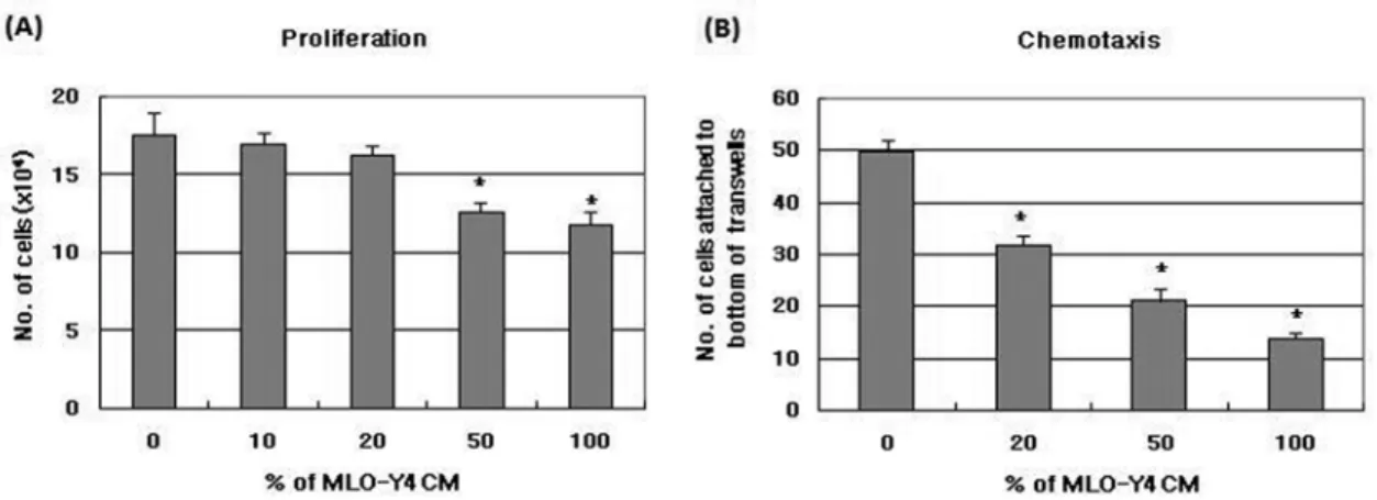
골세포가 조골세포 증식 및 이동에 영향을 주는지 알 아보기 위하여 MLO-Y4 세포와 조골세포주인 2T3 세포 를 이용하여 proliferation assay와 chemotaxis assay를 실시하였다. MLO-Y4 CM은 낮은 농도 (10, 20 %)에서 는 2T3 세포의 세포증식에 영향을 미치지 못하였으나 높 은 농도 (50, 100 %) 에서는 증식을 억제하였고 (Fig. 2A), 용량의존적으로 2T3 세포의 이동을 억제하였다 (Fig. 2B).
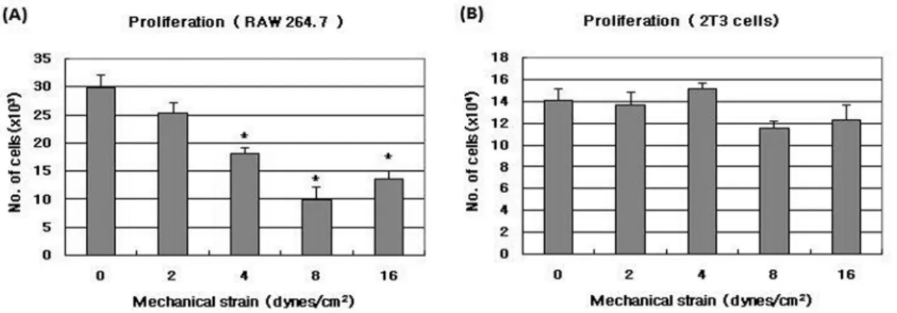
Fluid flow shear stress가 골개조에 미치는 골세포의 작용을 변화시키는지 알아보기 위하여 MLO-Y세포에 2, 4, 8, 16± 0.6 dynes/cm2의 pulsatile fluid flow를 가한 후 24시간 동안 조건배양액을 모아 파골세포 전구세포와 조골세포의 증식과 이동에 미치는 영향을 알아보았다.
MLO-Y4 세포에 의한 파골세포 전구세포 증식 증가 효 과는 pulsatile fluid flow shear stress에 의하여 감소되 었으며 4, 8, 16 dynes/cm2 에서 대조군에 비하여 유의하 게 감소하였다 (Fig. 3A). 조골세포 증식의 경우에는 shear stress를 가한 후의 MLO-Y4 CM이 대조군과 차이를 보 이지 않았다 (Fig. 3B). 파골세포 전구세포의 이동은 MLO-Y4 세포에 fluid flow shear stress를 가한 후 수 집한 조건배양액에 의하여 shear stress를 가하지 않은 대 조군에 비하여 감소되는 경향을 보였으며, 16 dynes/cm2 shear stress를 가한 경우에 대조군보다 유의하게 감소하 였다 (Fig. 4A). MLO-Y4 세포에 fluid flow shear stress를 가한 후 조건배양액을 가지고 조골세포의 이동
을 측정한 결과 모든 범위에서 조골세포의 이동이 감소 하였다 (Fig. 4B). 이는 골세포가 기계적 장력에 반응하 여 파골세포 전구세포의 증식과 이동을 조절하여 골흡수 를 억제할 수 있음을 보여준 결과라 생각된다.
MLO-Y4 세포에서 2, 8 dynes/cm2 fluid flow shear stress를 가한 경우 M-CSF 의 발현을 대조군과 큰 차이 를 보이지 않았으나 파골세포의 분화를 유도하는 것으로 알려진 RANKL의 발현은 대조군에 비하여 감소하였다.
또한 파골세포 분화 억제물질인 OPG 발현은 2, 8 dynes/
cm2 의 fluid flow shear stress를 가한 경우 모두 대조 군에 비하여 증가하였다 (Fig. 5).
Fig. 1. The effect of MLO-Y4 conditioned medium (CM) on proliferation (A) and transmigration (B) of MOCP-5 cells. (A) Proliferation assay; MOCP-5 cells were seeded 2000 cells/well in 48 well plates. After 6hrs, control media or MLO-Y4 CM was added. Cultures were stopped after 3 days and cell number was counted. (B) Chemotaxis assay; The migration of MOCP-5 cells was assayed using transwell. The control media or MLO-Y4 CM were placed in the lower wells. MOCP-5 cells (50,000) were placed in the transwell (upper well). The plates were incubated for 4 hrs, and cells attached bottom of the transwell were fixed and stained with crystal violet. The migrating cells were counted using reflection microscope and image analysis Results were presented with mean± S.E. (N=4) and statistical differences were ana- lyzed using the Student t-test. *p<0.05, compared to control (0 % CM).
Fig. 2. The effect of MLO-Y4 conditioned medium (CM) on proliferation (A) and transmigration (B) of 2T3 cells. (A) Proliferation assay;
2T3 cells were seeded 20,000 cells/well in 12 well plates. After 6hrs, control media or MLO-Y4 CM was added. Cultures were stopped after 3 days and cell number was counted. (B) Chemotaxis assay; The migration of 2T3 cells was assayed using transwell. The control media or MLO-Y4 CM were placed in the lower wells. 2T3 cells (50,000) were placed in the transwell (upper well). The plates were incubated for 4 hrs, and cells attached bottom of the transwell were fixed and stained with crystal violet. The migrating cells were counted using reflection microscope and image analysis Results were presented with mean± S.E. (N=4). *p<0.05, compared to control (0 % CM)
고 찰
골개조는 골조직 특정부위로 파골세포 전구세포가 이동, 분화되어 골흡수(targeted removal)가 유도됨으로 시작된 다. 파골세포에 분화에 대한 조골세포의 기능과 분자적 기전을 잘 알려져 있으나 이러한 골개조 기시(initiation) 의 분자적 기전에 관하여는 알려진 바가 없다. 골세포는 골조직의 lacunae내에 존재하며 이 부위는 석회화되지 않 고 fluid가 흐르고 있어 골세포에 영양분을 공급하므로 경조직내에서 세포가 생존하게 된다. 이런 골세포의 경조 직 내의 위치는 골세포가 골조직에 가해지는 기계적 자 극을 감지하고 반응하여 기계적 자극에 의한 골조직 변
화의 세포수준의 기전에 주요한 중심세포로서 역할을 할 수 있을 것이라는 가정을 할 수 있게 한다. 또한 골세포 는 canaliculi를 통해 골조직 표면에 위치한 조골세포, 파 골세포 전구세포등과 연결되어 여러 가지 신호를 주고 받을 수 있을 것으로 생각되어진다. 본 연구에서는 MLO- Y4 세포를 이용하여 골세포가 조골세포 및 파골세포 전 구세포 모집과 증식에 영향을 미치는지 조사하였다. MLO- Y4세포 조건배양액은 파골세포 전구세포 세포주인 MOCP- 5, 및 RAW264.7 세포의 증식 및 이동을 증가시켰다. 이 는 정상적인 골세포가 파골세포 전구세포에 신호를 보내 골파괴를 조절할 수 있음을 보여주는 결과이다. 골조직에 피로가 가해지고 연속적인 microcrack이 생성되면 micro- Fig. 3. The effect of pulsatile fluid flow shear stress on cell proliferation of osteoclast precursor cells (A) and osteoblastic cells (B) induced by MLO-Y4 conditioned medium. After MLO-Y4 cells had been subjected to pulsatile fluid - flow shear stress at 0, 2, 4, 8, 16 ± 0.6 dynes/
cm2 , 5 Hz for 2 hrs, the MLO-Y4 conditioned medium was collected. RAW 264.7 cells were seeded 2000 cells/well in 48 well plates and2T3 cells were seeded 20,000 cells/well in 12 well plates. After 6hrs, control media or 20 % MLO-Y4 CM after exposed to pulsatile fluid flow was added. Cultures were stopped after3 days and cell number was counted. Results were presented with mean ± S.E. (N=4). * P<
0.05compared to control (0 dynes/cm2).
Fig. 4. The effect of pulsatile fluid flow shear stress on cell migration of osteoclast precursor cells (A) and osteoblastic cells (B) induced by MLO-Y4 conditioned medium. After MLO-Y4 cells had been subjected to pulsatile fluid - flow shear stress at 0, 2, 4, 8, 16± 0.6 dynes/
cm2, 5 Hz for 2 hrs, the MLO-Y4 conditioned medium was collected. The migration of RAW 264.7 cells and 2T3 cells was assayed using transwell. The control media or 20 % MLO-Y4 CM after exposed to pulsatile fluid flow were placed in the lower wells. RAW 264.7 cells or 2T3 cells (50,000) were placed in the transwell (upper well). The plates were incubated for 4 hrs, and cells attached bottom of the transwell were fixed and stained with crystal violet. The migrating cells were counted using reflection microscope and image analysis. Results were presented with mean± S.E. (N=4). * P< 0.05 compared to control (0 dynes/cm2).
damage가 되고, 이러한 microdamage의 회복은 골세포의 생존과 관계가 있어 상처입거나 세포사멸 골세포가 micro- damage의 회복신호로 골흡수를 시작하는 신호를 보낸다 는 보고가 있었으며 (Lanyon, 1993), Gu 등은(2005) 쥐 두개골 조직배양 (rat calvaria organ culture)을 이용한 연구에서 골세포가 골흡수를 억제하고 있으며 골세포의 사멸로 인해 이러한 골흡수억제 작용이 끝나고 골흡수가 시작된다고 보고하였다. 본 연구에서는 세포사멸상태의 골세포가 아닌 정상 골세포도 골흡수를 유도하는 신호를 파골세포 전구세포에 보낼 수 있음을 보여주어 Gu등과 는 다른 결과를 보였다. 두개골 조직배양시에는 골세포뿐 아니라 조골세포등의 다른 골조직의 세포들로 영향을 주 어 복합적인 결과를 보였을 것으로 생각된다. M-CSF는 파골세포 전구세포의 생존에 반드시 필요하고 그 증식을 촉진하는 물질로 알려져 있으며 (Komada 등, 1991) MLO-Y4에 의해 세포막에 존재하는 유형과 분비되는 유 형의 M-CSF가 생성되며 MLO-Y4 세포에 의한 파골세 포 분화 유도에 기여함이 보고되었다 (Zhao 등, 2002).
그러므로 본 연구 결과인 MLO-Y4 세포에 의한 파골세 포 전구세포 증식 촉진에 M-CSF가 관여할 것이라 생각 되며 이는 M-CSF 항체를 이용한 blocking assay를 통 해 확인하였다 (Ko 등, 2002). MLO-Y4 세포에서 생성
되며 monocyte, 파골세포 계통의 세포에 화학주성 효과 가 있는 M-CSF, transforming growth factor β (TGF- β), monocyte chemoattractant protein 3 (MCP-3), macrophage inflammatory protein 2 (MIP-2), vascular endothelial growth factor (VEGF) (Engsing 등, 2000;
Zheng 등, 1994; Fuller 등 1993; Fuller 등, 1995) 등 이 MLO-Y4 세포에 의한 파골세포 전구세포의 화학주 성을 담당할 것으로 생각되나 골세포에 의한 파골세포 전구세포 증식과 이동을 매개하는 정확한 물질을 알아내 기 위한 계속적인 연구가 필요하다. 또한 세포사멸상태의 골세포가 파골세포 전구세포의 증식과 모집에 미치는 역 할을 조사하고 정상 골세포와 그 영향을 비교하는 연구 가 필요하며, 이때 작용하는 물질에 관한 연구도 진행되 어야 할 것이다.
MLO-Y4 세포의 조골세포의 증식과 이동의 대한 영향 을 관찰한 결과 높은 농도의 MLO-Y4 CM에서 조골세 포의 증식과 이동을 감소시킴을 알 수 있었다. 이는 골 세포의 조건배양액이 골수 mesechymal stem cell의 증 식과 분화를 모두 증가시켰다는 Heino 등(2004)의 결과 와는 일치하지 않으나 이는 사용한 조골세포가 본 실험 에서 사용된 2T3 cell에 비하여 그 분화도가 낮은 골수 mesechymal stem cell로서 본 실험과 다른 결과를 보인 것으로 생각된다.
골세포의 세포주위 액체 (pericellular fluid)에 가해지는 shear stress를 골세포가 감각하고 반응함으로써 골조직에 가해지는 기계적 장력이 전하여지고 골개조에 영향을 미 칠 것이라는 여러 선행된 연구가 있어 왔으며 pulsatile fluid flow (PFF) 가 골세포에 가해지는 기계적 장력의 크기와 시간을 비교적 생체내에서와 비슷하게 모방한다 는 보고들이 있다 (Klein-Nulend 등, 1995a). 이에 MLO- Y4 세포에 PFF를 가하고 MLO-Y4 세포의 파골세포 전 구세포와 조골세포에 대한 영향을 조사하였다. PFF shear stress를 가한 후의 MLO-Y4세포에 의한 조골세포 증식 은 대조군과 차이를 보이지 않았으며 조골세포의 이동은 모든 범위에서 감소하였다. 닭의 골세포와 조골세포를 이 용한 Vezeridis 등 (2006)의 연구에서는 골세포는 PFF에 노출 후 조골세포 증식을 억제시키고 조골세포 분화지표 인 알카리성 인산분해효소활성은 증가시켜 조골세포 분 화를 촉진하여 골형성을 증가시키는 신호를 보낼 것이라 고 보고하였다. 본 실험은 이와 완전히 일치하지 않으나 MLO-Y4 세포는 PFF에 노출되지 않은 상태에서 조골세 포의 증식을 억제시킴을 알 수 있었다. 또한 파골세포 전 구세포 증식 증가 효과는 PFF shear stress에 의하여 감 소되었으며 파골세포 전구세포의 이동은 shear stress를 가하지 않은 대조군에 비하여 감소되는 경향을 보였다.
이는 골세포가 기계적 장력에 반응하여 파골세포 증식, 이동을 감소시켜 골흡수를 억제할 수 있음을 보여주는 결과이다. 골흡수를 담당하는 파골세포는 조골세포에 의 Fig. 5. The effect of pulsatile fluid flow shear stress on M-CSF,
OPG and RANKL mRNA expression in MLO-Y4 cells. After MLO-Y4 cells had been subjected to pulsatile fluid - flow shear stress at 0, 2, 8± 0.6 dynes/cm2 , 5 Hz for 2 hrs, total RNA was extracted from cells. cDNA was prepared by use of a Superscript II system. Sequence of the primers for PCR described in Material and Methods. Amplification was carried out for 20-27 cycles under sat- uration, each at 95oC, 1 min; 55oC, 30s; 72oC, 1 min. After ampli- fication, 20µl of each reaction mixture was analyzed by 2 % agarose gel electrophoresis and the bands were visualized by ethid- ium bromide staining. The PCR products for M-CSF, OPG, RANKL and β- actin were 362, 503, 568 and 635 bp respectively.
하여 분화가 진행되며 그 과정을 조골세포에서 생성하는 RANKL와 파골세포 전구세포 세포막의 RANK 간의 상 호작용으로 일어나며 분화 억제인자인 OPG 또한 조골세 포가 생성하는 것이 알려져 있다 (Yasuda 등, 1998;
Simonet 등, 1997). MLO-Y4 세포는 세포의 접촉에 의 해 파골세포의 형성과 활성을 유도할 수 있고, M-CSF 과 RNAKL을 생성함이 보고되어 (Zhao 등, 2002), PFF 에 의한 MLO-Y4 세포에서의 M-CSF, RNAKL, OPG 의 mRNA 발현의 변화를 관찰하였다. 2 및 8 dynes/cm2 의 PFF 에 노출된 MLO-Y4 세포에서 RANKL의 발현 은 감소하였고 OPG 발현은 증가하였다. 이는 You등의 (2007) 보고와 일치하는 것으로 기계적 장력에 의하여 골 세포가 파골세포 전구세포의 분화를 감소시켜 골흡수를 억제시킬 수 있음을 보여주었다. 이상의 결과로 골세포가 골조직의 특정부위로 파골세포 전구세포를 모으고 그 증 식을 조절함으로 골개조를 필요한 특정부위에서 시작되 게 하는 기능이 있으며 또한 파골세포 전구세포 및 조골 세포에 여러 신호를 보내 증식 및 분화를 조절할 수 있 으며 기계적 장력에 반응하여 골흡수를 억제하는 신호를 보냄으로 골개조를 조절할 것으로 생각된다.
감사의 글
이 논문은 정부(교육인적자원부)의 재원으로 한국학술진 흥재단의 지원을 받아 수행된 연구임 (R04-2004-000- 10146-0)
참 고 문 헌
Ajubi NE, Klein-Nulend J, Nijweide PJ, Vrijheid-Lammers T, Alblas MJ, Burger EH. Pulsating fluid flow increases prostaglandin production by cultured chicken osteocytes- A cytoskeleton-dependent process. Biochem Biophys Res Commun. 1996;225:62-8.
Burr DB, Robling AG, Turner CH. Effects of biomechanical stress on bones in animals. Bone. 2002;30(5):781-6.
Chen W, Li YP. Generation of mouse osteoclastogenic cell lines immortalized with SV40 large T antigen. J Bone Miner Res. 1998;13:1112-23.
Engsig MT, Chen QJ, Vu TH, Pedersen AC. Therkidsen B, Lund LR, Henriksen K, Lenhard T, Foged NT, Werb Z, Delaisse JM. Matrix metalloproteinase 9 and vascular endothelial growth factor are essential for osteoclast recruitment into developing long bones. J Cell Biol.
2000;151(4):879-89.
Fuller K, Owens JM, Jagger CJ, Wilson A, Moss R, Chambers TJ. Macrophage colony-stimulating factor stimulates survival and chemotactic behavior in isolated osteoclasts. J Exp Med. 1993;178(5):1733-44.
Fuller K, Owens JM, Chambers TJ. Macrophage inflammatory protein-1 alpha and IL-8 stimulate the motility but suppress the resorption of isolated rat osteoclasts. J Immunol.
1995;154(11): 6065-72.
Ghosh-Choudhury N, Windle JJ, Koop BA, Harris MA, Guerrero DL, Wozney, JM, Mundy GR. Harris SE.
Immotalized murine osteoblasts derived from BMP-2-T- antigen expressing transgenic mice. Endocrinology.
1996;137(1) : 331-9.
Gu G, Mulari M, Peng Z, Hentunen TA, Vaananen HK. Death of osteocytes turns off the inhibition of osteoclasts and triggers local bone resorption. Biochem Biophys Res Commun. 2005;335:1095-101.
Heino TJ, Hentunen TA, Vaananen HK. Conditioned medium from osteocytes stimulates the proliferation of bone marrow mesenchymal stem cells and their differentiation into osteoblasts. Exp Cell Res. 2004;294: 458-68.
Hsu H, Lacey DL. Dunstan CR, Solovyev I, Colombero A, Timms E, Tan HL, Elliott G, Kelley MJ, Sarosi I, Wang L, Xia XZ, Elliott R, Chiu L, Black T, Scully S, Capparelli C, Morony S, Shimamoto G, Bass MB, Boyle WJ. Tumor necrosis factor receptor family member RANK mediates osteoclast differentiation and activation induced by osteo- protegerin ligand. Proc Natl Acad Sci USA. 1999;96:3540-45.
Kamioka H, Honjoa T, Takano-Yamamoto T. A three- dimensional distribution of osteocyte processes revealed by the combination of confocal laser scanning microscopy and differential interference contrast microscopy. Bone.
2001;28:145-9.
Kato Y, Windle JJ, Koop BA, Mundy GR, Bonewald LF.
Establishment of an osteocyte-like cell line, MLO-Y4. J Bone Miner Res. 1997;12:2014-23.
Klein-Nulend J, van der Plas A, Semeins CM, Frangos JA, Nijweide PJ, Burger EH. Sensitivity of osteocytes to biomechanical stress in vitro, FASEB J. 1995a;9:441-5.
Klein-Nulend J, Semeins CM, Ajubi NE, Nijweide PJ, Burger, EH. Pulsating fluid flow increases nitric oxide (NO) synthesis by osteocytes but not periosteal fibroblasts- Correlation with prostaglandin upregulation. Biochem Biophys Res Commun. 1995b;217:640-8.
Ko S, Harris SE, Dallas M, Bonewald LF. A potential role for osteocytes in recruiting osteoclast precursors to targeted bone sites. J Bone Mir Res. 2002;17 (suppl): s353.
Komada H, Nose M, Niida S, Kasai S, Yamamoto A. Essential role of macrophage colony-stimulating factor in the osteoclast differentiation supported by stromal cells. J Exp Med. 1991;173: 1291-4.
Lanyon LE. Osteocytes, strain detection, bone modeling and remodeling. Calicf Tissue Int. 1993;53(Suppl 1): S102-S6, Lean JM, Jagger CJ, Chambers TJ, Chow JWM. Increased
insulin-like growth factor I mRNA expression in rat osteocytes in response to mechanical stimulation. Am J Physiol Endocrinol Metab. 1995;268: E318-E27.
Mason DJ, Suva LJ, Genever PJ, Patton AJ, Steuckle S, Hillam RA, Skerry TM. Mechanically regulated expression of a neural glutamate transporter in bone: A role for excitatory amino acids as osteotropic agents? Bone.
1997;20: 199-205.
Matsumoto T, Nakayama K, Kodama Y, Fuse H, Nakamura T, Fukumoto S. Effect of mechanical unloading and reloading on periosteal bone formation and gene expression in tail- suspended rapidly growing rats. Bone. 1998;22: 89S-93S.
Rawlinson SCF, Mosley JR, Suswillo RFL, Pitsillides AA, Lanyon LE. Calvarial and limb bone cells in organ and monolayer culture do not show the same early responses to dynamic mechanical strain. J Bone Min Res. 1995;10: 1225-32.
Simonet WS, Lacey DL, Dunstan CR, Kelley M, Chang MS, Luthy R, Nguyen HQ, Wooden S. Osteoprotegerin : a novel secreted protein involved in the regulation of bone density.
Cell . 1997;89:309-19.
Sun Y-Q, McLeod KJ, Rubin CT. Mechanically induced periosteal bone formation is paralleled by the upregulation of collagen type one mRNA in osteocytes as measured by in situ reverse transcript-polymerase chain reaction. Calcif Tissue Int. 1995; 57:456-62.
Tomkison A, Reeve J, Shaw RW, Noble BS. The death of osteocytes via apoptosis accompanies estrogen withdrawal in human bone. J Clin Endocrinol Met. 1997;82(9):3128-35.
Turner CH, Forwood MR, Otter MW. Mechanotransduction in bone: Do bone cells act as sensors of fluid flow? FASEB J.
1994; 8:857-78.
Verborgt O, Gibson GJ, Schffler MB. Loss of osteocyte integrity in association with microdamage and bone remo- deling after fatigue in vivo J Bone Min Res. 1999;15(1) : 1-8.
Vezeridis PS, Semeins CM, Chen Q, Klein-Nulend J.
Osteocytes subjected to pulsating fluid flow regulate osteoblast proliferation and differentiation. Biochem Biophys Res Commun. 2006;348(3):1082-8.
Weinbaum S, Cowin SC, Zeng Y. A model for excitation of
osteocytes by mechanical loading induced fluid shear stresses. J Biomech. 1994;27:339-60.
Westbroek I, Ajubi NE, Alblas MJ, Semeins CM, Klein- Nulend J, Burger EH, Nijweide PJ. Differential stimulation of prostaglandin G/H synthase-2 in osteocytes and other osteogenic cells by pulsating fluid flow. Biochem Biophys Res Commun. 2000;268 : 414-9.
Winkler DG, Surtherland MK, Geoghegan JC, Yu C, Hayes T, Skonier JE. Osteocyte control of bone formation via sclerostin, a novel BMP antagonist. EMBO J. 2003;22; 6227-76.
Yasuda H, Shima N, Nakagawa N, Yamaguchi K, Masahiko K, Mochizuki S, Tomoyasu A, Yano K, Goto M, Murakami A, Tsuda E, Morinaga T, Higashio, K, Udagawa N, Takahashi N, Suda T. Osteoclast differentiation factor is a ligand for osteoprotegerin/osteoclastogenesis -inhibitory factor and is identical to TRANCE/RANKL. Proc Natl Acad Sci USA. 1998;95:3597-602.
You L, Temiyasathit S, Lee P, Kim CH, Tummala P, Yao W, Kingery W, Malone AM, Kwon RY, Jacobs, CR. Osteocytes as mechanosensors in the inhibition of bone resorption due to mechanical loading. Bone. 2008;42(1): 172-9.
Zhao S, Kato Y, Zhang Y, Harris S, Ahuja S, Bonewald LF.
MLO-Y4 osteocyte-like cells support osteoclast formation and activation without the addition of 1,25(OH)2D3. J Bone Miner Res. 2002; 17:2068-79.
Zheng MH, Fan Y, Wysocki SJ, Lau AT, Robertson T, Beilharz M, Wood DJ, Papadimitriou JM. Gene expression of transforming growth factor-beta 1 and its type II receptor in giant cell tumors of bone. Possible involvement in osteo- clast-like cell migration. Am J Pathol. 1994;145(5):1095-104.