- 61 -
R eceived R e v i s e d A ccepted
: July 10, 2018 : August 5, 2018 : August 7, 2018
+Corresponding author: Soon Young Kwon
123, Jeokgeum-ro, Gojan-dong, Danwon-gu, Ansan-si, Gyeonggi-do, Republic of Korea, 15355
Tel: +82-31-412-4920, Fax: +82-31-412-5174 E-mail: entkwon@korea.ac.kr
대한두경부종양학회지, 제34권 제2호, 2018. pp.61-64 Korean Journal of Head & Neck Oncology, Vol.34, No.2
https://doi.org/10.21593/kjhno/2018.34.2.61 ISSN 1229-5183(Print) / ISSN 2586-2553(Online)
갑상선을 침범한 기관암 환자 1례
임강현1⋅정용준1⋅한문수1⋅이주한2⋅김영식2⋅오경호1⋅권순영1+
고려대학교 의과대학 안산병원 이비인후-두경부외과학교실1, 고려대학교 의과대학 안산병원 병리학교실2
A case of tracheal cancer with thyroid invasion
Kang Hyeon Lim, MD1, Yong Jun Jeong, MD1, Mun Soo Han, MD1, Ju Han Lee, MD, PhD2, Young Sik Kim, MD, PhD2, Kyung Ho Oh, MD, PhD1, Soon Young Kwon, MD, PhD1+
Department of Otolaryngology-Head and Neck Surgery, College of Medicine, Korea University Ansan Hospital, Ansan, Korea1 Department of Pathology, College of Medicine, Korea University Ansan Hospital, Ansan, Korea2
= Abstract =
Malignant lesion of the trachea predominantly results from direct spread of adjacent tumors, whereas primary tracheal malignancies are rarely observed. Tracheal tumors are usually diagnosed late on account of delayed specific symptoms: dyspnea, stridor, coughing and hemoptysis. Primary tracheal tumors, although very rare, may extend into the thyroid gland and present as a thyroid mass. Surgery, followed by adjuvant radiotherapy, is the treatment of choice. A case of primary tracheal cancer with thyroid invasion is reported, and a review of the literature is presented.
Key W ords : Primary tracheal cancer, Thyroid invasion, Squamous cell carcinoma
서 론
기관(trachea)에 존재하는 대부분의 악성종양은은 폐와 식도, 후두, 갑상선 같은 주변 구조물의 악성종양이 직접 침윤(direct invasion)하여 나타나고, 원발성 기관암(tracheal cancer)은 드물다.1,2)원발성 기관암의 발생률은 연간 인 구 100,000명 당 0.1명으로 보고되었고, 이는 모든 악성 종양의 0.02%-0.04%에 해당한다.2)조직학적으로는 편평 세포암종(squamous cell carcinoma)이 가장 흔한 형태이 며, 원발성 기관암의 44.8%-54.5%를 차지한다.1,3,4)한편, 갑상선에 생기는 편평세포암종은 빠르게 증식하는 특성
이 있어 주변조직을 침범하는 경우가 많다.5,6) 갑상선과 경부 기관(cervical trachea)은 해부학적으로 인접해 있기 때문에 원발성 갑상선암이 기관을 침범하는 경우는 흔하 며, 경부 기관에서 발생한 악성종양이 갑상선을 침범하 기도 한다.7) 하지만 임상소견 및 영상검사만으로 원발 부위를 식별하는데 어려움이 있을 수 있다.4)본 연구에 포함된 증례도 갑상선을 침범한 기관암이지만, 수술 전 임상적 진단 시에는 기관을 침범한 갑상선 편평세포암종 으로 생각되었던 경우이다. 저자들은 갑상선을 침범한 기관암 증례 1례를 문헌고찰과 함께 보고하고자 한다.
증 례
77세 여자가 6개월 전부터 만져지는 전경부 종물 및 3주 전부터 점차 악화되는 흡기성 호흡곤란 증상으로 본 원 이비인후과 외래를 방문하였다. 문진 상 특이 병력은 없었고, 흡연력도 없었다. 내원 당시 시행한 신체진찰 상 에서는 전경부의 약 5cm 크기로 촉지되는 종물이 있어
- 62 - A
B
C
D
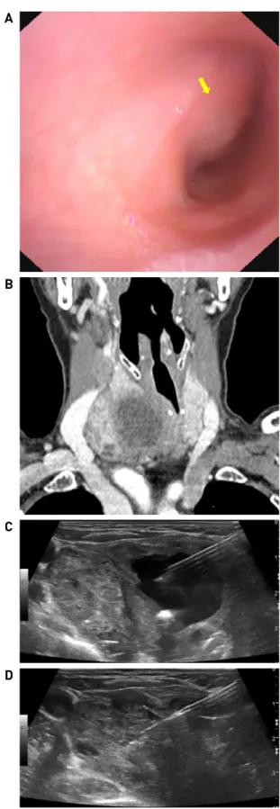
Fig. 1. (A) A lesion, occupying about 50% of the lumen (arrow) was observed in the cervical trachea with a flexible laryngoscope. The abnormal finding of mucosa was not observed. (B) A 4.5 cm sized heterogeneous mass, revealed an unclear margin with the right thyroid parenchyma, and trachea deviation to the left were shown in computed tomography. (C) A 3.3 cm cystic portion in the center of the mass was observed in ultrasonography. (D) A fine needle as- piration was performed in the remaining solid portion, after collapsing the central cyst.
갑상선 비대 혹은 종양이 의심되었으며 압통은 없었다.
흡기성 호흡곤란의 원인 감별을 위해서 굴곡 후두 내시경 을 시행하였다. 내시경상 양측 성대의 움직임은 정상이 었으나 경부 기관 내강이 우측에서 50% 정도 좁아져 있 었고, 기관 점막으로의 침윤은 관찰되지 않았다(Fig. 1A).
추가적인 평가 및 치료를 위하여 입원하여 갑상선 기 능검사와 경부 전산화단층촬영 및 초음파 유도 세침흡인 검사를 시행하였다. 전산화단층촬영에서 우측 윤상연골 외측에서 4.5cm 크기의 불균일한 조영증강을 보이면서 경부기관을 좌측으로 편위 시키는 종물이 확인되었고, 해당 종물은 우측 갑상선 실질과의 경계가 불명확하였다 (Fig. 1B). 또한, 우측 경부 구역 III, IV와 좌측 경부 구역 III에서 다발성 림프절 비대소견이 관찰되었다. 초음파 검사에서는 경부 종물 내부에 3.3cm 크기의 낭성 병변이 확인되어 세침흡인으로 낭을 허탈(collapse) 시킨 이후 남은 고형질 부위에서, 그리고 지방문(fatty hilum)이 소 실되어 경부전이가 의심되는 우측 경부림프절에서 세침 흡인 및 세포검사를 실시하였다(Fig. 1C and D). 경부 종 물에서 시행한 세포검사에서는 비정형 편평세포(atypical squamous cells)가 확인되었지만, 우측 경부림프절에서는 악성세포가 확인되지 않았다. 갑상선기능검사는 정상이 었다.
입원 후 일련의 검사를 시행하던 중 환자의 호흡곤란 이 급격히 악화되어 기관절개술을 시행하였고, 이때 경 부 종물에 대한 절개생검도 같이 시행하였으며, 조직검 사 결과에서 편평세포암종이 확인되었다. 전신 양전자 방출촬영에서는 우측 경부 종물과 양측 경부 림프절(우 측 구역 III, IV 및 좌측 구역 III)에서 포도당 섭취율이 증가되어 있었으나 원격 전이 소견은 없었다.
이상의 검사 결과들을 종합하였을 때, 경부 기관을 침 범한 갑상선 편평세포암 및 양측 경부림프절 전이를 임 상적으로 의심하였고, 전신마취 하에 근치절제술 및 양 측 변형-근치경부절제술을 시행하였다. 수술 중 우측 갑 상선을 침범한 장경 5cm의 종괴와 함께 종양이 기관 내 강으로 침윤된 소견이 관찰되었다. 이에 해당 종괴와 함께 갑상선 우엽 전체와 좌엽의 일부, 그리고 기관에 침윤된 부위를 포함한 3cm 가량의 경부 기관을 충분한 절제연을 확보하며 일괄로 절제한 후, 기관 단단문합술(tracheal end-to-end anastomosis)을 시행하였다. 수술 후 방사성 요 오드 치료에 효과가 없을 것이므로 좌측 갑상선엽의 일 부는 보존하였다. 현재까지 갑상선 편평세포암종의 희 귀성 때문에 수술 범위에 대한 합의점이 존재하지는 않 으나 갑상선 기능 보존을 고려하였을 때, 갑상선 전 절제 술이 충분한 절제연을 확보하여 갑상선의 일부를 보존하
- 63 - A
B
Fig. 2. (A) Microscopic finding of the specimen showed squamous cell carcinoma, which originate from the upper portion of the trachea, was attached to the thyroid gland (×12.5, H-E staining). (B) The more magnified specimen revealed well-differ- entiated squamous cell carcinoma with keratinization (×200, H-E staining).
A
B
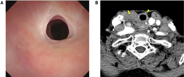
Fig. 3. (A) The cervical tracheal lumen was observed to be patent in a flexible laryngoscope a year after the surgery. (B) Follow-up computed tomography after a year showed diffuse soft tissue infiltrations at the right thyroid operative bed (arrow) and left anterior side of the trachea (arrowhead), but those are likely to be radiation-induced changes as having not changed for a year.
는 갑상선 아전 절제술(subtotal thyroidectomy) 보다 뚜렷 한 이득이 없을 것이라 생각하였다.
병리결과에서 경부 종괴는 5.0x3.7cm의 고분화 편평세 포암종(well-differentiated squamous cell carcinoma)으로 확인되었으며, 현미경으로 보았을 때 기관에서 기원하 여 갑상선 조직에 유착되어 있는 소견이 관찰되어 임상 적 진단과는 달리 기관암종의 갑상선 침범으로 최종 진 단하였다(Fig. 2). 수술 절제연은 모두 음성이었으나 좌 측 절제연이 종물과 매우 가까운 것으로 확인되었고, 절 제된 양측 경부의 111개의 림프절로의 전이는 없었다.
환자는 술 후 방사선치료(누적 선량 66Gy)를 시행 받았 고, 특이 합병증 없이 12개월 째 외래 추적 관찰 중이다.
술 후 6개월 및 12개월에 시행한 경부 전산화단층촬영과 정기적인 신체진찰 및 내시경검사상에서 재발의 증거는 보이지 않고 있으며(Fig. 3A and 3B), 갑상선호르몬 수치 는 정상(TSH 2.91μIU/mL, Free T4 0.99ng/dL, T3 33.7ng/dL) 으로 유지되고 있다.
고 찰
성인에서 기관에 발생하는 원발성 종양의 90%는 악성 신생물이다.1,2)하지만 기관에 발생하는 종양 자체가 매 우 드물어 관련 역학이나 치료 방법, 예후와 관련된 자료가 제한되어 있으며, WHO 분류(World Health Organization Classification)에서도 기관의 종양에 대한 독립된 조직 분 류 체계가 없다.1,2)다만, 두경부 종양 WHO 분류에서 하 인두 및 후두의 종양과 함께 기관의 종양을 포괄하여 조직학적으로 분류하였고, 기관에서는 편평세포암종과 샘낭암종(adenoid cystic carcinoma)이 70%이상을 차지하 며 편평세포암종이 가장 흔하다.2)
기관암은 호흡곤란과 객혈, 기침 등의 증상을 유발할 수 있으나, 초기에는 대부분 증상이 없어서 진단이 늦어 지는 경우가 많다.8,9) 기관암의 임상양상은 발생위치와 크기, 조직학적 아형에 따라 다양하게 나타날 수 있다.10) 경부 기관암의 경우 상기도 폐색으로 인해 흡기성 천명
- 64 - 및 협착음, 체위성 호흡곤란이 나타날 수 있고, 기관점막의 자극이나 궤양으로 인해 기침, 객혈이 발생할 수 있다.11) 편평세포암종의 경우에는 주로 기관 점막을 침범하여 점막 자극 증상을 일으키고 샘낭암종은 주로 점막하 침 윤을 일으켜서 상기도 폐색으로 인한 호흡곤란이 초기 임상 증상으로 알려져 있다.11,12) 그러나 본 증례에서는 기관의 편평세포암종임에도 환자는 기관 점막의 자극 증상을 호소하지 않았으며 굴곡 후두 내시경 상에서 기 관 점막 내부로 종양의 침범이 저명하지 않았다.
기관벽을 침범한 갑상선암은 자주 볼 수 있으며, 기관 내강까지 침범하는 경우도 있다.7)특히 갑상선 암종의 0.2%에서 1.1%를 차지하는 편평세포암종은 빠르게 증식 하는 특성 때문에 진단 시 주변조직으로의 침윤을 동반한 경우가 흔하다.5,6) 빈도가 높진 않으나 원발성 기관암의 경우에도 갑상선을 침범하는 경우가 있다.13)원발성 기 관암 환자를 후향적으로 분석한 한 연구에서는 기관암의 54.5%(30/55)가 편평세포암종이었으며, 약 18%(10/55)의 환자에서 인접한 구조물로 직접 침윤을 보였고, 약 7%
(4/55)에서 갑상선의 침윤소견을 보였다.4)
기관암의 치료 방법에는 수술과 방사선치료, 근접방사 선치료(brachytherapy), 내시경적 중재법(interventional en- doscopy), 항암치료 등이 있다.8,14)기관암 환자의 약 50%
에서 수술적 치료가 선택될 수 있고, 기관의 구획절제술 (segmental resection)이 가장 효과적인 치료법으로 알려져 있으며, 수술 후 3년 및 5년 생존률은 편평세포암종에서 각각 80% 및 20%로 나타났다.15)또한, 수술 후 방사선요 법이 방사선 단독 치료 또는 항암방사선치료 보다 기관암 환자의 생존율 면에서 유의하게 효과적이다.15)
흔치 않은 갑상선을 침범한 기관암 1례를 보고하였다.
내시경상에서 경부 기관이 편위되어 있고 기관 점막의 침윤소견이 없다는 점에서, 임상양상이 전형적인 기관 평 편세포암종에 부합하지 않아 갑상선암이 기관을 침범한 것으로 생각되었으나, 병리결과에서는 기관에서 기원한 암종이 갑상선 조직에 유착되어 있는 소견을 보여 최종적 으로는 기관암종이 갑상선을 침범한 것으로 확인되었다.
갑상선 종양을 평가할 때에는 종양이 다른 기관(organ)에 서 전이되었거나 주변 구조물에서 발생한 암이 갑상선을 침범했을 가능성도 고려해야 할 것으로 생각된다.
References
1) Honings J, Van Dijck JA, Verhagen AF, Van Der Heijden HF, Marres HA. Incidence and treatment of tracheal cancer: a na- tionwide study in the Netherlands. Ann Surg Oncol. 2007;14(2):
968-976.
2) Junker K. Pathology of tracheal tumors. Thorac Surg Clin. 2014;
24(1):7-11.
3) Urdaneta AI, Yu JB, Wilson LD. Population based cancer regis- try analysis of primary tracheal carcinoma. Am J Clin Oncol.
2011;34(1):32-37.
4) Li W, Ellerbroek NA, Libshitz HI. Primary malignant tumors of the trachea. A radiologic and clinical study. Cancer. 1990;66(5):
894-899.
5) Rosa M, Toronczyk K. Fine needle aspiration biopsy of three cases of squamous cell carcinoma presenting as a thyroid mass:
cytological findings and differential diagnosis. Cytopathology.
2012;23(1):45-49.
6) Cook AM, Vini L, Harmer C. Squamous cell carcinoma of the thyroid: outcome of treatment in 16 patients. Eur J Surg Oncol.
1999;25(6):606-609.
7) Felson B. Neoplasms of the trachea and main stem bronchi.
Semin Roentgenol. 1983;18(1):23-37.
8) Meyers BF, Mathisen DJ. Management of Tracheal Neoplasms.
Oncologist. 1997;2(4):245-253.
9) Li Y, Peng A, Yang X, Xiao Z, Wu W, Wang Q. Clinical manifes- tation and management of primary malignant tumors of the cer- vical trachea. Eur Arch Otorhinolaryngol. 2014;271(2):225-235.
10) Shadmehr MB, Farzanegan R, Graili P, Javaherzadeh M, Arab M, Pejhan S, et al. Primary major airway tumors; management and results. Eur J Cardiothorac Surg. 2011;39(5):749-754.
11) Sherani K, Vakil A, Dodhia C, Fein A. Malignant tracheal tu- mors: a review of current diagnostic and management strategies.
Curr Opin Pulm Med. 2015;21(4):322-326.
12) Gaissert HA. Primary tracheal tumors. Chest Surg Clin N Am.
2003;13(2):247-256.
13) Weber AL, Grillo HC. Tracheal tumors. A radiological, clinical, and pathological evaluation of 84 cases. Radiol Clin North Am.
1978;16(2):227-246.
14) Schneider P, Schirren J, Muley T, Vogt-Moykopf I. Primary tra- cheal tumors: experience with 14 resected patients. Eur J Cardiothorac Surg. 2001;20(1):12-18.
15) Abbate G, Lancella A, Contini R, Scotti A. A primary squamous cell carcinoma of the trachea: case report and review of the literature. Acta Otorhinolaryngol Ital. 2010;30(4):209.