랫드 심근세포유래 H
9
C2
세포주에서의 망간화합물의 산화적스트레스 유도작용박은정·박광식
# 동덕여자대학교(Received March 24, 2008; Revised June 2, 2008)
Induction of Oxidative Stress by Mananese Chloride in Cultured H
9C
2Cells
Eun-Jung Park and Kwangsik Park
#College of Pharmacy, Dongduk Women’s University, 23-1, Wolgok-dong, Seongbuk-gu, Seoul 136-714, Korea
Abstract
— Manganese is a naturally occurring element which is widespread in the environment. Also, manganese is an essential trace element and plays a key role in important biological reactions catalyzed by enzymes. However, exposure to high levels of manganese can cause toxicity in neurone and inhalation system, also damage in various tissues. We inves- tigated the toxicity induced by manganese compound (MnCl
2) in cultured rat cardiomyocytes. Treatment of manganese to cultured cardiomyocyte led to cell death, reactive oxygen species (ROS) increase, and cytosolic caspase-3 activation. The ROS increase was related with the decreased level of glutathione. Expressions of ROS related genes such as heme oxy- genase-1, thioredoxin reductase, and NADH quinone oxidase were significantly induced in manganese treated cells. These results suggest that manganese induce oxidative stress and apoptosis in cardiomyocytes, and may be the one of risk factors to cause heart dysfunction in vivo.
Keywords □
manganese chloride, cytotoxicity, oxidative stress, cardiomyocytes
망간은지구생성초기부터자연에존재하는미량원소로서
,
1)제폭제
,
살균제,
촉매제,
건전지,
화학비료등산업분야에서다양 한용도로이용될뿐만아니라체내에서일어나는생화학적반 응을촉매하는필수적인원소이다.
1,2)즉,
망간은아미노산,
지질,
단백질
,
탄수화물대사를포함한다양한생리학적과정의정상적 인작동을위해필요하며또한면역계의작동,
세포의에너지조절
,
뼈와결체조직의성장및혈액응고등에중요한역할을담당 하고있다.
이밖에도신경전달체의합성과대사에관련된효소 뿐만아니라항산화효소인superoxide dismutase(SOD)
를포함한다양한효소의보효소로서작용하는것으로알려져있다
.
그 러나망간은망간광산에서일하는광부들에게서추체외로증후 군(extra-pyramidal syndrome)
이라불리는신경독성이동정된150
여년전부터과다노출시신경독성을일으키는물질로알려 지게되었다.
3)망간은우리가섭취하는거의모든식품에저농도로함유되어 있다
.
그러나지금까지보고된역학연구자료를통해서도알수 있듯이대부분의망간중독사례는광부,
제련업자,
용접공,
건전지용베터리공장의근로자등직업적으로고농도의망간을흡 입하는근로자들에게서발생되었으며
,
식품에함유된저농도의 망간을장기간섭취함으로인해발생된사례는거의보고된바 없다.
독일의한건전지용베터리공장의공기중망간농도는 큐빅미터당4~40
µg
이었으며,
이곳에서근무하고있는근로자 들의혈액(3~26
µg/
l)
과머리카락(0.4~50 mg/kg)
에서는일반인보다높은농도의망간이검출되었다
.
4)이밖에도많은역학연 구자료가근로자들에게서나타나는신경계및호흡기계의기능 이상이고농도의망간노출과의미있는상관관계가있다고보고 하고있다.
5-10)체내조직이나기관은유입된독성물질이과량인경우정도의 차이는있으나
,
모두독성영향을받을수있으며,
망간의경우도주대상기관인뇌나신경계를비롯해다양한조직에대한독 성이발표된바있다
.
즉,
망간의과다한조직내축적은운동항 진증,
만성파킨슨질환과유사한신경증상,
그리고망간증과같은#본논문에관한문의는저자에게로
(
전화) 02-940-4522 (
팩스) 02-940-4195
(E-mail) [email protected]
신경행동학적이상외에도
,
3)수정및생식기능을감소시키고,
11)간및신장에도독성을나타내는것으로알려져있다
.
12)그러나아직까지망간이질병을발생시키는메카니즘에대한 연구는미흡한상황이며
,
특히어느정도의농도에서심근독성 이발현될수있는지에대한연구는거의보고된바없다.
이에본연구자는랫드의심근세포에서유래한
H
9C
2세포주 에망간을처리함으로써망간의독성정도및독성발현기전을세 포수준에서연구해보고자하였다.
재료 및 방법
시약
Dulbecco's Modified Eagle's Medium(DMEM)
은GIBCO Invitrogen(Seoul, Korea)
제품을 구입하였고, 3-(4,5-Dimethyl- thiazol-2-yl)-2,5-Diphenyltetrazolium Bromide(MTT), Dichloro- fluorescein-diacetate(DCFH-DA), 4',6-diamidino-2-phenylindole (DAPI), reduced glutathione,
및ortho-phthaldialdehyde
등은Sigma-Aldrich(St. Louis, MO, USA)
제품을 구입하였다. Caspase-3 assay kit
는R&D systems Inc.(Minneapolis, MN, USA)
에서, RNA isolation kit(RNAgents
®)
및Genomic DNA purification kit
는Promega Company(Madison, WI, USA)
에서 구입하였으며AccuPower RT/PCR Premix
는(
주)
바이오니아(Daejeon, Korea)
에서구입하였다.
세포배양
랫드의심근에서유래한
H
9C
2cell line
은한국세포주은행으로부터분양받아
10% FBS
를함유하는DMEM
배지를이용하여37
oC, 5% CO
2배양기에서계대배양하였다.
세포독성
계대배양한세포를트립신처리하여회수한후무혈청배지 로
2
회세척하고, 96 well plates
에2×10
4~5×10
3cells/m
l씩노출시간별로분주하여
37
oC, 5% CO
2배양기에서24
시간동안부 착시킨후200, 400, 800, 1600
µM
의망간화합물(MnCl
2)
을처리하고각각
24, 48, 72, 96
시간씩노출시켰다.
노출시간이종료되면
2 mg/m
l의MTT
용액을well
당40
µl씩넣고배양기내에 서4
시간정도반응시켰다.
배지를제거하고DMSO
를well
당150
µl씩넣어준후30
분정도가볍게흔들어주고540 nm
에서흡광도를측정하였다
.
각농도당4 well
씩3
회이상시험하였으 며각각의흡광도를대조군의흡광도와비교하여백분율로표시 하였다.
세포내ROS측정
세포를
12 well plate
에2×10
5cells/m
l로분주하여24
시간동안부착시킨후
200, 400, 800, 1600
µM
의망간화합물(MnCl
2)
을
24
시간씩노출시켰다.
이어서DCFH-DA
용액을가하여37
oC
에서
30
분동안반응시킨후1 M NaOH
를넣어세포를분해시키고일정량을검정
96 well plate
으로옮겨480 nm(Ex.)
및530 nm(Em.)
에서형광도를측정하였다.
단백질정량은Lowry assay
로실행하였으며
,
결과는단백질(
µg)
당흡광도의변화로계산한것을대조군의백분율로나타내었다
.
또한ROS
를생성하는세 포의형광이미지를제작하기위해8 chamber slide
에세포를2×10
3cells
로분주하여24
시간동안부착시킨후각시료를농도별로
24
시간동안처리하였다.
계속해서DCFH-DA
를37
oC
에 서30
분동안반응시키고PBS
로세척한후형광현미경으로촬영하였다
.
13,14)세포내GSH측정
세포를
6 well plate
에1×10
6cells/m
l농도로분주하여24
시간 동안 부착시키고
200, 400, 800, 1600
µM
의 망간화합물(MnCl
2)
을24
시간동안처리하였다.
트립신으로세포를회수한후
PBS
로세척하고, 1% perchloric acid
를넣어얼음위에서10
분간방치하였다
. KH
2PO
4/EDTA
로희석한1 mg/m
l의ortho- phthaldialdehyde
를 넣어30
분간 실온에서 반응시키고350 nm(Ex)
과420 nm(Em)
에서형광도를측정한후대조군에대한 백분율로나타내었다.
13,14)단백질정량은제조회사의프로토콜에따라
BCA protein assay
로실행하였다
.
15)Caspase-3 Activity측정
세포를
5×10
5cells/m
l로분주하여24
시간동안부착시키고200, 400, 800, 1600
µM
의망간화합물(MnCl
2)
을24
시간동안처 리한후,
트립신으로회수하여세포용해완충액을이용하여용해시켰다
.
이렇게얻어진 세포용해물에caspase-3
특이기질인DEVD-pNA
와37
oC
에서30
분동안반응시킨후, 405 nm
에서발색정도를측정하고대조군에대한백분율로나타내었다
.
단백질정량은제조회사의프로토콜에따라
BCA protein assay
로실행 하였다.
DNA분절시험
세포를
1×10
6cells/m
l로분주하여24
시간동안부착시킨후200, 400, 800, 1600
µM
의망간화합물(MnCl
2)
을96
시간동안처리하였다
.
트립신처리하여회수한세포를PBS
로세척하고Genomic DNA purification kit(Promega, Madison, WI, USA)
를이용하여제조회사의프로토콜에따라DNA
를추출하였으며
,
추출된DNA
는ethidium bromide(10 mg/m
l)
를0.02%
함유하는
1.5%
아가로스겔에전기영동하여분절정도를확인하였다
.
DAPI염색
8 chamber slide
에세포를2×10
3cells
로분주하여24
시간동 안부착시키고200, 400, 800, 1600
µM
의망간화합물(MnCl
2)
을24
시간동안처리하였다. 4% paraformaldehyde
를넣고5
분간고정시킨후염색액이잘침투할수있도록
0.1% Triton X-100
을 처리하고DAPI(4',6-diamidino-2-phenylindole)
용액으로염색하여세포핵의변화를형광현미경으로관찰하였다
.
16)PCR기법을이용한유전자발현분석
세포를
6 cm petridish
에분주하여24
시간동안부착시키고200, 400, 800, 1600
µM
의망간화합물(MnCl
2)
을24
시간동안처 리하였다. RNA isolation kit
를이용하여제조회사의프로토콜에따라
RNA
를추출하고UV spectrophotometer
를이용하여추출 한RNA
의양을정량한후,
동량의RNA
를이용해RT-PCR
과PCR(Polymerase Chain Reaction)
을실행하였다. PCR
에사용된primer
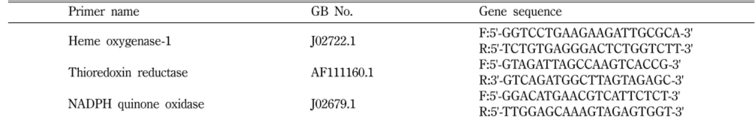
의염기서열은Table I
과같다.
통계처리
실험결과는평균
±SD
로표현하였으며,
통계적의미는Student- t tests
를이용하여분석하였다.
결과 및 고찰
망간화합물은지각이나물
,
혹은대기중에다양한물리적,
화학적형태로존재하며
,
체내에서일어나는다양한효소적반응 의보조인자로서필수적인역할을담당한다.
그러나고농도의망간에장기간지속적으로노출됨으로써체내에축적되는망간의 양이증가하는경우신경계및호흡기계에손상을야기할수있 다
.
4)더나아가고농도의망간을함유하는분진이나흄에장시간노출되는작업장노출의경우기관지를통해흡수된망간은 혈행을타고전신으로이동해다양한장기에영향을미칠수있 을것으로생각되며심혈관계또한그목표장기가될수있다
.
12)이에본연구에서는망간에의한심근독성이어느정도의농도 에서발현되며
,
어떠한기전을통해독성이일어나는지에대해 랫드에서유래한심근세포주를이용하여세포수준에서평가해 보고자하였다.
먼저
,
망간이H
9C
2세포주에미치는독성을평가하기위하여200, 400, 800, 1600
µM
의망간화합물(MnCl
2)
을단계희석하여 각각24, 48, 72, 96
시간씩처리한결과Fig. 1
과같이농도의존 적인세포독성을관찰할수있었으며, 1600
µM
로24
시간처리한경우의세포생존율은약
84.5%
로감소하였고, 96
시간의세포 생존율은15.7%
정도로낮아졌다.
Rovetta
등은17) 간(HepG2, human hepatoblastoma),
신장(MDCK, Madine-Darby canine kidney cell line)
신경교(GL15, human glioblastoma),
신경(SHSY5Y, human neuroblastoma)
에 서유래한4
종의세포주에0.1~1000
µM
의망간화합물(MnCl
2)
을처리함으로써망간화합물에대한각조직의
IC50
값을산출 하고이를비교·분석하였는데, 24
시간을처리한결과신경세포 인SHSY5Y
세포주의IC50
값이500
µM
로가장낮았고 다른세포주의
IC50
값은700~800
µM
로거의유사하였다.
따라서 Table I −Primer be used to investigate change of gene expression
Primer name GB No. Gene sequence
Heme oxygenase-1 J02722.1 F:5'-GGTCCTGAAGAAGATTGCGCA-3'
R:5'-TCTGTGAGGGACTCTGGTCTT-3'
Thioredoxin reductase AF111160.1 F:5'-GTAGATTAGCCAAGTCACCG-3'
R:3'-GTCAGATGGCTTAGTAGAGC-3'
NADPH quinone oxidase J02679.1 F:5'-GGACATGAACGTCATTCTCT-3'
R:5'-TTGGAGCAAAGTAGAGTGGT-3'
Fig. 1 −
Effect of Mn on the viability of H
9C
2cell line. Cells were
treated with MnCl
2for 24, 48, 72 and 96 hr. The indicated
concentrations represent Mn
2+concentration calculated
from MnCl
2added to the media. Cell viability was assessed
by MTT assay. Data represented as the percentage of the
control group. Cell viability was greatly reduced in a dose-
and time- dependent manner by Mn. Results represent the
means of three separated experiments, and error bars
represent the standard error of the mean. Except the
treated events with 200
µM Mn for 24, 48 hr, all events
indicate a statistically significant difference (** : p<0.01)
from the control group.
랫드의심근세포에서유래한
H
9C
2세포주의경우는이들세포 주에비해덜민감한것으로판단된다.
호흡을통해인체에유입된대기중유해물질은일반적으로 체내에반응산소종
(Reactive oxygen species)
을발생시킴으로써인체에독성을나타내는것으로알려져있다
.
그리하여심근 세포에서나타난세포독성과반응산소종의발생과의상관관계를 살펴보고자200, 400, 800, 1600
µM
의망간화합물을24
시간씩처리한후기질인
DCFH-DA
를이용하여형광강도의변화를측정해본결과
Fig. 2
와3
에서보는바와같이형광강도가농도의존적으로증가하였으며
, 1600
µM
로처리한경우대조군에비해약
2
배정도증가하는것을알수있었다.
이러한결과는동 일한세포주에비소화합물(As
2O
3)
을처리한경우약98%
정도의세포생존율을나타낸
0.5 ppm
의농도에서ROS
가약2
배정도증가하였고
,
18)사람의자궁경부암세포주(uterine cervical carcinoma)
에서유래한Hela
세포에1 mM
의망간화합물을처리한경우
ROS
의발생이약6.5
배정도증가하였다19)는기존의연구결과와비교한다면
ROS
의발생또한상대적으로낮은것으로 평가되었다.
일반적으로세포내반응산소종의증가는세포내에서항산화물 질로작용하는환원형글루타치온의감소를동반하는것으로알 려져있다
. 200, 400, 800, 1600
µM
의망간화합물을24
시간씩처리한후얻어진세포용해물과형광기질인
ortho-phthaldialdehyde
를반응시킨결과
Fig. 4
에서보는바와같이세포내환원형글루타치온함량이
200
µM
부터농도의존적으로감소하였으며, 1600
µM
로24
시간처리한경우에는대조군의61%
정도로감소 하는것을관찰할수있었다.
또한,
망간화합물이랫드의심근세포주에 야기하는 일련의 산화적 스트레스 과정은
heme
oxygenase-1, thioredoxin reductase, NADPH quinone oxidase
Fig. 2 −
Effect of Mn on ROS production in H
9C
2cell line. Cells grown in confluent were pre-treated with MnCl
2, washed with phosphat buffered saline, and then incubated with DCFH-DA 40 mM. At the end of DCFH-DA incubation, the cells were lysed with NaOH and fluorescence of aliquot was measured. Results represent the means of three independent experiments, and error bars represent the standard error of the mean. All group indicate a statistically significant difference (** : p<0.01) from the control group.
Fig. 4 −
Effect of Mn on the level of intracellular reduced glutathione (GSH). Fluorometric method using o -phthaldialdehyde was used to measure the intracellular GSH. Results represent the means of three independent analysis. GSH was calculated as nmol of glutathione per mg of protein and then was represented as the percentage of the control group. Asterisks indicate a statistically significant dif- ference (* : p<0.05, **: p<0.01) from the control group.
Fig. 3 −
Qualitative characterization of ROS generation by DCFH-
DA staining using fluorescence microscopy. Cells grown in
confluent were pre-treated with MnCl
2, and then loaded
with 40 mM DCFH-DA. After washing with PBS, cells
were visualized by fluorescent microscopy (×200).
등기존의연구자료를통해산화적스트레스와깊은상관성이있 는것으로알려진여러유전자의발현을뚜렷하게증가시키는것 을확인할수있었다
(Fig. 5).
Milatovic
등은세포수준에서망간의독성을관찰한경우,
세 포내에흡수된망간은미토콘드리아에선택적으로축적되어미 토콘드리아의주요기능인산화적인산화를억제시키고활성산소 종의발생을증가시켰다고보고하였으며,
1)Crossgrove
등은고 농도의망간이Ca
++채널을차단한다고보고하였다.
20)본연구에서망간에의한
ROS
의발생이비교적낮게측정된것은아마도망간에의한세포독성이또다른경로를통해유도될수있 기때문인것으로추정된다
.
아울러본연구에서는망간화합물에의한세포독성이
apoptosis
Fig. 8 −
Increase of chromosome condensation by Mn with DAPI staining. Cells were treated with the indicated concen- trations of Mn for 24 hr. DAPI solution was applied to the cultured cells in 8 chamber slides, and the slides were incubated for 10 min in the dark at 37
oC and the images of nuclei were made by fluorescent microscope.
Fig. 5 −
Effect of Mn on the induction of oxidative stress-related genes. Cells were treated with the indicated concentrations of Mn for 24 hr. mRNA transcription was detected by RT- PCR analysis using respective primers described in Table I. (A): heme oxygenase-1, (B): thioredoxin reductase, (C):
NADPH quinone oxidase.
Fig. 6 −
Effect of Mn on the caspase-3 activity. Cells were treated with the indicated concentrations of Mn for 24 hr. Caspase- 3 activity was measured using a colorimetric caspase-3 specific substrate. After the reaction, the chromophore pNA was determined at 405 nm. Results represent the means of four separated experiments, and error bars represent the standard error of the mean. All group indicate a statistically significant difference (p<0.01) from the control group.
Fig. 7 −
DNA fragmentation by Mn on agarose gel electrophresis.
Cells were treated with the indicated concentrations of Mn
for 24 hr. DNA laddering was performed using genomic
DNA extractions which were prepared from control and
Mn-treated cells. DNA fragmentation was shown by
electrophoresis using 1.5% agarose gel.
에의해야기되는지혹은
necrosis
에의해야기되는지를확인하 기위하여caspase cascade
의최종단계에해당하는caspase-3
활성의증가와
DNA
분절,
핵내크로마틴의응축여부를관찰해보았다
. 200, 400, 800, 1600
µM
의망간화합물을각각24
시간씩 처리한후얻어진세포용해물에기질인DEVD-pNA
를반응시킨후발색정도를비교한결과
Fig. 6
에서보는바와같이caspase- 3
의활성이농도의존적으로증가하였으며1600
µM
로처리한 경우대조군에비해3.4
배정도증가하는것을관찰할수있었다
.
또한, 400, 800, 1600
µM
의망간화합물을각각24
시간씩처리한후추출한
DNA
를이용하여DNA
분절여부를관찰한결과DNA
분절정도가농도의존적으로증가하였으며(Fig. 7), 400, 800, 1600
µM
의망간화합물을24
시간씩처리한세포의핵내에서
breakage
와크로마틴응축이뚜렷하게관찰되었다(Fig. 8).
이 러한현상은망간화합물을PC12
세포주에처리한Hirata
의연구결과와
Hela
세포에처리한Oubrahim
의연구결과에서도확인할수있었다
.
19,21)결 론
산업장등에서고농도의망간화합물을함유하는분진이나흄 이호흡을통해근로자들의체내에유입되는경우
,
어느정도의 농도에서심장기능이손상될수있는지를세포수준에서관찰해 보고자랫드의심근세포에서유래한H
9C
2세포주를이용하여연 구한결과200
µM
로24
시간처리한경우독성이관찰되기시작 하였으며,
이러한세포독성은세포내ROS
의증가와환원형글 루타치온의감소를동반하는산화적스트레스기전에의하여일 어났다.
이러한결과는망간의비정상적축적이심부전,
심근경 색등심질환의위험인자로작용할수있음을암시하는것이라 고생각된다.
참고문헌
1) Milatovic, D., Yin, Z., Gupta, R. C., Sidoryk, M., Albrecht, J., Aschner, J. L. and Aschner, M. : Manganese induces oxidative impairment in cultured rat astrocytes. Toxicol. Sci.
98, 198 (2007).
2) Liao, S. L., Ou, Y. C., Chen, S. Y., Chiang, A. N. and Chen, C. J. : Induction of cyclooxygenase-2 expression by manganese in cultured astrocytes. Neurochem. International.
50, 905 (2007).
3) Aschner, M., Guilarte, T. R., Schneider, J. S. and Zheng, W. : Manganese : Recent advances in understanding its transport and neurotoxicity. Toxicol. Appl. Pharmacol.
221, 131 (2007).
4) http://toxnet.nlm.nih.gov, Manganese compounds, Environmental fate & Exposure.
5) Bowler, R. M., Roels, H. A., Nakagawa, S., Drezgic, M., Diamond, E., Park, R., Koller, W., Bowler, R. P., Mergler, D., Bouchard, M., Smith, D., Gwiazda, R. and Doty, R. L. : Dose-effect relationships between manganese exposure and neurologic neuropsychological and pulmonary function in confined space bridge welders. Occup. Environ. Med.
64, 167 (2007).
6) Roels, H., Lauwerys, R., Buchet, J. P., Genet, P., Sarhan, M. J., Hanotiau, I., de Fays, M., Bernard, A. and Stanescu, D. : Epidemiological survey among workers exposed to manganese : effects on lung, central nervous system, and some biological indices. J. Ind. Med.
11, 307 (1987).
7) Roels, H. A., Ghyselen, P., Buchet, J. P., Ceulemans, E. and Lauwerys, R. R. : Assessment of the permissible exposure level to manganese in workers exposed to manganese dioxide dust. J. Ind. Med.
49, 25 (1992).
8) Lucchini, R., Apostoli, P., Perrone, C., Placidi, D., Albini, E., Migliorati, P., Mergler, D., Sassine, M. P., Palmi, S. and Alo, L. : Long-term exposure to "low levels" of manganese oxides and neurofunctional changes in ferroalloy workers.
Neurotoxicology
20, 287 (1999)
9) Lucchini, R., Selis, L., Folli, D., Apostoli, P., Mutti, A., Vanoni, O., Iregren, A. and Alessio, L. : Neurobehavioral effects of manganese in workers from a ferroalloy plant after temporary cessation of exposure. Scand. J. Work. Environ. Health
21, 143 (1995).
10) Myers, J. E., Thompson, M. L., Ramushu, S., Young, T., Jeebhay, M. F., London, L., Esswein, E., Renton, K., Spies, A., Boulle, A., Naik, I., Iregren, A. and Rees, D. J. : The nervous system effects of occupational exposure on workers in a south african manganese smelter. Neuro. Toxicol.
24, 885 (2003).
11) Elbetieha, A., Bataineh, H., Darmani, H. and Al-Hamood, M. H. : Effects of long-term exposure to manganese chloride on fertility of male and female mice. Toxicol. Lett.
119, 193 (2001).
12) Crossgrove, J. and Zheng, W. : Manganese toxicity upon overexposure. NMR Biomed.
17, 544 (2004).
13) Elbekai, R. H. and El-Kadi, A. O. S. : The role of oxidative stress in the modulation of aryl hydrocarbon receptor- regulated genes by As
3+, Cd
2+and Cr
6+. Free Radical Biol.
Med.
39, 1499 (2005).
14) Fotakis, G., Cemeli, E., Anderson, D. and Timbrell, J. A. : Cadmium chloride-induced DNA and lysosomal damage in a hepatoma cell line. Toxicol. In Vitro.
19, 481 (2005).
15) Brenner, A. J. and Harris, E. D. : A quantitative test for copper using bicinchoninic acid. Anal. Biochem.
226, 80 (1995).
16) Dhar-Mascareno, M., Carcamo, J. M. and Golde, D. W. :
Hypoxia-reoxygenation induced mitochondrial damage and
apoptosis in human endothelial cells are inhibited by vitamin
C. Free Radic Biol Med.
38, 1311 (2005).
17) Rovetta, F., Catalani, S., Steimberg, N., Boniotti, J., Gilberti, M. E., Mariggio, M. A. and Mazzoleni, G. : Organ-specific manganese toxicity : a comparative in vitro study on five cellular models exposed to MnCl
2. Toxicol. In Vitro.
21, 284 (2007).
18) Park, E. J. and Park, K. S. : Gene expression profiles of cultured rat cardiomyocytes (H9C2 cells) in response to arsenic trioxide at subcytotoxic level and oxidative stress. J.
Health Sci.
52, 512 (2006).
19) Oubrahim, H., Stadtman, E. R. and Chock, P. B. : Mitochondria play no roles in Mn(II)-induced apoptosis in HeLa cells. PNAS
98