2010년도 춘계 학술발표회 논문집 대한방사선방어학회
122_http://www.karp.or.kr
Identification of Protein Markers in Intestine and Brain of Irradiated Mice by Proteomic Analysis
Young-Bin Lim, Bo-Jeong Pyun, Hae-June Lee, Sang-Rok Jeon and Yun-Sil Lee
*Laboratory of Radiation Effect, Korea Institute of Radiological and Medical Sciences, Seoul, Korea
E-mail: [email protected] Key words : Proteomics, Transaldolase, γ-ray, Phosphoglycerate kinase 1
Introduction
Several bioassays such as cytogenetics, mutations, cell survival, clonogenicity, and cell transformation, have been used for decades to aid in the assessment of health risk following exposure to radiation. However, the currently available assays do not directly reveal molecular mechanisms involved in response to radiation making the estimation of long tern health risks uncertain. Consequently, there are increasing efforts in developing more sensitive and rapid molecular methodologiesboth at the genomic and proteomic levels for the identification of biomarkers of exposure to radiation.
Material and Method
Female C57BL/6 mice (7 weeks of age) were used in this study. All were purchased from SLC (Hamamatsu) and kept at 22 ± 2℃ and 50 ± 10%
humidity with a 12 h light-dark cycle and free access to food and water. Whole body irradiation (1 Gy) was performed using a 137Cs γ-ray source (Atomic Energy of Canada) at a dose rate of 3.51 Gy/min. At 1 day after irradiation, the subject mice were sacrificed, followed by quickly harvesting tissue from the brain, lung, spleen and
intestine that were subsequently stored in liquid nitrogen. 2D-MS assay with the tissues were then performed. To confirm 2D-MS data, Western blotting was employed.
Results
Proteins were solubilized from various tissues including brain, lung, spleen, and intestine sham exposed or exposed to 1 Gy of irradiation for 1 day.
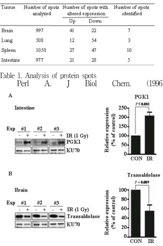
These proteins were separated using 2-DE. Protein spots were then chosen for MS only if they showed greater than two-fold expression changes compared with the average percent volume values from irradiated and matched normal tissue, could be removed from the gel without excising other nearby proteins. Most of the spots did not meet these criterions, leaving 252 of the 3532 available for identification (table 1). MS analysis showed only 25 from 252 spots matched a single protein with above 95% of protein confidence index. The total number of spots for brain was 7, 3 for lung, 10 for spleen, and 5 for intestine. In order to confirm significant differences between the control and IR treated sample in 2-DE analysis, ten identified proteins with available commercial antibodies were selected for immunoblotting.
However, Only 5 of 10 proteins showed similar
2010년도 춘계 학술발표회 논문집 대한방사선방어학회
대한방사선방어학회_123
Figure 2. Expression of PGK1 and TA1 in various tissues exposed to IR
Table 1. Analysis of protein spots
Figure 1. Altered expression of PGK1 and TA1 by IR expression pattern in all three independent
experiments compared to MS analysis data. The 5 proteins were heat shock protein 5, heat shock protein 90kDa beta, heat shock protein 1, transaldolase 1 [1,2], and phosphoglycerate kinase 1.
Since heat shock protein 5, heat shock protein 90 kDa beta, and heat shock protein 1 are belong to heat shock protein family, which is a well known IR dosimeter, we focused on transaldolase 1, and phosphoglycera kinase 1. The immunoblotting, presented in Fig. 1a, confirms the up-regulation of PGK1 in IR-treated intestine of all animals examined (p ≤ 0.001). As shown in Fig. 1B, IR negatively regulated expression of transaldolase 1 in brain (p = 0.009). As shown in Fig. 2A, IR upregulated expression of PGK1 only in intestine, whereas in other tissues the expression was not affected at all. IR showed no effect on expression of transaldolase 1 in lung, spleen, and intestine, but strongly downregulated its expression in brain.
Conclusion
Our data have shown that PGK1 was specifically upregulated in irradiated mouse intestine and TA1 was down-regulated in irradiated mouse brains without their alterations in other tissues. Based on these data, we suggest that TA1 and PGK1 might be candidates for tissue specific biomarkers of IR exposure.
Reference
[1] Banki K, Colombo E, Sia F, Halladay D, Mattson DH, Tatum AH, Massa PT, Phillips PE, Perl A. J. Exp. Med. (1994) 180(5):
1649-63.
[2] Banki K, Hutter E, Colombo E, Gonchoroff NJ,
Perl A. J Biol Chem. (1996)
271(51):32994-3001.