축구선수의 진행형 요부안정화운동이 복횡근 두께와 하지근육 피로지수에 미치는 영향
이준희1․박승규1․강정일1․양대중1․김제호2, 3․정용식2, 3
1세한대학교 보건대학 물리치료학과․2세한대학교 대학원 물리치료학과․3목포중앙병원
The Effect of Progressive Lumbar Stability Exercise on the Transversus Abdominis Muscle Thickness and Lower extremity muscle Fatigue Index in Soccer Players
Joon-Hee Lee1․Seung-Kyu Park1․Jeong-Il Kang1․Dae-Jung Yang1․Je-Ho Kim2, 3․Yong-Sik Jeong2, 3
1Department of Physical Therapy, College of Health, Sehan University, Yeongam, Korea
2Department of Physical Therapy, Graduate School of Sehan University, Yeongam, Korea
3Jung-Ang General Hospital, Mokpo, Korea
Received 31 July 2012; Received in revised form 16 August 2012; Accepted 27 September 2012
ABSTRACT
This study aimed to assess the effects of progressive lumbar stability exercises and lumbar stability exercises on changes in the transversus abdominis muscle thickness and lower extremity muscle fatigue index in soccer players. Ten subjects were assigned to undergo training in each of the 2 groups, namely, the progressive lumbar stability exercise group and lumbar stability exercise group.
Each intervention session lasted for 30 min, and 4 sessions were conducted in a week for 6 weeks for soccer players of S. University in Jeonnam, Korea. Changes in the transversus abdominis muscle thickness and lower extremity muscle fatigue index were measured using ultrasound and surface electromyogram. The results of the ultrasound measurement for the transversus abdominis muscle thickness indicated that progressive lumbar stability exercises were more effective than lumbar stability exercises. The results of the lower extremity muscle fatigue index measurements using surface electromyogram indicated that the fatigue index decreased in the progressive lumbar stability exercise group. Progressive lumbar stability exercise is believed to have put more workload during the shaking of the limbs, leading to increased stability and increased efficiency of the lower extremity muscle, thereby decreasing the fatigue index. Therefore, progressive lumbar stability exercises can be an effective measure for preventing injuries and improving the game performance of sports players by increasing the transversus abdominis muscle thickness and decreasing the lower extremity muscle fatigue index.
Keywords : Lumbar Stability Exercise, Transversus Abdominis, Fatigue Index, Active Straight Leg-Raising Test
Ⅰ. 서 론
요부 안정화는 중립위치에서 요부와 골반의 움직임을 조절 하는 능력으로 정의 된다(Jull, Richardson & Toppenburg, 1993).
Corresponding Author : Yong-Sik Jeong
Department of Physical Therapy, College of Health, Sehan University, 72 Sanho-ri, Samho-eup, Yeongam-gun, Jeollanam-do, Korea
Tel : +82-61-280-3092 / Fax : +82-61-469-1317 E-mail : [email protected]
본 논문은 2012년도 세한대학교 교내 연구지원에 의하여 쓰여진 것임.
요부 안정화 훈련은 근력을 강화시키며 근육의 조절 능력과 균형을 회복시켜 요부와 골반의 안정성을 증가 시킨다(Lee, 1994; Kim, Hyong & Kim, 2008). 재활훈련이나 운동선수에게 적 용한 안정화운동프로그램은 근력, 지구력, 신경근 기능을 증진 시켜 근육조절 능력을 향상시킨다(Davidson, Hubley-Kozey, 2005).
안정화운동과 유사 동의어로는 체간 안정화(trunk stabilization), 요 부안정화(lumbar stabilization), 중심부 강화운동(core strengthening), 운동조절훈련(motor control training) 등의 용어로 사용되고 있다 (Akuthota 2004). 체간근육들은 요부 안정화에 중요한 역할을 하
고(Cholewicki, 1997), 동적인 활동과 체중 부하 시 체간의 근육 들을 순차적으로 동원시켜 안정성을 제공한다(Hodges, 1999). 부 하에 대해 체간이 흔들릴 때 수동 및 능동적인 체간 구조들은 신경시스템 통제하에 몸통 안정화에 관여 한다(Panjabi, 1992).
골반의 불안정성이 있는 환자는 심부 안정화에 관여하는 복횡 근(transversus abdominis muscle)를 중점적으로 강화시켜야 한다 고 하였다(Richadson & Jull, 1995).
복횡근은 요부 안정성 조절에 가장 중요한 근육 중 하나이 고(Pietrek, Sheikhzadeh & Nordin 2000), 연속적인 동작 시 복압 에 관여한다(Cresswell, Oddsson & Thorstensson, 1994). 즉 요부 의 안정화에 기여한다고 하였다(Cholewicke, Juluru, Radebold, Panjabi & McGill, 1999). 하지만 단일 근육 하나 만으로 중요한 안정성을 제공한다고 할 수 없으며, 안정성는 움직이는 동안에 지속적인 부하를 받거나 자세에 따라 다르다고 하였다(Kavcic, Grenier & McGill, 2004b).
충분한 요부의 안정화는 척추 주변의 모든 근육들에 참여가 필요하며(Cholewicke & McGill, 1996), 척추 주변근육의 동시 활 동이 추체관절의 안정성을 향상시킨다고 하였다(Cholewicki, Juluru & McGill, 1999). Kavcic, Grenier와 McGill(2004a)은 체간 의 안정성을 증가시키기 위한 근육들의 수축수준을 정량화하였 고 체간근육의 동시수축 훈련이 척추의 불안정성 치료와 예방 에 사용된다고 하였다. McGill과 Karpowicz(2009)의 연구에 따 르면 몸통 웅크리기(curl-up), 측면 교량(side bridge)자세, 새 사 냥개(bird dog)자세에서 팔, 다리를 움직여 요부에 부하를 증가 시킨 진행형 요부안정화운동이 요부안정화운동 보다 요부 주변 과 하지 근육에 보다 큰 근 활성도를 나타내었다.
최근 들어 운동 전문가들이 시행하는 스포츠 컨디션닝 프로 그램에 있어서 요부안정화의 중요성이 점차 강조되고 있는 추 세이다(Kim & Lee, 2010).
축구선수들의 요부주변 근력약화는 요부손상과 함께 대퇴 또는 하퇴 근력의 감소로 인하여 경기력 저하의 원인이 될 수 있다(Yun, 2000). 특히 축구는 다른 경기와 달리 골과 직접적으 로 관련된 슈팅으로 경기의 승패가 좌우되기 때문에 가장 필요 한 것이 하지의 근력이다(Kim, 2004). 또, 현대축구는 대인방어 의 개념으로 바뀌면서 더욱 더 강인한 체력을 요구하고 있다 (Kim, Jin, Jun & Jung).
축구 경기의 체력적 요인은 적절한 트레이닝으로 그 능력을 향상시킬 수 있으므로 많은 노력과 연구가 진행되고 있다(Kim
& Mo, 2007).
요부안정화운동을 수행한 선수는 수행하지 않은 선수와 비 교하여 기능적인 동작 수행에 보다 좋은 결과를 보였으며(Tes, McManus & Masters, 2005), 운동선수에게 적용한 요부-골반 운 동프로그램은 하지의 운동능력에 기여한다고 하였다(Baker, 1999). 안정화되어 있지 않은 요부-골반은 효율적인 근력을 발
휘하지 못한다고 하였다 (Faccioni, 1994).
따라서 심부근육의 약화는 스포츠 선수의 수행능력을 감소시 키거나 손상 위험을 초래할 수 있다고 하였으며(Thomas, Nesser &
William, 2009), 하지근육의 피로는 균형전략의 변화로 동적인 움 직임에서 근육이나 인대의 손상위험도를 높인다고 하였다 (Adlerton, Moritz & Moe-Nilssen, 2003).
근피로는 생리학적으로 근육에 충분한 산소가 공급되지 않거 나 혈액 중 젖산 비율이 증가할 때 근육내 pH의 증가에 의해 근 력 생산이 감소되는 것으로 설명된다(Stanley, Wayne & Jane, 2006; Yoon, Chae & Kang, 2010). 근전도를 활용한 근피로의 발생 여부는 측정된 근전도 신호를 파워스펙트럼을 통해 중앙주파수 (median frequency; MF)나 평균 주파수(mean edge frequency, MEF) 의 주파수 영역대가 낮아지는 현상을 평가하여 나타낼 수 있다 (Basmajian & Luca, 1985; Yoon, Chae & Kang, 2010). 다른 연구들 에서는 등척성수축 동안에 신경근 활성과 피로도를 측정하고 있 으나, 등척성수축 동안의 평가는 근 활성도 증가와 피로도를 정확 하게 반영하지 못한다고 하였다(Mark & Lawrence, 2006).
선행 연구들에서 안정화운동 훈련 후 체간 근육의 근 활성도 에 관한 연구는 많이 보고되었으나, 표면 근전도를 이용한 복횡근 의 활성도는 정확히 측정하기 어렵다고 보고하였다(Ian, Stokes, Sharon & Henry, 2003).
안정화운동 후 심부근육 두께변화에 대한 연구는 부족하며 또 한 근력에 관한 연구는 많으나 피로도 변화에 대한 연구는 부족한 실정이다. 요부안정화 훈련 후 복횡근 두께 변화와 하지 근육의 피로도 변화를 알아본다면 경기력 향상과 부상방지를 위한 훈련계 획에 도움이 될 것이다.
본 연구에서는 진행형 요부안정화운동과 요부안정화운동을 훈 련한 후 초음파 영상을 이용하여 축구선수들의 복횡근 두께 변화 율을 측정하고, 표면 근전도를 이용하여 하지근육의 피로지수에 변화를 분석하고자 한다.
Ⅱ. 연구방법
1. 연구 대상자
본 연구를 위해 S대학교 축구부 남자선수 20명을 피험자로 선정하였다.
피험자는 진행형 요부안정화운동군(n=10)과 요부안정화운동 군(n=10)으로 구성하였으며, 연령은 20~24세 미만으로 고관절, 슬관절, 발목관절에 수술 경험이 없으며 최근 3개월 동안 부상 의 경험이 없고 다른 내 ·외과적 질환을 가지고 있지 않은 선 수로 무작위임의 선정하였으며 연구대상자의 일반적 특징은
<Table 1>과 같다.
Characteristics PLSE (n=10) LSE (n=10)
Number 10 10
Age(years) 20.50±0.70 21.1±0.70
Height(cm) 176.30±6.88 176.5±4.76
Body weight(kg) 69.80±6.51 70.80±4.44 Values aremean±SD
PLSE : Progressive Lumbar-Stability Exercise LSE : Lumbar Stability Exercise
Table 1. General characteristics in the progressive lumbar stability exercise group and lumbar stability exercise group
2. 실험방법
본 연구는 대학축구선수를 대상으로 진행형 요부안정화운동 (n=10)과 요부안정화운동(n=10)을 1일 30분, 주 4회, 6주 동안 실시 하였다. 운동은 각 그룹에 물리치료사 1명씩 배정하여 트레이닝 시켰다. 진행형 요부안정화운동 프로토콜은 McGill과 Karpowicz(2009) 의 연구를 근거로 몸통 웅크리기(curl-up), 측면 교량(side bridge)자 세, 새 사냥개(bird dog)자세에서 상지와 하지에 움직임을 통해 몸 통에 부하를 가중시키는 운동으로 구성하였다(Figure 1).
Figure 1. Progressive lumbar stability exercise(A, B, C) and lumbar stability exercise(D, E, F)
요부안정화운동 프로토콜은 치료 원칙에 따라 척추 중립을 인 식하는 것을 목적으로 다양한 자세에서 시행하는데, 본 연구에서는 McGill(2007)이 기술한 몸통 웅크리기(curl-up), 측면 교량(side bridge)자세, 새 사냥개(bird dog)자세로 구성하였다(Figure 1).
1) 측정도구
(1) 초음파 이미지 측정
복횡근의 두께변화를 측정하기 위해 MyLab 25 GOLD (Esaote, Italy)를 이용하였고 직선형 프로브 12MHz 변환기로 측 정하였고, 초음파 영상이 화면의 약 60-70%는 근육층들로 채울 수 있도록 깊이(depth)를 조절하였다. 잠재적으로 피험자에게 학 습되거나 근육 피로도와 연관된 순서 효과를 없애기 위해 오른 쪽과 왼쪽 측정은 무작위 순서로 진행되었고, 변환기는 능동하 지직거상 검사가 시행되는 동안 엉덩뼈능선 상부의 복부벽에서 전 -가쪽면과 겨드랑이선의 수직면에 위치하였다(Teyhen, Williamson
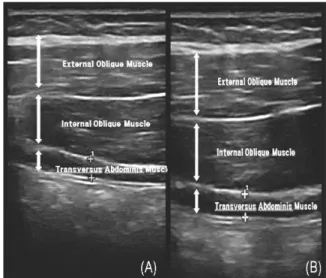
& Carlson 2009). 측정 시 호흡의 영향을 줄이기 위해 호기 (expiration)를 마치고 영상을 수집하였다. 휴식 시 복횡근의 두 께 값은 정상 호기 후 측정되었다. 수축 시 복횡근의 두께 값은 능동직거상검사 수행 후 10초 유지 후 측정되었다. 복횡근의 측정은 3회 반복 측정하였으며, 근피로를 최소화하기 위해 30 초간 휴식을 취한 후 휴식기의 근 두께 측정을 실행하였고, 한 명을 대상으로 오른쪽 왼쪽을 3회 반복 측정하여 평균값을 측 정치로 이용하였다(Figure 2). 측정된 근육의 두께변화의 공식은 근육 두께에 변화율(%)={수축기의 근육두께(cm) - 휴식기의 근 육두께(cm)} / 휴식기의 근육두께(cm) X 100을 이용하여 백분 율로 정규화(normalized)시켰다.
Figure 2. Transversus abdominis ultrasound image (A. Resting, B. Active)
(2) 표면 근전도 측정
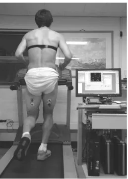
하지근육의 피로지수를 측정하기 위해 무선 근전도(FREEEMG
300, BTS, Italy)을 이용하였다. 피험자는 경사도가 0인 트레드 밀에서 각자 70%의 운동 강도에 해당하는 목표 심박수로 20분 동안 지속하여 뛰었다. 70%의 운동 강도에 해당하는 목표 심박 수는 최대 심박수에 0.7을 곱하여 계산하였으며(Ko, 2004), 최대 심박수는 220에서 대상자의 나이를 뺀 값으로 정하였다. 주파 수 대역은 20∼500 Hz로 설정하여 필터과정을 거쳤다. 20분 달 리는 동안 근전도 신호를 수집하였다(Figure 3).
Figure 3. Measurement of surface EMG
Muscle Location
vastus lateralis approximately 3 to 5cm above the patella, on anoblique angle just lateral to mid-line.
vastus medialis approximately 6cm above the patella, on anoblique angle just medial to mid-line.
biceps femoris between the trochanter and the back of the knee.
Table 2. Location of electrodes
수집된 근전도 신호중에서 목표 심박수에 도달하는 시간을 고려하여 운동 시작 후 5분을 제외한 전체 운동시간을 근전도 신호로 전환(rectification)하고, 적분(integration)한 뒤 RMS값을 구한 뒤 중간 주파수를 구하였다.
이 중앙주파수 값을 사용하여 피로지수(fatigue index)를 알아 보았다. 피로지수(fatigue index)는 초기 중앙주파수에서 마지막 중앙주파수를 뺀 값을 초기 중앙 주파수로 나누어 계산하였다 (Ko, 2004). 초기 중앙주파수 값은 운동시작 5분을 제외하고 초 기 3분 동안의 신호를 이용하였고 후기 중앙 주파수값은 후기 3분 동안의 신호를 이용하여 구하였다.
표면근전도 신호에 대한 피부저항을 감소시키기 위하여 부 착부위 털을 제거하고 가는 사포로 3~4회 문질러 피부 각질층 을 제거한 후, 소독용 알코올로 피부를 깨끗이 하였다. 하지의 피로도를 측정하기 위해 양측 외측광근(vastus lateralis), 내측광 근(vastus medialis), 대퇴이두근(biceps femoris)에 부착하여 피로 도를 측정하였고 측정부위는 Table 2와 같다.
3. 자료분석
모든 통계처리는 SPSS win 17.0을 이용하였으며 운동방법에 따 른 각 복횡근의 두께변화 및 하지 근육의 피로도를 비교하기 위하 여 공분산분석(analysis of covariance: ANCOVA)을 실시하였으며, 통계학적 유의성을 검정하기 위한 유의수준은 α=.05로 하였다.
Ⅲ. 결 과
1. 운동 방법에 따른 복횡근 두께 비교
두 그룹의 능동하지직거상 검사 동안의 복횡근(transversus abdominis muscle) 두께변화는 훈련 전·후 %백분화 하여 비교하 였다. 오른쪽 복횡근의 두께변화는 진행형 요부안정화운동 전 4.70±2.62 mm에서 운동 후 11.20±2.31 mm 로 요부안정화운동 군은 훈련 전 4.79±2.26 mm에서 훈련 후 7.27±2.98 mm로 유의한 차이가 나타났다(p<.01). 왼쪽 복횡근의 두께변화는 진행형 요부안 정화운동군 훈련 전 6.81±3.52 mm에서 훈련 후 14.140±2.62 mm로 요부안정화운동군은 훈련 전 6.81±3.52 mm에서 훈련 후 10.77±3.52 mm로 훈련 방법에 따른 복횡근의 두께 변화율은 유의한 차이가 났다(Table 3)(p<.05).
PLSE (n=10) LSE (n=10)
F p
pre-test post-test pre-test post-test
Right 4.70±2.62 11.20±2.31 4.79±2.26 7.27±2.98 8.510 .010**
Left 6.81±3.52 14.14±2.62 6.81±3.52 10.77±3.52 5.737 .028*
Values are mean±SD, *p< .05, **p< .01 PLSE : Progressive lumbar-Stability Exercise LSE : Lumbar-Stability Exercise
Table 3. Comparison of transverse abdominis muscle chang muscle thickness of each groups by exercise method. (unit: mm)
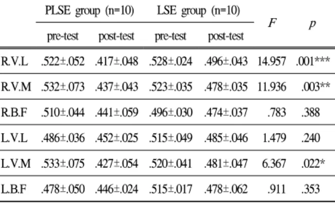
2. 운동 방법에 따른 피로지수(fatigue index)의 비교 두 그룹 간의 피로지수 비교는 훈련 전․후 트레드밀 위에 서 달리는 동안 오른쪽과 왼쪽의 내측광근, 외측광근, 대퇴이두 근을 측정하였다. 오른쪽 외측광근(vastus lateralis)의 피로지수
변화는 진행형 요부안정화운동군의 훈련 전 .522±.052에서 훈련 후 .417±.048으로 감소하였고, 요부안정화운동군의 훈련 전 .528±.024 에서 훈련 후 .496±.043으로(p<.001)유의한 차이를 나타냈다. 오 른쪽 내측광근(Vastus medialis)의 피로지수 변화는 진행형 요부안정 화운동군의 훈련 전 .532±.073에서 훈련 후 .437±.043으로 감소하였 고, 요부안정화운동군의 훈련 전 .523±.035에서 훈련 후 .478±.035 으로(p<.01)유의한 차이를 나타내었다. 왼쪽 내측광근의 피로지수 변화는 진행형 요부안정화운동군의 훈련 전 .510±.044에서 훈련 후 .441±.059으로 감소하였고, 요부안정화운동군의 훈련 전 .520±.041 에서 훈련 후 .481±.047으로(p<.05) 유의하게 감소하였다. 그러나 오른쪽 대퇴이두근(biceps femoris), 왼쪽 외측광근(vastus lateralis), 왼쪽 대퇴이두근(biceps femoris)에서는 수적 감소는 있었으나 통계 적인 유의차는 나타나지 않았다(Table 4).
PLSE group (n=10) LSE group (n=10)
F p
pre-test post-test pre-test post-test
R.V.L .522±.052 .417±.048 .528±.024 .496±.043 14.957 .001***
R.V.M .532±.073 .437±.043 .523±.035 .478±.035 11.936 .003**
R.B.F .510±.044 .441±.059 .496±.030 .474±.037 .783 .388 L.V.L .486±.036 .452±.025 .515±.049 .485±.046 1.479 .240 L.V.M .533±.075 .427±.054 .520±.041 .481±.047 6.367 .022*
L.B.F .478±.050 .446±.024 .515±.017 .478±.062 .911 .353 Values are mean±SD, *p< .05, **p< .01, ***p< .001
PLSE : Progressive lumbar-Stability Exercise LSE : Lumbar-Stability Exercise
R.V.L : Right Vastus Lateralis, R.V.M : Right Vastus Medialis, R.B.F : Right Biceps Femoris, L.V.L : Left Vastus Lateralis, L.V.M : Left Vastus Medialis, L.B.F : Left Biceps Femoris.
Table 4. Comparison of muscle fatigue index of each groups by exercise method
Ⅳ. 논 의
과거에는 요부안정화운동이 주로 물리치료실에서 요통환자 들에게 국한되어 사용되어졌으나 현재는 개인뿐만 아니라 스포 츠 선수들에게도 광범위하게 적용되고 있다(Krabak & Kennedy, 2008; Kim & Lee, 2010). 본 연구는 6주간의 진행형 요부안정화 운동이 요부안정화운동과 비교하여 복횡근의 근육 두께 변화와 하지근육 피로지수에 미치는 영향을 알아보고자 하였다. McGill 과 Karpowicz(2009)는 몸통 웅크리기(curl-up), 측면 교량(side bridge)자세, 새 사냥개(bird dog)자세에서 팔·다리를 흔드는 동 작과 유지하는 동작을 비교한 결과 팔·다리를 흔드는 동작 그 룹에서 몸통 근육과 하지근육의 활성도에 유의한 증가를 나타 내었다. 또한 Lee(2010)는 네발기기 자세에서 팔들기, 다리들기, 그리고 팔과 다리들기의 세 가지 동작을 트레이닝하면서 다열
근, 복횡근, 외복사근의 근활성도를 분석하였다. 그 결과 더 힘 든 동작으로 진행할수록 근활성도가 증가하는 것을 나타내었 다. Kim, Park, Kang과 Yang(2010)는 대학축구선수들에게 팔다 리를 흔드는 동적 요부안정화운동과 정적 요부안정화운동을 4 주 동안 실시한 후 근전도를 이용하여 몸통근육의 근활성도을 비교한 결과 동적 운동군에서 근활성도가 유의하게 증가하였 다. Cresswell, Oddsson과 Thorstensson(1994)등의 연구에서도 사 지의 균형을 저해하는 외적 하중은 몸통근육의 활동을 증가시 킨다고 하였다. 본 연구와 측정도구에 차이는 있었지만 팔·다리 를 움직여서 요부에 부하를 증가 시키는 진행형 요부안정화운동 이 복횡근의 활성도에 더 효과적임을 알 수 있었다. Hong(2007) 은 초음파 영상을 이용하여 주 3회 6주간 안정화운동을 시행 한 결과, 3주까지는 복횡근의 두께변화가 관찰되지 않았으나 6 주후부터 통계학적으로 유의한 변화를 보였다. 이 연구에서 적 용한 몸통 웅크리기(curl-up), 측면 교량(side bridge)자세, 새 사 냥개(bird dog) 자세에서 과도한 골반의 전만을 방지하고 팔, 다 리의 움직임으로부터 몸통을 고정 시켜야 하기 때문에 복횡근 의 두께변화가 나타난 것으로 사료된다. McMeeken, Beith, Newham, Milliqan과 Critchley(2004)은 복횡근의 두께변화와 근 활성도의 변화는 상관관계가 있다고 하였다. 그러므로 안정화 운동에 따른 복횡근의 두께변화는 움직임의 효율성을 증진시킬 것으로 생각된다.
근피로는 균형능력의 저하 및 손상의 큰 요인으로 작용하며 (Rahnama, Reilly & Lees, 2002), 근피로가 과도하면 생리학적인 적응보다는 스트레스를 유발시켜 전반적인 근기능과 운동수행 능력을 저하시키게 된다. Rahnama, Reilly & Lees(2006) 는 축구 경기와 같은 고강도 운동에서의 하지근육 피로는 전기적인 근 육 활성도를 감소시킴으로써 부상의 주요한 요인이 될 수 있다 고 하였다. Yuk, Hwang과 Kwon(2006)은 운동선수들의 경기 성 적을 높이기 위하여 피로도를 줄이는 컨디셔닝의 중요성을 강 조 하였다. 그리고 피로에 의한 슬괵근의 원심성 수축력 감소 는 근염좌를 일으키고, 이런 상태에서 강한 원심성 수축은 심 한 근손상을 일으킨다.(Verrall, Slavotinek & Barnes, 2001). 슬괵 근은 보행주기의 마지막 유각기에서 하퇴의 원심성 수축을 통 해 감속하는 역할을 한다(Stanton & Purdam, 1989). 이런 메카니 즘은 전력질주 하는 축구선수들의 슬괵근 염좌에 주원인이 될 수 있다(Woods, Howkins, Hulse & Hodson. 2002).
Stanton, Reaburn과 Humphries(2004)의 연구에서는 22명의 운 동선수를 대상으로 일반적인 훈련과 스위스볼을 이용한 요부 안정화훈련의 효과를 비교한 결과, 그룹 간에 최대산소섭취량 에서 통계적 유의성은 없었다. Stray Pedersen, Magnu -ssen, Kuffel과 Seiler의 연구에 따르면 축구선수을 대상으로 슬링을 이용한 안정화 훈련군이 일반군에 비하여 최대 발차기 속도에 유의한 증가를 보였다. Sato와 Mokha(2009)의 연구에서 요부안 정화 훈련을 받은 그룹이 일반 훈련만 받은 그룹에 비해 5000
m 달리기 수행시간에 유의한 차이를 나타내었다. 본 연구에서 는 두 그룹 모두 훈련 전·후 트레드밀에서 70%의 강도로 달리 는 동안 하지근육을 근전도로 측정한 결과 중앙주파수가 감소 하는 즉 피로도가 감소했다는 것을 알 수 있었다. 이는 요부안 정화운동이 하지근육의 피로지수에 영향을 주는 것으로 사료되 며 일반적 요부안정화운동보다는 진행형 요부안정화운동이 에 너지 사용에 더 효율적이라는 것을 알 수 있었다. 그 이유는 진 행형 요부안정화운동이 팔·다리를 흔드는 동안 요부에 더 많은 부하를 주어 체간에 안정화를 증진시키고 하지근육에 효율성을 증가시켜 피로지수를 감소시킨 것으로 생각된다.
V. 결 론
본 연구는 축구선수들을 대상으로 진행형 요부안정화운동과 요부안정화운동 프로그램을 6주간 실시한 후 복횡근의 두께변 화와 하지 근육의 피로지수를 측정하여 운동의 효과를 분석한 결과 다음과 같은 결론을 얻었다.
첫째, 두 그룹의 훈련 전·후에 능동직하지거상 검사를 하는 동안 복횡근에 대한 초음파 영상분석을 실시하여 근 휴식시와 작용시에 두께변화를 측정한 결과 진행형 요부안정화훈련이 요 부안정화훈련과 비교하여 오른쪽(p<.01)과 왼쪽(p<.05)의 복횡근 두께변화에 더 효과적임을 증명하였다.
둘째, 두 그룹의 훈련 전·후로 트레이드밀에서 70%의 운동 강도로 달리는 동안 근전도를 이용하여 하지근육의 피로지수를 측정하였으며, 분석결과 진행형 요부안정화훈련이 요부안정화 훈련과 비교하여 오른쪽 외측광근(p<.001), 오른쪽 내측광근 (p<.01), 왼쪽 내측광근(p<.05)에서 유의성 있는 차이를 나타내 었다. 그러나 오른쪽 대퇴이두근, 왼쪽 외측광근, 왼쪽 대퇴이 두근에서의 수적감소는 있었으나 통계상의 유의성은 없었다.
즉 진행형 요부안정화훈련이 하지근육 피로도를 감소시키는데 더 효과적임을 증명하였다.
결론적으로 진행형 요부안정화운동이 복횡근의 두께증가와 하 지근육 피로지수를 감소시켜 부상을 최소화 하고 효율적인 힘을 요구하는 축구선수들에게 도움을 줄 것으로 생각된다. 향후, 본 연구를 바탕으로 진행형 요부안정화운동이 축구의 기술력과 경기 후 근육 피로도에 미치는 영향에 대한 연구가 필요할 것이다.
참고문헌
Adlerton, A.K., Moritz, U., & Moe-Nilssen, R.(2003). Force plate and accelerometer measures for evaluating the effect of muscle fatigue on postural control during one-legged
stance. Physiotherapy Research International, 8(4), 187-199.
Akuthota, V., & Nadler, S. F.(2004). Core strengthening. Archives of Physical Medicine and Rehabilitation, 85(3), 86-92.
Basmajian, J. V., & De Luca, C. J.(1985).Muscle alive.Baltimore:
Williams and Wilkins. 125-127.
Baker, D. A.(1999). Comparison of lower abdominal strength and lumbo-pelvic stabilisation capabilities between rugby league players participating in the national versus state and city based leagues. Strength & Conditioning Coach, 7(3), 3-7.
Cholewicki, J., Juluru, K., & McGill, S. M.(1999). Intra -abdominal pressure mechanism for stabilizing the lumbar spine.
Journal of Biomechanics, 32(1), 13-17.
Cholewicki, J., Juluru, K., Radebold, A., Panjabi, M. M., & Mc Gill, S. M.(1999). Lumbar spine stability can be augmented with an abdominal belt and/or increased intra-abdominal pressure. European Spine Journal, 8, 388-395.
Cholewicki, J., & McGill, S. M.(1996). Mechanical stability of the in vivo lumbar spine: implications for injury and chronic low back pain.Clinical Biomechanics, 11,1-15.
Cresswell, A. G., Oddsson, L., & Thorstensson, A.(1994). The influence of sudden perturbations on trunk muscle activity and intra-abdominal pressure while standing. Experimental Brain Reserch, 98(2), 336-341.
Davidson, K. L., & Hubley-Kozey, C. L.(2005). Trunk muscle resopnses to demands of an exercise progression to improve dynamic spinal stability.Archives of Physical Medicine and Rehabilitation, 86, 217-223.
Faccioni, A.(1994). The role of mid-torso in maximizing sprint performance.Strength and Conditioning Coach, 2(2), 6-10.
Hodges, P. W., & Richardson, C. A.(1999). Altered trunk muscle recruitment in people with low back pain with upper limb movement at different speeds.Archives of Physical Medicine and Rehabilitation, 80, 1005-12.
Hong, S. L.(2007). The effects of lumbo-pelvic stabilization exercise with real-time ultrasound imaging on deep lumbar muscle activities in athletes with chronic back pain.
Journal of Korea Sport Research, 18(2), 563-572.
Ian, A. F., Stokes, A., Sharon, M., & Henry, B.(2003). Surface EMG electrodes do not accurately record from lumbar multifidus muscles. Clinical Biomechanics, 18(1), 9-13.
Jull, G., Richardson, C., & Toppenburg, R.(1993). Towards a measurement of active muscle control for lumbar stabilisation.
Australian Journal of Physiotherapy, 39, 187-193.
Kavcic, N., Grenier, S., & McGill, S. M.(2004a). Determining the stabilizing role of individual torso muscles during rehabilitation exercises. Spine, 29, 1254-1265.
Kavcic, N., Grenier, S., & McGill, S. M.(2004b). Quantifying tissue loads and spine stability while performing commonly prescribed low back stabilization exercises.
Spine, 29(10), 2319-2329.
Kim, H. S., Hyong, I. H., & Kim, E. Y.(2008). The effects of trunk stabilization exercise on the isometric muscle power and muscle activation in chronic low back pain.
Korean Journal of Sport Biomechanics. 18(4), 115-124.
Kim, J. H., Park, S. K., Kang, J. I., & Yang, D. J.(2010). Effects of lumbar stability exercise program on trunk, lower extremity of muscle activity and balance in soccer player. The Korean Society of Physical Therapy, 22(5), 25-31.
Kim, K. J.(2004). Training and periodizing programs of soccer players. International Journal of Coaching Science. 6(1), 115-127.
Kim, K. H., & Lee, S. C.(2010). Dynamic stability effect of applicable core and neruomuscular training for 12 weeks.
Korean Journal of Sport Biomechanics, 20(1), 101-108.
Kim, Y. K., Jin, S. Y., Jun, T. W., & Jung, S. T(2000). A Fitness profiles of the professional soccer players in korea. The Korean Society of Sports Medicine, 18(1), 83-91.
Kim, Y. J., & Mo, A. N.(2007). Analysis of low-leg activation and movement of soccer players during kicking action by applying kinesiotaping. Korean Journal of Sport Biomechanics, 17(2), 131-143.
Ko, E. H., Choi, H. S., Kim, T. H., Roh, J. S., & Lee, K.
S.(2004). Effect of the fatigue to insole types during treadmill exercise.Korean Academy of University Trained Physical Therapists, 11(2), 17-26.
Krabak, B., & Kennedy, D. J.(2008). Functional rehabilitation of lumbar spine injuries in the athlete. Sports Medicine and Arthroscopic Review, 16(1), 47-54.
Lee, H. O.(2010). Activation of trunk muscles during stabilization exercises in four-point kneeling. The Korean Society of Physical Therapy, 22(5), 33-38.
Lee, H. W.(1994). Progressive muscle synergy and synchronization in movement patterns: an approach to the treatment of dynamic lumbar instability. Journal of Manual Manipulative Therapy, 2, 133-42.
Mark, H., & Lawrence, S.(2006).Exercise Metabolism.2nd edition.
Human Kinetics Publication, United states: Champaign, Inc.
McGill, S. M.(2007).Low back disorders: evidenced- based preven -tion and rehabilitation. 2nd ed. Champaign, Human Kinetics, 226-234.
McGill, S. M., & Karpowicz, A.(2009). Exercises for spine stabilization: motion/motor patterns, stability progressions, and clinical technique. Archives of Physical Medicine and Rehabilitation. 90(1), 118-26.
McMeeken, J. M., Beith, I. D., Newhm, D. J., Milligan, P., &
Critchley, D. J.(2004). The relationship between EMG and change in thickness of transversus abdominis.
Clinical Biomechanics, 19(4), 337-342.
Panjabi, M. M.(1992). The stabilizing system of the spine. Part I.
Function, dysfunction, adaptation, and enhancement.
Journal of Spinal Disorders & Techniques,5(4), 383-38 Pietrek, M., Sheikhzadeh, A., & Nordin, M.(2000). Biome -chanical
modeling of intra-abdominal pressure generation should include the transversus abdominis.Journal of Biomechanics, 33, 787-90.
Rahnama, N., Reilly, T., Lees, A.(2002). Injury risk associated with playing actions during competitive soccer. British Journal of Sports Medicine, 36, 354-359.
Rahnama, N., Reilly, T., & Lees, A.(2006). Electromyography of selected lower-limb muscles fatigued by exercise at the intensity of soccer match-play.Journal of Electromyography and Kinesiology, 16,257-263.
Richardson, C. A., & Jull, G. A.(1995). Muscle control-pain control what exercises would you prescribe?Manual Therapy, 1, 2-10.
Stanley, P. B., Wayne, C. M., & Jane, M. E(2006). Exercise physiology; basis of human movement in health and disease, Philadelphia: Lippincott Williams & Wilkins.
Santo, K., & Mokha, M.(2009). Does core strength training influence running kinetics, lower extremity stability, and 5000-M performance in runners? Journal of Strength &
Conditioning Reserach, 23(1), 133-140.
Stanton, R., Reaburn, R. R., & Humphries, B.(2004). The effects of short-term swiss ball training on core stability and running economy. Journal of Strength & Conditioning Reserach, 18, 522-528.
Stanton, P., & Purdam, C.(1989). Hamstring injuries in sprinting:
the role of eccentric exercise. Journal of Orthopaedic Sports Physical Therapy, 343-349.
Stray Pedersen, J. I., Magnussen, R., Kuffel, E., & Seiler, S.(2006).
Sling exercise training improves balance, kicking velocity and torso stabilization strength in elite soccer players.
Medicine & Science in Sport & Exercise. 38(5), S234.
Teyhen, D. S., Williamson, J. N., & Carlson, N. H.(2009).
Ultrasound characteristics of the deep abdominal muscles during the active straight leg raise test. Archives of Physical Medicine and Rehabilitation, 90(5), 761-7.
Thomas, W., Nesser., & William, L.(2009). The relationship between core strength and performance in division 1 female soccer players. American Society of Exercise Physiologists, 12(2), 21-28.
Tse, M. A., McManus, A. M., & Masters, R. S.(2005). Develo -pment and validation of a core endurance intervention program: implications for performance in college-age rowers.The Journal of Strength & Conditioning Reserach, 19(3), 547-52
Verrall, G. M., Slavotinek, J. P., & Barnes, P. G.(2001). Clinical risk factors for hamstring muscle strain injury: a prospective study with correlation of injury by magnetic resonance imaging. British Journal of Sports Medicine, 35, 435-440.
Woods, C. R., Howkins, M., Hulse, A., & Hodson, A.(2002). The football association medical research programme: an audit of injuries in professional football analysis of preseason injuries. British Journal of Sports Medicine, 36, 436-441.
Yuk, J. Y., Hwang, H. J., Kwon, M. G.(2006). Influence of leg muscle fatigue on the balance ability. Journal of Korea Sport Research, 17(5), 643-648.
Yun, Y. H.(2000). A comparison of characteristics by clooege football player's of each position isokinetic trunk strength. Unpublished Master's Thesis, Graduate School of Youngnam University.
Yoon, C. J., Chae, W. S., & Kang, N. J.(2010). Comparative analysis of fatigue on muscle activities and physiological variables during ergometer test. Korean Journal of Sport Biomechanics, 20(3), 303-310.