I. INTRODUCTION
The purposes of palatoplasty are to separate
the oral and nasal cavities, to provide a functional velopharyngeal mechanism for adequate speech development and Eustachian tube function,
연구개내근성형술 및 서골피판을 동반한 2개 점막성골막판을 이용한 완전 양측성 구개열 환자의 치험례
이의룡, 서병무, 정필훈
서울대학교 치의학대학원 구강악안면외과, 치학연구소, BK21 Korea
ABSTRACT
Case Report: Repair of Complete Bilateral Cleft Palate Using Two-Flap Palatoplasty with Intravelar Veloplasty and Vomer Flap
Ui-Lyong Lee, Byoung-Moo Seo, Pill-Hoon Choung
Department of Oral and Maxillofacial Surgery, Seoul National University, Dental Research Institute, BK21 Korea
구개열이 있으면 언어장애 음식섭취의 어려움, 구개범장근의 기능장애로 인한 이관의 개폐기능부 전으로 중이의 액체고임, 부정교합등 여러 문제가 생길 수 있다. 따라서 구개성형술은 갈라진 경구개 와 연구개를 막아주며 동적인 연구개를 만들어 주어 충분한 구개인두폐쇄를 하여 정상적 발음을 하 는데 그 목표가 있다. 그 외에도 음식물을 정상적으로 섭취할 수 있고 중이염 및 난청을 일으킬 수 있는 기능을 개선시키고 정상적인 교합을 만들어주는데 있다. 위의 목표를 이루기 위해 지금까지 많 은 수술 방법이 개발되었고 개선되어 왔다. 하지만 아직도 가장 효과적인 수술방법, 수술시기에 대하 여 논쟁거리가 되고있다. 언어를 분명하게 하려면 연구개는 인두벽에 닿기 위해 후상방으로 올라가 고 인두의 후벽과 측벽은 올라온 연구개에 닿으려고 수축함으로써 비인두와 구인두 사이의 공간이 좁아지게 됨으로써 가능하다. 따라서 발음이 정확하려면 비인두괄약(nasopharyngeal sphincter)을 합리적으로 만들어주어 비인두와 구인두를 분리해 주어야 한다. 비인두괄약을 조성해 주는 방법에는 구개범거근이 괄약기능을 할수 있도록 연구개내근성형술(intravelar veloplasty)을 시행하여 양편구 개범거근을 횡위로 옮겨 연결하여 올림근 걸이(levator muscle sling)을 만드는 방법, 구개 연조직을 후방으로 밀어 구개 길이를 연장하는 방법, 인두 피판술을 하는 방법등이 있다. 구개범거근의 주행방 향과 부착이 잘못되어 있는 것으로, 정상에서는 구개범거근이 횡으로 주행하여 연구개의 정중봉선 (median raphe)에 부착하는 데 반해 구개열에서는 구개범거근이 전방으로 주행하여 개열 가까이에 있는 구개열 후연과 골선 개열연에 부착되어 있고 구개인두근과 구개수근이 연구개를 그냥 지나쳐 직접 구개열 후연에 붙는다. 저자등은 완전 양측성 구개열을 연구개내근성형술 및 서골피판을 동반 한 2개 점막성골막판을 이용한 구개성형술로 수술을 시행하여 다소의 지견을 얻었기에 문헌고찰과 함께 보고하는 바이다
Key words : 완전양측성구개열, 구개범거근, 구개범장근, 구개인두폐쇄, 비인두괄약
and to minimize any detrimental effect on dentomaxillofacial growth. Many techniques have been described to accomplish these goals, but there is still controversy over the type, timing, and sequence of cleft palate repair1). The two-flap palatoplasty was originally described by Janusz Bardach in Poland in 19672). Bardach described a technique of freeing mucoperiosteal flaps from the cleft margins only, arguing that the arch of the cleft would provide the length needed for central closure. The original technique is probably most applicable in relatively narrow clefts. The more extensive two-flap palatoplasty is a modification of the Langenbeck technique, extending the relaxing incisions along the alveolar margins to the edge of the cleft3). This designs flaps are entirely dependent on the circulation from the palatine vessels but also much more versatile in terms of their placement. In a complete unilateral cleft, the flap from the greater segment can be shifted across the cleft and closed directly behind the alveolar margin. This virtually eliminates fistulas in the anterior hard palate.
Another controversial aspect of palate repair is the manner in which the musculature of the velum is repaired. How one defines intravelar veloplasty is quite variable; most surgeons consider this process to encompass the aggressive repositioning of the muscle bundles of the soft palate toward the midline and posterior when compared with their original position4). Cutting has described a technique of veloplasty that includes division
of the tensor palatini tendon and repositioning of the muscle at the hamulus5). This method requires an extensive dissection of the levator muscle, freeing it from both nasal and oral mucosa. Both Cutting and Sommerlad have proposed“re-repair”of the levator muscle when primary palatoplasty still results in velopharyngeal insufficiency.6 Intravelar veloplasty is an essential part of this closure in the most successful reports5,7).
We hypothesized that to improve functional outcome, a more extensive dissection and repositioning of the levator muscle would be necessary. In present study, we report a case of repairing complete bilateral cleft palate using 2-flap palatoplasty with intravelar veloplasty and vomer flap.
II. CASE REPORT
A Cambodian 5-year old male child was brought from Cambodia to Korea through
‘Beautiful Change Project’managed by Korean Association of Research and Charity for Craniofacial Deformity for palatoplasty.
Physical examination revealed a complete bilateral cleft palate and vomer near the cleft margin (Figure 1).
The placement of the incisions in designing the two palatal mucoperiosteal flaps was influenced by the width of the cleft and the availability of vomerine mucosa. Care was taken to ensure that
Figure 1. Veau class IV, a complete bilateral
cleft of the hard and soft palates. Figure 2. Markings for Bardach two-flap palatoplasty with vomer flap.
Figure 3. An incision was made along the free
margin of vomer. Figure 4. Levator muscle dissection from the posterior palatine border and nasal mucosa.
Figure 5. Vomer flap allows closure of the nasal layer mucosa in a bilateral cleft of the
palate.
Figure 6. Both mucosal segments are shifted across the cleft and closed directly behind the alveolar margin. This virtually eliminates fistulas in the anterior hard palate.
there is sufficient mucosa at the edge of the cleft to enable it to be turned over to create the nasal lining (Figure 2).
Using the no. 15 blade, full-thickness relaxing incisions were made through the mucoperiosteum down to the bone of
the maxilla. Subperiosteal undermining was then carried out anterolaterally, working toward the midline, where the bony margin of the cleft was identified.
The incision along the cleft margin was made with a no. 12 blade, allowing separation of the oral lining from the nasal lining. Mucoperiosteal flaps were elevated by subperiosteal dissection on the maxilla and palatine bone. The palatine arteries were identified and preserved. Then, subperiosteal undermining was carried along the posterior edge of the hard palate and onto the medial aspect of the pterygoid plate. Subperiosteal undermining was also carried out on the nasal surface of the palatine bones and maxilla, mobilizing the nasal mucosa off the palatal shelves. The nasal lining was then freed from the palatal shelves using a right-angled curved periosteal dissector.
The anterior vomer flap, starting at the anterior edge of the cleft and extending to the junction of the hard and soft palate, was used in this case to close the
nasal lining. It provides additional tissue to minimize tension during closure of the nasal lining (Figure 3).
Complete dissection of the muscle from the nasal and oral lining allowed transverse muscle retrodisplacement.
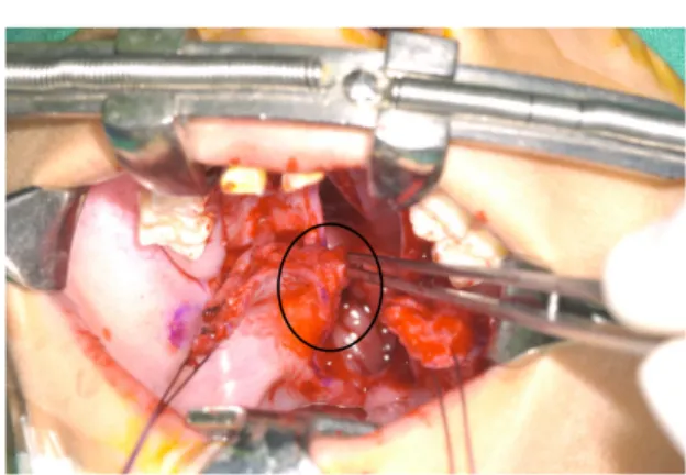
After careful sharp dissection of the levator muscle from the posterior palatine border, the blue nasal mucosa was identified. The levator muscle was separated completely from the nasal mucosa by careful sharp dissection until the hamulus and medial pterygoid plate were visualized laterally (Figure 4).
This dissection extended to the posterior border of the velum and laterally to the superior constrictor, resulting in a complete skeletonization of the levator muscle that was freely mobilized and easily attained a transverse orientation.
Closure begined with the nasal lining, which was sutured to the vomer flaps with 5-0 vicryl (Figure 5). Posteriorly, a 4-0 vicryl suture was placed in the uvular tissue that had been dissected earlier. The muscles were then closed as a separate layer using 3-0 vicryl. The palatal mucosa was then closed with interrupted chromic 4-0 vicryl sutures, catching the nasal lining over the hard palate to approximate the two layers of closure. The edgesof the mucoperiosteal flaps
weretacked to the edges of the palate; there was a minimal area of bare bone at the edges (Figure 6).
And then prefabricated palatal splint was delivered to hard palate to protect the raw surface.
III. DISCUSSION
Although great advances in treatment of cleft lip and palate have been made over the past 50 years ; in developing countries, due to social stigmas and unavailability of special medical facilities and personnels to the majority of the population, there is an increasing incidence of patients coming late for repair of cleft lip and palate. Recently, many humanitarian organizations have sent surgery teams to developing countries to treat patients with untreated congenital disabilities, tumors, burns, and traumas8). From 2002, Korean Association of Research and Charity for Craniofacial Deformity has sent charity operation teams to various developing countries and brought cleft lip and palate victims from those countries to Korea for cleft lip and palate operations. In present report, the 5-year old child with cleft palate was brought from Cambodia through ‘Beautiful Change Project’managed by Korean Association of Research and Charity for Craniofacial Deformity.
Regardless of the technique employed, the goals of cleft palatoplasty remains the same:9)
1. Closure of the palate, functionally separating the oral and nasal cavities
2. Development of normal speech with velopharyngeal competence and the absence of compensatory misarticulations
3. Normal facial development 4. Normal occlusal development
5. Normal nasal and nasopharyngeal airway patency
Palatoplasty techniques have undergone many innovations in the 150 years since Le Monnier10). Variations in these techniques have been aimed at adding length to the soft palate to reduce the incidence of VPI, reducing the incidence of fistula formation, decreasing the adverse effects on midfacial growth, and, in the most recent decades, accomplishing a functional muscular reconstruction of the soft palate to maximize its potential in terms of achieving normal velopharyngeal function.
The principle variations on the two-flap palatoplasties, as they now commonly referenced, are the Veau-Wardill-Kilner pushback, the von Langenbeck, and the Bardach two-flap palatoplasty9). The Bardach two-flap palatoplasty involves the creation of two axially patterned mucoperiosteal flaps pedicled on the greater palatine neurovascular bundles3). Using two-flap palatoplasty, access and visibility for the nasal repair and velar muscular reconstructions were relatively easy to perform in this case. Once the nasal layer and muscular reconstructions are complete, the flaps are medialized and annealed in the midline.
Palatal fistulas, epithelialized openings within the repair between the mouth and nasal cavity, have significant functional consequences. Avoidance of palatal fistulas in the treatment of cleft palates is critical, because nearly 50 percent of children with fistulas require re-operation11). The incidence of palatal fistulas after cleft repair ranges from 12 to 45 percent12-16). Bradon et al.17) reported the low fistula rate of 3.4 percent with the two-flap palatoplasty technique. An ongoing study from the University of Florida and Sao Paolo, Brasil seeks to evaluate the differences in outcome between the Furlow and two-flap palatoplasty procedures using four surgeons, standardized speech evaluations, a long-term follow-up (approximately 10 years), and enough patients to satisfy a power analysis with the variables at hand. The study remains unpublished in its full form at this time, but preliminary data suggest that many of the variables studied showed no difference in outcome, with the exceptions that the fistula rate is higher in the Furlow group and the hypernasal quality of speech is higher in the two-flap palatoplasty group9). Velopharyngeal insufficiency following palate repair is characterized by a typical hypernasal speech that may require secondary palatal surgery for correction, and results from poor function of the soft palate1). Rates of velopharyngeal insufficiency vary in the reported literature. Recently, Salyer et al.3) reported that 6.7% of patients with unilateral cleft lip and palate required secondary
velopharyngeal surgery after classic 2-flap palatoplasty. Intravelar veloplasty suggested by Kriens in 1969 was an big advancement on previous soft palatoplasties18). Kriens’innovation was to restore the levator sling and palatal musculature at the midline where they normally meet. This is accomplished by dissecting the anteriorly malpositioned muscle bundles from the posterior edge of the hard palate and repositioning them in the midline.
This technique is widely used today, though there is much variability among surgeons in how the musculature is dissected and repositioned1). In 1989, March et al. published results from a prospective study that compared the effects of intravelar veloplasty and traditional side to side techniques on velopharyngeal insufficiency. They found that repositioning of the levator muscles during primary palatoplasty was no better at improving velopharyngeal insufficiency than the side to side veloplasty19). Some practitioners have suggested that use of an operating microscope to perform palate repair, which allows for improved lighting, visualization of the muscle fibers results in improved outcomes5). Sommerlad reported on low-up and observed with 10 years of follow-up and observed that velopharyngeal insufficiency rates have decreased from 10.2 percent to 4.6 percent.
He attributes this improvement to radical dissection and repositioning of the velar musculature5). In this case with a bilateral, complete cleft palate, further technical considerations were required for closure of the
cleft. Because the vormer was not attached to either free edge of the hard palate and the cleft gap was too wide for direct approximation of nasal mucosal edges, a vomerine flap was used for closure of the nasal mucosa. An incision was made along the free margin of vomer, which was exposed in the cleft gap;
two septal mucosal flaps were raised, creating the vomer flaps. These flaps were then used to bridge the gap to the nasal mucosa recruited from the underside of the medial edge of the hard palate. The two-flap palatoplasty combined with with a vomer flap results in a four-flap palatoplasty. The vomer flap has been used particularly for wider bilateral clefts since the 1920s; more recently, its use has been advocated as a standard repair for all clefts18).
The choice of technique for cleft palate repair is often surgeon dependent. The goals of palatoplasty, however, should be same, and they contain separation of the oral and nasal cavity, with restoration of velopharyngeal sphincter, allowing normal speech. Careful interdisciplinary postoperative follow-up is important to observe and potentially correct fistula formation, Velopharyngeal insufficiency, maxillary growth deficiency, and sleep apnea1).
REFERENCES
1. van Aalst J A, Kolappa KK, Sadove M.
MOC-PSSM CME article: Nonsyndromic cleft palate. Plast Reconstr Surg 2008;
121(1 Suppl):1-14.
2. Bardach J, Kelly KM, Salyer KE. A comparative study of facial growth following lip and palate repair performed in sequence and simultaneously: an experimental study in beagles. Plast Reconstr Surg 1993;91:1008-1016.
3. Salyer KE, Sng KW, Sperry EE. Two- flap palatoplasty: 20-year experience and evolution of surgical technique.
Plast Reconstr Surg 2006;118:193-204.
4. Kriens OB. Fundamental anatomic findings for an intravelar veloplasty.
Cleft Palate J 1970;7:27-36.
5. Sommerlad BC. A technique for cleft palate repair. Plast Reconstr Surg 2003;112:1542-1548.
6. Sommerlad BC, Henley M, Birch M, Harland K, Moiemen N, Boorman J G.
Cleft palate re-repair-a clinical and radiographic study of 32 consecutive cases. Br J Plast Surg 1994;47:406-140.
7. Andrades P, Espinosa-de-los-Monteros A, Shell DHt, Thurston TE, Fowler JS, Xavier ST, et al. The importance of radical intravelar veloplasty during two-flap palatoplasty. Plast Reconstr Surg 2008;122:1121-1130.
8. Abenavoli FM. Operation Smile humanitarian missions. Plast Reconstr Surg 2005;115:
356-357.
9. Fonseca. Oral and maxillofacial surgery.
2 ed. St. Luis: Saunders;2009.
10. Millard. Alveolar and palatal deformities.
Boston: Little Brown;1980.
11. Rohrich RJ, Gosman AA. An update on
the timing of hard palate closure: a critical long-term analysis. Plast Reconstr Surg 2004;113:350-352.
12. Abyholm FE, Borchgrevink HH, Eskeland G. Palatal fistulae following cleft palate surgery. Scand J Plast Reconstr Surg 1979;13:295-300.
13. Amaratunga NA. Occurrence of oronasal fistulas in operated cleft palate patients. J Oral Maxillofac Surg 1988;
46:834-838.
14. Cohen SR, Kalinowski J, LaRossa D, Randall P. Cleft palate fistulas: a multivariate statistical analysis of prevalence, etiology, and surgical management. Plast Reconstr Surg 1991;
87:1041-1047.
15. Emory RE, Jr., Clay RP, Bite U,
Jackson IT. Fistula formation and repair after palatal closure: an institutional perspective. Plast Reconstr Surg 1997;
99:1535-1538.
16. Rohrich RJ , Byrd HS. Optimal timing of cleft palate closure. Speech, facial growth, and hearing considerations.
Clin Plast Surg 1990;17:27-36.
17. Wilhelmi BJ, Appelt EA, Hill L, Blackwell SJ. Palatal fistulas: rare with the two- flap palatoplasty repair. Plast Reconstr Surg 2001;107:315-318.
18. LaRossa D. The state of the art in cleft palate surgery. Cleft Palate Craniofac J 2000;37:225-228.
19. Marsh JL, Grames LM, Holtman B.
Intravelar veloplasty: a prospective study.
Cleft Palate J 1989;26:46-50.
교신 저자
정필훈, 서울대학교 치의학대학원 구강악안면외과학교실 서울시 종로구 연건동 275-1 우편번호 110-768/
Tel: 02-2072-3477/ e-mail: [email protected]