관동맥
11
0
0
전체 글
(2) 서. 론. 대상 및 방법. 관동맥질환에서 스텐트가 풍선성형술에 비하여 재협. 관동맥 Palmaz-Schatz 스텐트 재협착 조직 표본. 착율을 낮추고, 임상적 경과를 향상시킬 수 있다고 알. 29개의 관동맥표본들(LAD 14, LCX 5, RCA 10)이. 려져 있지만 아직까지도 재협착율은 6개월에 15~22%. 관동맥내 Palmaz-Schatz 스텐트가 삽입된 후 재협착. 1-3). 2)3). 이 발생한 25명의 환자(연령:59 13, 남/여:18/7). 와 동물실험에서의 연구결과4)에 따르면 이러한 재협착. 에서 directional coronary atherectomy(Simpson. 기전은 스텐트 자체의 반동작용(recoil)이나 혈관의 re-. Coronary Atherocath, Devices for Vascular Inter-. modeling보다는 신생내막조직(neointima)의 스텐트내. vention, Redwood City, CA, USA)를 이용하여. 부로의 생성(ingrowth)이 중요하게 생각되어진다. 인. Massachusetts General Hospital(Boston, MA, USA). 체의 스텐트재협착의 병리학적 연구에 의하면 myxoid. 심장내과에서 1995년 8월부터 1996년 11월 까지스. tissue의 발현이 종종 관찰되었으며,5) 세포의 증식율은. 텐트 시술후 평균 5.7±5.4개월 후 생검되었다. 생검조. 에 이른다고 알려져 있다.. 인체에서의 임상연구. 6)7). 8). 직은 즉시 methyl Carnoy’s 고정액(60% methanol,. 에서는 약 25%의 평활근 세포가 PCNA 양성으로 높. 30% chloroform, 10% glacial acetic acid)에 2~3일. 다고 하여 아직 논란의 여지가 있다.. 간 조직고정후 paraffine으로 처리하였다. 동맥경화반. 매우 낮다고 보고된 연구들. 이 있는 반면 다른 연구. 본 연구는 스텐트 재협착과정에서 스텐트내부로의. 제거술의 적응증은 전 예에서 혈관조영술상 재협착병. 세포의 이동 및 침투과정이 신생내막형성에서 필수적. 변이 관찰되었는데, 17예는 재발되는 불안정형 협심증,. 인 기전일 것이라는 가설하에 이러한 세포의 이동에. 5예는 운동부하검사에서 양성소견, 3예는 급성 심근경. 밀접하게 관계하는 요소들의 발현양상을 면역조직화학. 색증등이었다.. 적 병리분석을 통하여 살펴보고자 하였다. 이러한 세 포의 침투 및 이동기전은 아직 확실히 알려져 있지 않 9-12). 지만 MMP(matrix metalloproteinases),. 병리조직검사. uPA. 조직학적 염색은 paraffin 처리된 조직의 5 μm두께. (urokinase type plasminogen activator),9)13)14) PD-. 의 연속적 조직슬라이드에 시행하였다. Hematoxylin. GF (platelet derived growth factor) β receptor,15)16). & Eosin 염색을 하여 일반적인 조직의 형태를 분석하. hyaluronan17-19)등의 역할이 중요하게 보고된 바 있. 였다. 면역 세포화학염색(immunocytochemical sta-. 다. MMP의 subgroup에는 적어도 9가지가 알려져 있. ining)은 AEC(3-amino-9 ethylcarbazole:DAKO,. 9). 이 중 MMP1과 MMP9이 인체의 특발성 동맥. Carpenteria, CA, USA) 또는 DAB(3, 3’-diamino-. 경화반에서 보고된 바 있다.10)11) Hyaluronan의 합성. benzidine tetrahydrochloride, Sigma, St.Louis, MO). 에 관련된 HAS(hyaluronan synthase)는 인체에서. 를 chromogen으로 이용하여 시행하였다. 간략히 정리. 는데,. 20). hsHAS1 hsHAS2 두가지가 알려져 있으며. 아직. 하면 deparaffinization 후 endogenous peroxidase를. 인체의 동맥경화반에서의 발현정도에 대해서는 잘 알. 0.3% H2O2로 차단후 10% 정상염소혈청(normal goat. 려져 있지 않다. TGF(transforming growth factor)-. serum)을 이용하여 비특이적 항체의 결합을 억제 후. β1은 혈관조직에서 hyaluronan외에도 proteoglycan. 각각의 일차 항체(primary antibody)에 실온에서 2시. 및 collagen 세포외기질의 생성에 영향을 미칠 수 있. 간동안 incubation, biotinylated goat anti rabbit IgG. 5)21-23). 다고 알려진 바 있다.. 본 연구에서는 이러한. (Link:DAKO, Carpinteria, CA, USA)에 실온에서. growth factor 및 cytokine들의 인체에서 동맥경화반. 30분, conjugated peroxidase(DAKO) 30분 incubation. 제거술(atherectomy)을 이용한 스텐트 재협착조직에. 후 DAB 또는 AEC를 chromogen으로 이용하였으며,. 서의 발현양상을 면역조직화학적 방법을 통하여 분석. counterstaining은 Hematoxylin 또는 methyl green. 하고자 하였다.. 을 이용하였다. Antigen retrieval technique24)을 이용 시에는 0.3% H2O2 용액으로 endogenous peroxi-. 229.
(3) dase를 차단 후 10 mM sodium citrate pH 6.0 용액. 스텐트시술후 기간경과에 따라 감소하는 경향을 보였. 내에 조직슬라이드를 담근 채로 microwave(800 Watt). 다. 특히 스텐트 삽입 후 초기 6개월 미만의 기간에서. 내에서 10분간 열처리한 후 실온에서 식힌 후 이후의. 그 이후의 경우에 비하여 빈번하게 관찰되는 경향을 보. 염색과정을 계속 진행하였다.. 였는데 통계적 유의수준에는 미치지 못하였다(17/21. 사용된 항체는 다음과 같다. 1) 인체의 TF-β1에. vs 3/8, p=0.067). 이러한 myxoid tissue가 스텐트. 대한 토끼의 polyclonal affinity purified antibody:. 삽입 후 초기에 풍부히 관찰되어지며 시술후 9개월까. Leslie Gold교수(New York University Medical Center,. 지도 부분적으로 관찰되어지는 반면에 세포의 밀도가. New York, NY, USA)의 친절한 기증에 의하여 사용. 낮은(48~320 세포/mm2) 저세포밀도부위(cell poor. 25). 되었음. 2) 인체의 hsHAS1 에 대한 토끼의 polyclonal. area)의 관찰이 총 29표본중 17(59%) 표본에서 관찰. affinity purified antibody는 Heldin교수(Uppsala Uni-. 되어졌다(Fig. 1). 이러한 저세포밀도부위는 스텐트 삽. versity, Uppsala, Sweden)의 친절한 기증에 의하여. 입 후 4개월 이후의 기간에서 4개월 미만의 기간에 비. 사용되었음 3) uPA에 대한 monoclonal antibody. 하여 유의하게 빈번하게 관찰되어졌다(13/16 vs 4/13,. (product# 3689:American Diagnostica, Greenwich,. p<0.01). 세포의 밀도는 매우 다양한 양상을 보이는데. CT, USA) 4) MMP1에 대한 mouse monoclonal. 세포밀도가 높은 조직과 세포밀도가 매우 낮은 저세포. antibody clone 41-1E5(cat# IM35L:Oncogene. 밀도부위가 혼재되어 있으며, 대부분의 세포들이 α-. Research Products, Cambridge, MA, USA) 5) MMP9. actin 염색에 양성인 것으로 혈관 평활근세포가 수적인. 에 대한 mouse monoclonal antibody clone 56-2A. 면에서 주된 세포임을 알 수 있다(사진 생략).. (cat #IM37L:Oncogen Research Products) 6) PDGF 수용체에 대한 토끼의 polyclonal affinity purified an-. TGF-β1. tibody(cat#sc-339:Santa Cruz Biotech, Santa Cruz,. TGF-β1의 발현은 조사한 총 20표본 중 16표본에. CA, USA). 항체의 활성도를 확인하기 위하여 인체의. 서 관찰되었는데, 대부분의 경우 세포질(cytoplasm)에. 유방선세포암(breast ductal cell carcinoma) 조직과 원. 서 발견되었으며 스텐트 삽입 후 기간에 무관하게 발현. 숭이(baboon)의 폐동맥조직을 대조군으로 이용하였다.. 이 관찰되었다(Table 1). 두 표본에서는 세포외기질 에서 발현되었는데 모두 인접조직편의 해당부위에서. 통계방법. Movat 염색과 fibrin 염색에서 fibrin을 포함하는 혈전. 통계치는 평균±표준편차로 표시하였으며, 스텐트시술. 부위가 관찰되었으므로 이러한 부위에서의 TGF-β1. 후 기간에 따른 저세포밀도 발현 빈도의 비교는 Chi-. 은 혈소판에서 생성되었을 가능성이 많다.26) 현미경시. Square test를 이용하였고, myxoid tissue의 발현빈도. 야(100×)에서 전체 세포의 5% 이상의 세포질에서 발. 의 비교는 Fisher’s exact test를 이용하였으며 통계. 현된 경우는 11표본으로 이중 5예에서 myxoid tissue. 적 유의 수준은 p<0.05로 정하였다.. 에서 성상세포들의 세포질에서 TGF-β1의 발현이 현 저하게 나타남을 관찰할 수 있었으며(Fig. 2), 2예는. 결. 과. 저세포밀도부위 내의 성상세포 또는 방추세포(spindle cell) 등의 세포질에서도 발현을 관찰할 수 있었다.. 관동맥 스텐트 재협착조직의 형태학적 특징. TGF-β1의 발현정도는 스텐트 삽입 후의 기간과 특. 재협착조직의 Hematoxylin과 Eosin 염색에 의한 형. 별한 관련성을 발견할 수 없었다. 원숭이 폐동맥조직에. 태학적 분석에 의하면 우선적으로 myxoid tissue가 흔. 서는 TGF-β1은 내피세포의 세포질에서 발현이 확인. 하게 관찰되어지는데, myxoid tissue는 Fig. 1에서 보. 되었다(사진 생략).. 는 바와 같이 성상세포(stellate-shaped cell)들 주변 에 proteoglycan이 풍부한 세포외기질로 구성되어 있. HAS1. 다. 이러한 myxoid tissue가 Table 1에서 보는 바와. HAS1 발현양상은 대조군 표본인 유방의 ductal cell. 같이 총 29표본 중 20(69%) 표본에서 발견되었으며,. carcinoma조직내부의 carcinoma cell nest의 세포질. 230. Korean Circulation J 1999;29(2):228-238.
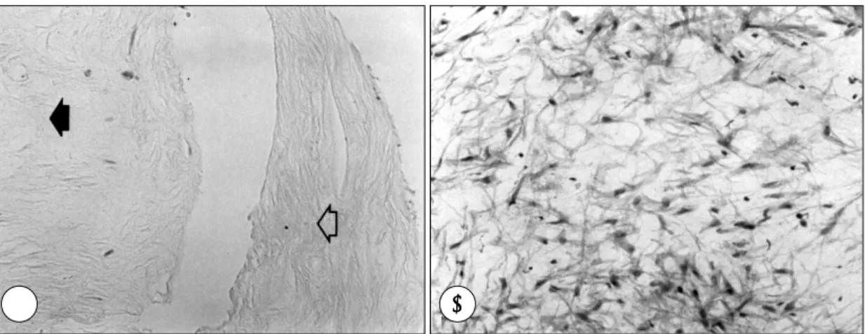
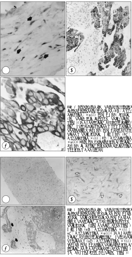
(4) A. B. Fig. 1. Hematoxylin and eosin staining of atherectomized coronary arterial in-stent restenotic tissues showing two kinds of tisse specimen:cell poor area (1A) and myxoid tissue(1B). Specimen in 1A shows two kinds of cell poor area:one (open arrow) with dense extracellular matrix and the other (closed arrow) with loose extracellular matrix (magnification ×100). Specimen in B shows myxoid tissue containing many stellate-shaped cells embedded in a loose extracellular matrix (magnification ×200).. A. B. Fig. 2. Atherectomized coronary arterial in-stent restenotic specimen shows TGF-β1 labeled cells. This specimen shows myxoid tissue containing many stellate-shaped cells labeled (brown color:open arrow) with TGF-b1 (2A:magnification ×200;2B:magnification ×400).. 중 특히 세포막에서 양성 염색소견을 나타내었으며. 으며 이중 5예가 myxoid tissue내의 세포에서 발현되. (Fig. 3), 스텐트 재협착조직에서는 성상세포와 방추형. 었다(Table 1). 발현은 주로 방추형세포와 성상세포의. 세포 모두에서 관찰되어졌다. 조사한 총 13표본 중. 세포질에서 관찰되었다. 100× 현미경시야에서 5%이. HAS-1은 10예에서 관찰되었고, 이중 myxoid tissue. 상의 양성세포를 보인 경우가 3예 모두 myxoid tissue. 내에서 2예가 관찰되었다. 현미경 100배 시야에서 5%. 내에서 관찰되어졌다. uPA의 발현은 유방선세포암조직. 이상의 세포에서 HAS1의 발현이 4예에서 관찰되었다. 에서 암세포군집(tumor cell nest)과 혈관평활근세포. (Table 1). HAS-1의 발현정도는 스텐트삽입후 2개월. 에서 관찰되었다(Fig. 4).. 에서 23개월 후까지의 기간동안 기간의 차이에 따른 특별한 관련성은 발견할 수 없었다.. MMP MMP1의 발현은 조사된 16표본 중 8예에서 관찰되. uPA uPA의 발현은 조사된 15표본 중 6예에서 관찰되었. 었다(Table 1). 100× 현미경시야에서 5%이상의 양 성세포가 4예에서 관찰되어졌는데 그 중 2예에서는 231.
(5) Table 1. Histopathological characteristics of atherectomized human coronary arterial in-stent restenotic tissues Time (mo.). Vessel. Cell poor area. Myxoid tissue. PDGFRcb. 0.5. LCX. -. 1.9. LAD. -. 2. LAD. -. 2. LCX. +. 2.4. RCA. -. 2+. NA. NA. NA. NA. NA. NA. 2.4. LAD. -. 1+. 2+:IC. -. 1+. -. 1+. NA. TGF-β1. HAS1. MMP1. MMP9. uPA. -. EC. -. NA. NA. NA. NA. 2+. NA. NA. NA. NA. 2+, m. 2+, m. 1+. NA. 2+, m. NA. NA. NA. NA. 2+. NA. NA. 1+. -. 1+, m. 1+. 2.4. RCA. -. 2+. 2+:IC, m. NA. NA. NA. 2+, m. NA. 2.5. RCA. -. 1+. 2+:IC, m. NA. NA. NA. -. NA 2+, m. 2.6. LCX. -. 2+. 2+:IC, m. NA. 2+, m. NA. NA. 3. LCX. +. -. NA. NA. NA. NA. NA. NA. 3. LCX. +. -. -. 2+. -. -. -. -. 3.5. RCA. +. 1+. NA. NA. NA. NA. NA. NA. 3.6. RCA. -. 1+. 1+:IC. -. -. -. -. -. 4. LAD. -. 1+. 1+:IC. 1+. 1+, m. -. NA. 1+. 4.1. LAD. +. 1+. 1+:IC. 1+. 1+. 1+, m. NA. 1+, m. 4.3. LAD. +. -. 2+:IC. NA. -. NA. -. 2+. 4.5. LAD. +. 1+. NA. 1+. -. -. NA. -. 5. LAD. -. 1+. 2+:IC, m. NA. NA. NA. -. NA. 5. LAD. +. 1+. 1+:IC. 2+. -. -. -. -. 5.8. LAD. +. 1+. 2+:IC. 2+. -. -. -. 1+ 2+, m. 5.8. RCA. +. 2+. 2+:IC, m. 1+, m. 2+, m. 1+, m. 2+, m. 6. RCA. +. 1+. NA. NA. 2+. 2+, m. NA. 2+, m. 6.1. LAD. +. 1+. NA. 1+. -. -. NA. 1+. 6.9. LAD. -. -. -. NA. NA. NA. NA. NA. 9. RCA. +. 1+. -. NA. 2+. 1+. 1+, m. 2+,m. 11. LAD. +. -. 2+:IC. NA. NA. NA. -1. +. 11. LAD. +. -. 2+:IC. NA. NA. NA. -. NA. 23. RCA. +. -. -. NA. NA. NA. NA. NA. + 23 RCA 2+:IC, EC 1+ NA NA In general, 1+ stands for‘positively labeled cells in less than 5%’ , whereas 2+ stands for‘positively labeled cells ;NA stands for‘not available’ ;m stands for‘found in exceeding 5% of total cells in microscopic field (×100)’ ;in myxoid tissue, 1+ stands for‘myxoid tissue found in a focal area’ myxoid tissue’ , 2+ stands for‘myxoid ;in TGF-β1 (transforming growth factor β1) IC stands for‘intracellular localization’ tissue found abundantly’ , ;HAS1 stands for‘hyaluronan synthase 1’ ;MMP stands for and EC stands for‘extracellular localization’ ‘matrix metalloproteinases’ ;uPA stands for‘urokinase type plasminogen activator’ ;PDTF-RcB stands for ‘platelet derived growth factor β receptor’ .. myxoid tissue내의 성상세포들에서 풍부히 관찰되어. 포가 1예이었으며 myxoid tissue내에서 관찰되었다.. 졌다(Fig. 5). MMP1의 발현은 주로 방추형세포와 성 상세포 모두에서 관찰되었으며, 스텐트삽입후 기간경과. PDGF β 수용체. 에 따른 특별한 발현양상은 관찰할 수 없었다. MMP-. PDGF β 수용체의 발현은 조사한 총 17표본 중 12. 9의 발현은 조사된 13표본 중 4예에서 관찰되었으며. 예에서 관찰되었으며, 이 중 6예는 myxoid tissue내에. (Table 1), 이중 3예는 myxoid tissue에서 발견되었. 서 관찰되었다(Table 1). 100× 현미경시야에서 5%. 고(Fig. 6), 주로 방추형세포와 성상세포의 세포질에서. 이상의 양성세포를 보인 경우가 6예로 이 중 5예가. 관찰되었다. 100× 현미경시야에서 5%이상의 양성세. myxoid tissue내의 세포에서 발현되었다. 발현은 주로. 232. Korean Circulation J 1999;29(2):228-238.
(6) A. C. A. C. B. Fig. 3. Atherectomized coronary arterial in-stent restenotic specimen (3A:magnification:×400) shows HAS1 labeled (red color) spindle-shaped cells (closed arrow). Immunostaining of breast infiltrating ductal cell carcinoma as a positive control tissue for HAS1 shows nest of carcinoma cells labeled with HAS1 (3B: magnification ×100;3C:magnification ×400). Note the pericytoplasmic staining of HAS1 indicating this enzyme’s location at plasma membrane.. B Fig. 4. Atherectomized coronary arterial in-stent restenotic specimen shows uPA labeled cells. This specimen shows myxoid tissue containing many stellate-shaped cells labeled (brown color:open arrow) with uPA (4A : magnification ×100 ; 4B: magnification ×400). Immunostaining of breast infiltrating ductal cell carcinoma (4C:magnification ×200) for uPA shows positive cell cytoplasmic staining on tumor cell nest (open arrow) and on vascular SMCs (closed arrow).. 233.
(7) A. B. Fig. 5. Atherectomized coronary arterial in-stent restenotic specimen shows MMP1 labeled cells. This specimen shows myxoid tissue containing many stellate-shaped cells labeled (red color:open arrow) with MMP1 (5A: magnification ×200;5B:magnification ×400).. A. B. Fig. 6. Atherectomized coronary arterial in-stent restenotic specimen shows MMP9 labeled cells. This specimen shows myxoid tissue containing many stellate-shaped cells labeled (red color:open arrow) with MMP1 (6A: magnification ×200;6B:magnification ×400).. A. B. Fig. 7. Atherectomized coronary in-stent restenotic specimen shows PDGF β receptor labeled cells. This specimen shows myxoid tissue containing many stellate-shaped cells labeled (red color:open arrow) with PDGF β receptor (7A:magnification ×100;7B:magnification ×400).. 234. Korean Circulation J 1999;29(2):228-238.
(8) 방추형세포와 성상세포의 세포질에서 관찰되었다.. 경화반의 파손(rupture)에 관련될 가능성이 있는 것으 로 알려져 있다.9-13) MMP9과 MMP1은 각각 인체의. 고. 찰. 관동맥10)과 경동맥11) 동맥경화반에서의 세포에서 발현 이 보고되어진 바 있다. MMP-9은 collagen types IV,. 인체의 관동맥스텐트 재협착조직의 병리학적 분석연. V, XI, elastin, proteoglycans등을 분쇄시키며,9) 인체. 구결과들은 아직 제한적이지만 대체적으로 재협착조직. 의 경피적방법으로 시행된 반절제술에서 얻은 관동맥경. 내에서의 세포는 대부분이 평활근세포이며, PCNA또는. 화반에서(불안정형협심증 83% ;안정형협심증 75%). Ki67분석에 따르면 대부분 매우 낮은(0~1%) 세포증. macrophage, 혈관평활근, 임파구등에서 발견되나 정. 식율을 보고하고 있다.6)7) 그러나 한 연구8)에 따르. 상적인 내유동맥(internal mammary artery)에서는. 면 말초동맥내 스텐트 재협착조직에서 매우 높은(평균. 발현되지 않는다고 보고되었다.10) in vivo연구12)에서. 15.2%) 세포증식율을 보고하고 있는데 이러한 차이의. MMP9 mRNA가 풍선도자에 의한 손상된 백서의 동맥. 원인은 아직 확실하지 않다. 본 연구결과 재협착조직의. 혈관평활근에서 빠르면 6시간에 발현이 유도되어 3일. 세포밀도는 다양하며, 세포밀도가 매우 낮은 저세포밀. 후 정점에 이르며, MMP억제에 의한 평활근세포의 이. 도부위가 59%의 표본에서 관찰되었고, 이러한 저세포. 동이 손상후 초기에 강력히 억제됨이 알려져 있다.12). 밀도부위는 스텐트 삽입후 기간이 경과함에 따라서 점. MMP1은 collagen type Ⅰ과 type Ⅲ를 분해시키며,9). 차 증가하는 양상을 관찰할 수 있었다. 특히 myxoid. 인체의 경동맥의 동맥경화병변의 macrophage, 혈관평. tissue의 발현이 69%의 표본에서 관찰되어지는데, 이. 활근세포, 내피세포등에서 발견되었는데, 주로 대식세. 27). 조직은 PTCA후. 7). 또는 스텐트삽입후. 재협착조직에. 포가 밀집되어있는 lipid core 주변에서 현저히 관찰되. 27). 서 빈번히 관찰되었으며, 특발성 동맥경화병변 의 일. 었고, 동맥경화반 주변의 비후성 내피(intimal thick-. 부에서도 관찰되어졌는데 이러한 myxoid tissue의 생. ening)에서의 평활근 세포에서도 관찰되었으나, 정상적. 물학적인 기능은 아직 잘 알려져있지 않다.. 비후성 내피에서는 발현되지 않았다.11) MMP1과 동맥. PDGF는in vitro실험에서 혈관 평활근세포에 대한. 경화반의 출혈성 병변과 밀접한 상관성을 갖음이 발견. mitogen일 뿐 아니라 강력한 chemoattractant로 작용. 되어 MMP1이 동맥경화반의 불안정성에 관계될 가능. 15)28). PDGF는 PDGF-A와. 성이 제시된 바 있다.11) Plasmin은 대부분의 MMPs. PDGF-B의 두가지 종류의 peptide chain의 homo-. 에 대하여 강력한 활성화 작용을 하는 것으로 알려져. dimer 또는 heterodimer로 존재하며, 정상 성인조직. 있다.9)14) Plasminogen activator는 plasminogen을. 에서는 그 발현이 매우 낮거나 미미하지만 조직의 손상. plasmin으로 전환시키는데 촉매역할을 하여 MMPs를. 시 그 발현이 증가되는 것으로 알려져 있다. 또한 PD-. 활성화시키는데, uPA는 수용체와 결합되고 활성화됨으. GF에 대한 수용체는 α-subunit과 β-subunit의 두. 로써 국소적으로 단백분해활성을 일으키게된다.9) 동물. 가지가 알려져 있는데, 전자는 A또는 B chain과 결합. 생체실험에서는 혈관내피손상 후 내피로 이동하는 혈. 할 수 있는 반면 후자는 B chain만 결합이 이루어 질. 관평활근세포에서 uPA와 수용체 uPAR의 mRNA의. 하는 것으로 알려져 있다.. 15). 수 있다고 알려져 있다.. 특히 PDGF-BB는 백서의. 현저한 발현을 초기 5~8일에 관찰할 수 있었으며, 6. 평활근세포의 chemotaxis를 강력하게 유도할 수 있으. 주후에는 현저하게 감소함을 관찰하였다.13) 본 연구결. 며15) 이러한 PDGF의 세포의 이동에 대한 역할은. 과 MMP1, MMP9, uPA의 발현은 각각 50%, 31%,. PDGF수용체 중 receptor β를 통하여 일어남이 보고. 40%의 표본에서 발견되었으며, 스텐트삽입후 시간경. 되었다.16) 본 연구결과 PDGF β수용체의 발현은 조사. 과에 따른 일정한 양상을 보이지는 않았다. TGF-β가. 대상 표본의 71%에서 발현되었으며, 특히 이 발현이. uPA등의 proteases의 합성과 분비를 감소시키고 plas-. 비교적 풍부하게 관찰되었던 예의 대부분이 myxoid. minogen activator inhibitor의 발현을 증가시킴으로. tissue내에서 관찰됨은 흥미있는 결과이다.. 세포외기질의 단백분해(proteolysis)와 분쇄를 억제한. MMP와 plasminogen activator는 세포의 이동과정,. 다고 보고된 바 있는데,29)30) 이러한 관점에서 TGF-. 세포외기질의 분쇄 및 흡수에 중요한 역할을 하며 동맥. β1의 발현이 본 연구결과 80%의 표본에서 빈번하게 235.
(9) 발현되었음을 고려하면 이러한 조직내에서의 TGF-β. β1은 in vivo 및 in vitro 실험에서 세포외기질성분의. 1에 의한 uPA생성 억제의 가능성을 생각하여 볼 수. 유전자를 조절하는데 중요한 역할을 하는 것으로 알려. 있겠다. 아울러 MMP와 uPA의 발현이 이전의 동물실. 져 있다.5)21-23) TGF-β1에 전이된 돼지의 동맥에서,. 12)13). 험. 에서 비교적 내피손상후 초기 1주일 전후임을. proteoglycan, procollagen, collagen의 합성 증가와. 감안하면 본 연구에서의 대부분의 표본들이 스텐트시. 아울러 내막층의 비후가 관찰되었고,21) 풍선도자에 의. 술후 2개월 이후의 것으로 스텐트시술직후 1~2개월. 한 손상받은 백서의 동맥에서 손상후 6시간만에 TGF-. 내에 일어나는 현상을 파악하는데 한계가 있다고 본다.. β1의 mRNA가 증가하고 신생내막(neointima)의 평. 본 연구에서 PDGF β 수용체, uPA, MMP1, MMP9. 활근세포에서 TGF-β1의 발현이 현저하였음이 보고. 의 발현이 수적으로 증가한 경우의 50% 이상의 예가. 되었다.22)23) 또한 TGF-β1에 대한 중화항체(neu-. myxoid tissue에서 관찰되었음은 성상세포를 포함하. tralizing antibody)에 의하여 백서의 경동맥에 풍선도. 며 proteoglycan이 풍부한 myxoid tissue가 스텐트. 자에 의해 발생한 신생내피의 크기를 감소시킬 수 있다. 재협착과정에서 세포의 이동과정에 중요한 역할을 할. 는 보고가 있다.22) 아울러 TGF-β1은 hyaluronan의. 가능성이 있다. 또한 이전의 연구결과에서6) hyaluronan. 합성에 주요한 영향을 줄 수 있음이 in vitro study에서. 이 스텐트 재협착조직내의 세포외기질의 구성성분으로. 밝혀진 바 있으므로,5) TGF-β1은 세포외 기질의 형. 발견되었는데 hyaluronan은 동물에서 태아동맥관(fetal. 성외에도 hyaluronan의 생성에 따른 세포의 이동과정. 17). ductus arteriosus),. 우동맥 평활근세포(bovine ar-. 에 관계될 수 있다고 보여진다. 본 연구결과 TGF-β. terial smooth muscle cell)18)의 세포이동, 종양세포의. 1의 발현이 조사표본 20개중 80%에서 관찰되었으며. 19). 조직내 침투과정(invasion) 에서 중요한 역할을 하는. 대부분에서는 세포질에서 그 발현을 관찰할 수 있으나,. 것으로 알려져 있다. Hyaluronan의 생합성과정에 중요. 일부조직에서는 세포외기질에서 혈전과 fibrin을 관찰. 한 역할을 하는 hyaluronan synthase(HAS)는 세포. 할 수 있었던 부위에서 발현되었는데, TGF-β1이 혈. 의 원형질막(plasma membrane)에 존재함이 알려져. 소판에 의해서도 생성되어 유리될 수 있다는 연구보고26). 있으며, 인체에서는 적어도 hsHAS1과 hsHAS2 두가. 에 근거하면 혈전의 형성과정에서 혈소판에 의하여 유. 지가 밝혀져 있다.20) 본 연구에서 hsHAS1은 조사된. 리된 TGF-β1이 세포외기질의 형성을 촉진시킴으로. 표본 13개중 77%에서 스텐트시술후 2~23개월에 걸. 서 신생내막을 형성할 가능성이 있다. TGF-β1의 발. 쳐 발견되었는데, 그 발현은 방추형세포와 성상세포 모. 현이 현미경 시야에서 비교적 빈번하게 관찰되었던 11. 두에서 관찰되었다. Hyaluronan의 생합성이 세포외기. 예 중 5예에서 myxoid tissue를 관찰할 수 있음은 아. 질의 성분으로서 hygroscopic property 즉 많은 양의. 마도 myxoid tissue가 이러한 재협착과정에서 세포외. 물을 결합하는 특성을 갖고 혈관조직을 부풀릴 수 있으. 기질의 형성과정에서도 중요한 역할을 할 가능성이 있. 며, 이 부위로의 세포의 이동이 용이하게 됨으로써 동. 다고 생각되어된다.. 맥관(ductus arteriosus) 내막을 비후시키고 폐쇄시킬. 결론적으로 관동맥스텐트 재협착병변에서 흔히 발견. 수 있다는 가설이17) 스텐트 재협착 병변의 발생에도 적. 되는 myxoid tissue는 아마도 생물학적으로 세포의 이. 용될 수 있는지는 현재로선 아직 확실하지는 않다.. 동 및 세포외기질 형성과정에서 활발한 조직일 가능성. 본 연구결과에서 흥미로운 사실중의 한가지는 세포. 이 있으며, 아울러 세포외기질의 축적이 스텐트삽입후. 밀도가 매우 낮은 저세포밀도부위의 발견이며, 이는 스. 시간 경과에 따라서 점차로 증가하는 경향을 관찰할 수. 텐트 삽입후 시간의 경과에 따라서 그 빈도가 증가하는. 있었다.. 경향을 보였다. 이는 세포외 기질의 합성과 분쇄(de-. 요. gradation)의 균형이상 또는 apoptosis가 관계할 가. 약. 능성이 있다. 이전의 연구에 의하면 TGF-β1과 이의 mRNA 발현이 인체의 관동맥과 말초동맥에서 혈관평. 연구배경:. 활근세포에서 대부분 관찰되었으며, 특발성병변보다는. 인체의 관동맥 스텐트 재협착과정에서 스텐트의 반. 재협착조직에서 증가한다고 알려져 있으며,31) TGF-. 동(recoil)이나 혈관의 remodelling에 의한 효과보다는. 236. Korean Circulation J 1999;29(2):228-238.
(10) 신생내막의 스텐트 구조물(strut) 내부로의 새로운 형. ■ 감사문. 성과정이 중요한 기전으로 인식되어지고 있다. 그간의. 1997년도 대한순환기학회 신진 연구비의 지원으로 본 과제를 수행할 수 있게된 데에 대하여 먼저 순환기학회의 여러 선생 님들에게 감사의 말씀을 드리옵니다. 본 연구를 위해 헌신적으 로 도와주시고 가르쳐주신 Harvard University 심장내과의 Herman K Gold교수님, University of Washington 병리학교 실의 Stephen M Schwartz교수님, Michael A Reidy교수님, Thomas N Wight 교수님들께 존경과 감사를 드리고 싶습니 다. 또한 Sweden Uppsala University의 Paraskevi Heldin 교수님의 HA-S1 항체지원 그리고 New York University의 Leslie Gold교수님의 TGF-β1항체 지원 역시 본 연구에서 큰 도움이 된 데에 대하여 고맙게 생각합니다. 그리고 본 연구 의 수행과정에서 많은 협조와 지도 그리고 격려를 해주신 연 세대학교의 조승연 교수님, 조상호 교수님, 장양수 교수님, 송 영민 선생님, 울산대학교의 박승정 교수님과 허주령 교수님, 아주대학교의 최병일 교수님, 그리고 이화대학교의 신길자 교 수님께도 깊은 감사를 드립니다.. 연구에 의하면 이러한 조직에서 세포의 증식율이 매우 낮고 myxoid tissue가 빈번히 발견되었는데, 이러한 myxoid tissue의 생물학적 기능은 아직 잘 알려져 있 지 않다. 본 연구는 세포의 이동과 세포외기질 형성과 정이 스텐트 재협착에서 중요한 역할을 할 것으로 가설 을 세우고 이들 기전에 긴밀한 영향을 미치는 cytokine 및 growth factor들의 조직내 발현을 분석하고자 하였다.. 방 법: Palmaz-Schatz 스텐트 시술을 받고 재협착병변이 확인된 환자25명(연령:59±13, 남/여:18/7)의 환자 에서 경피적 atherectomy시술을 통한 29개의 표본 (LAD14, LCX 5, RCA 10)을 얻어서 면역세포화학적 방법을 이용하여 TGF-β1, HAS1, MMP1, MMP9,. REFERENCES. uPA, PDGF β receptor의 재협착 조직내에서의 발현. 1) Serruys PW, Jaegere PD, Kiemeneij F, Macaya C,. 을 조직학적으로 분석하였다.. 결 과: 성상세포들로 구성되는 myxoid tissue가 20개의 표. 2). 본에서 관찰되었으며, 스텐트 삽입 후 기간이 경과함에 따라서 감소하는 경향을 보였다. 또한 세포밀도가 매우 낮은 저세포 밀도부위가 17표본에서 관찰되었는데 그. 3). 빈도는 스텐트시술후 기간경과에 따라서 증가하였다(4 개월 미만13/16 vs 4개월 이후 4/13, p<0.01). 면역세 포화학적 방법에 의한 각각의 cytokine 및 growth factor의 발현정도는 다양하였는데, TGF-β1은 16/20. 4). 예에서, HAS1은 10/13예에서, MMP1은 8/16예에서, MMP9은 4/13예에서, uPA는 6/15예에서, PDGF β. 5). receptor는 12/17예에서 양성세포들을 관찰할 수 있 었으며, 특히 PDGF β receptor, TGF-β1, uPA 등 이 myxoid tissue내에서 비교적 풍부히 발견되었다.. 6). 결 론: 관동맥 스텐트 재협착병변에서 흔히 발견되는 myxoid tissue는 아마도 생물학적으로 세포의 이동 및 세포외. 7). 기질 형성과정에서 활발한 조직일 가능성이 있으며, 아 울러 세포외기질의 축적이 스텐트삽입후 시간 경과에. 8). 따라서 점차로 증가하는 경향을 관찰할 수 있었다.. 중심 단어 :스텐트 재협착・세포이동・세포외기질・ Myxoid tissue.. 9). Rutsch W, Heyndrickx G, et al. For the BENESTENT study group. A comparison of balloon expandable-stent implantation with balloon angioplasty in patients with coronary artery disease. N Engl J Med 1994;331:489-95. Hoffmann R, Mintz GS, Dussaillant GR, Popma JJ, Pichard AD, Satler LF, et al. Patterns and mechanism of in-stent restenosis. A serial intravascular ultrasound study. Circulation 1996;94:1247-54. Mudra H, Regar E, Klauss V, Werner F, Henneke K, Sbarouni E, et al. Serial follow-up after optimized ultrasoundguided development of Palmaz-Schatz stents. In-stent neointimal proliferation without significant reference segment response. Circulation 1997;95:363-70. Post MJ, Smet BJGL, Helm Y, Borst C, Kuntz RE. Arterial remodeling after balloon angioplasty or stenting in an atherosclerotic experimental model. Circulation 1997;96:996-1003. Haubeck H-D, Kock R, Fischer D-C, Leur EVD, Hoffmeister K, Greiling H. Transforming growth factor-β1, a major stimulator of hyaluronan synthesis in human synovial lining cells. Arthritis Rheum 1995;38:669-77. Chung I, Reidy MA, Schwartz SM, Wight TN, Gold HK. Enhanced extracellular matrix synthesis may be important for restenosis of arteries after stent deployment (abstract). Circulation 1996;94:I-349. Strauss BH, Umans VA, Suylen R, Feyter PJ, Marco J, Robertson GC, et al. Directional atherectomy for treatment of restenosis with coronary stents: Clinical, angiographic and histologic results. J Am Coll Cardiol 1992;20:1465-73. Kearney M, Pieczek A, Haley L, Losordo DW, Andres V, Schainfeldr, et al. Histopathology of in-stent restenosis in patients with peripheral artery disease. Circulation 1997;95:1998-2002. Dollery CM, McEwan JR, Henney AM. Matrix metalloproteinases and cardiovascular disease. Circ Res 1995;. 237.
(11) 77:863-8.. 10) Brown Dl, Hibbs MS, Kearney M, Loushin C, Isner JM.. 11). 12). 13) 14). 15). 16). 17). 18). 19) 20). Identification of 92-kD gelatinase in human coronary atherosclerotic lesions. Association of active enzyme synthesis with unstable angina. Circulation 1995;91:2125-31. Nikkari ST, O’Brien KD, Ferguson M, Hatsukami T, Welgus HG, Alpers CE. Interstitial collagenase (MMP-1) expression in human carotid atherosclerosis. Circulation 1995;92:2125-31. Bendeck MP, Zempo N, Clowes AW, Galardy RE, Reidy MA. Smooth muscle cell migration and matrix metalloproteinase expression after arterial injury in the rat. Circ Res 1994;75:539-45. Reidy MA, Irvin C, Lindner V. Migration of arterial wall cells. Expression of plasmingen activators and inhibitors in injured rat arteries. Circ Res 1996;78:405-14. Sperti G, van Leeuwen RTJ, Quax PHA, Maseri A, Kluft C. Cultured rat aortic vascular smooth muscle cells digest naturally produced extracellular matrix: Involvement of plasminogen-dependent and plasminogen-independent pathways. Circ Res 1992;71:385-92. Ferns GAA, Raines EW, Sprugel KH, Motani AS, Reidy MA, Ross R. Inhibition of neointimal smooth muscle accumulation after angioplasty by an antibody to PDGF. Science 1991;253:1129-32. Koyama N, Hart CE, Clowes AW. Different function of the platelet-derived growth factor-α and β receptors for the migration and proliferation of cultured baboon smooth muscle cells. Circ Res 1994;75:682-91. Boudreau N, Turley E, Rabinovitch M. Fibronectin, hyaluronan, and a hyaluronan binding protein contribute to increased ductus arteriosus smooth muscle cell migration. Dev Biol 1991;143:235-47. Savani RC, Wang C, Yang B, Zhang S, Kinsella MG, Wight TN, et al. Migration of bovine aortic smooth muscel cells after wounding injury. The role of hyaluronan and RHAMM. J Clin Invest 1995;95:1158-68. Knudson W. Tumor-associated hyaluronan. Providing an extracellular matrix that facilitates invasion. Am J Pathol 1996;148:1721-6. Weigel PH, Hascall VC, Tammi M. Hyaluronan synthases. J Biol Chem 1997;272:13997.. 238. 21) Nabel EG, Shum L, Pompili VJ, Yang Z, San H, Shu. 22) 23) 24). 25) 26). 27). 28) 29). 30). 31). HB, et al. Direct transfer of of transforming growth factor-β 1 gene into arteries stimulates fibrocellular hyperplasia. Proc Natl Acad Sci USA 1993;90:10759-63. Wolf YG, Rasmussen Rupslahti. Antibodies against transforming growth factor-ν1 supress intimal hyperplasia in a rat model. J Clin Invest J 1994;93:1172-8. Majesky MW, Lindner V, Twardzik DR, Schwartz SM, Reidy MA. Production of transforming growth factor-β1 during repair of arterial injury. J Clin Invest 1991;88:904-10. Shi SR, Gu J, Kalra KL, Chen T, Cote RJ, Taylor CR. Antigen retrieval technique: A novel approach to immunohistochemistry on routinely processed tissue sections. Cell Vision 1995;2:6-22. Asplund T, Brinck J, Sujuki M, Briskin MJ, Heldin P. Characterization of hyaluronan synthase from a human glioma cell line. Biochem Biophys Acta 1998;1380:377-88. Wakefield LM, Smith DM, Flanders KC, Sporn MB. Latent transforming growth factor-β from human platelets. A high molecular weight complex containing precursor sequences. J Biol Chem 1988;263:7646-54. Riessen R, Isner JM, Blessing E, Loushin C, Nikol S, Wight TN. Regional differences in the distribution of the proteoglycans biglycan and decorin in the extracellular matrix of atherosclerotic and restenotic human coronary arteries. Am J Pathol 1994;144:962-74. Ross R. The pathogenesis of atherosclerosis: A perspective for the 1990s. Nature 1993;362:801-9. Laiho M, Saksela O, Andreasen PA, Keski-Oja J. Enhanced production and extracellular deposition of the endothelial-type plasminogen activator inhibitor in cultured human lung fibroblasts by transforming growth factor-β. J Cell Biol 1986;103:2403-10. Keski-Oja J, Raghow R, Sawdey M, Loskutoff DJ, Postlethwaite AE, Kang AH, et al. Regulation of mRNAs for type-1 plasminogen activator inhibitor, fibronectin, and type I procollagen by transforming growth factor-β. J Biol Chem 1988;263:3111-5. Nikol S, Isner JM, Pickering G, Kerney M, Leclerc G, Weir L. Expression of transforming growth factor-β1 is increased in human vascular restenotic lesions. J Clin Invest 1992;90:1582-92.. Korean Circulation J 1999;29(2):228-238.
(12)
수치

관련 문서
