• 검색 결과가 없습니다.

Epizootiological characteristics of viable bacteria and fungi in indoor air from porcine, chicken, or bovine husbandry confinement buildings JVS

N/A
N/A
Protected

Academic year: 2022

Share "Epizootiological characteristics of viable bacteria and fungi in indoor air from porcine, chicken, or bovine husbandry confinement buildings JVS"

Copied!
8
0
0

로드 중.... (전체 텍스트 보기)

전체 글

(1)

J Vet Sci 2016, 17(4), 531-538ㆍhttps://doi.org/10.4142/jvs.2016.17.4.531 JVS

Received 19 Feb. 2016, Revised 13 Apr. 2016, Accepted 8 Jun. 2016

*Corresponding author: Tel: +82-2-2258-7364; Fax: +82-2-532-3820; E-mail: [email protected]

Journal of Veterinary Scienceㆍⓒ 2016 The Korean Society of Veterinary Science. All Rights Reserved.

This is an Open Access article distributed under the terms of the Creative Commons Attribution Non-Commercial License (http://creativecommons.org/licenses/

pISSN 1229-845X eISSN 1976-555X

Epizootiological characteristics of viable bacteria and fungi in indoor air from porcine, chicken, or bovine husbandry confinement buildings

Katharine Roque

1

, Gyeong-Dong Lim

1

, Ji-Hoon Jo

1

, Kyung-Min Shin

1

, Eun-Seob Song

1

, Ravi Gautam

1

, Chang-Yul Kim

1

, Kyungsuk Lee

2

, Seungwon Shin

3

, Han-Sang Yoo

3

, Yong Heo

1

, Hyoung-Ah Kim

4,

*

1

Department of Occupational Health, College of Medical and Public Health Sciences, Catholic University of Daegu, Gyeongsan 38430, Korea

2

National Institute of Agricultural Sciences, Rural Development Administration, Wanju 55365, Korea

3

Department of Infectious Diseases and BK21 PLUS, College of Veterinary Medicine, Seoul National University, Seoul 08826, Korea

4

Department of Preventive Medicine, College of Medicine, The Catholic University of Korea, Seoul 06591, Korea

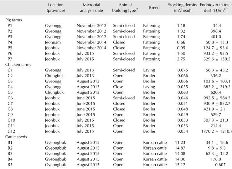
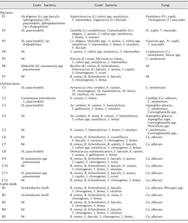
Microorganisms found in bioaerosols from animal confinement buildings not only foster the risk of spreading diseases among livestock buildings, but also pose health hazards to farm workers and nearby residents. This study identified the various microorganisms present in the air of swine, chicken, and cattle farms with different kinds of ventilation conditions in Korea. Microbial air samples were collected onto Petri dishes with bacterial or fungal growth media using a cascade impactor. Endotoxin levels in total dust were determined by the limulus amebocyte lysate kinetic QCL method. Prevalent Gram-positive bacteria were Staphylococcus (S.) lentus, S. chromogenes, Bacillus (B.) cereus, B.

licheniformis, and Enterococcus faecalis, while the dominant fungi and Gram-negative bacteria were Candida albicans and Sphingomonas paucimobilis, respectively. Considering no significant relationship between the indoor dust endotoxin levels and the isolation of Gram-negative bacteria from the indoor air, monitoring the indoor airborne endotoxin level was found to be also critical for risk assessment on health for animals or workers. The present study confirms the importance of microbiological monitoring and control on animal husbandry indoor air to ensure animal and worker welfare.

Keywords: Gram-negative bacteria, airborne microorganisms, cattle, chickens, swine

Introduction

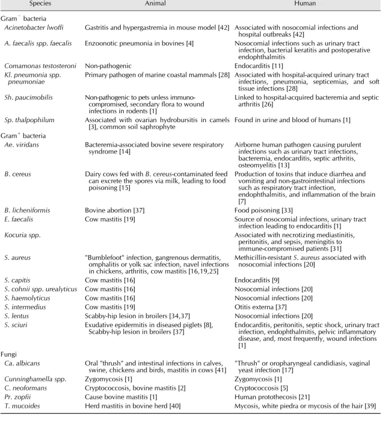
Because of the rapid growth of intensive livestock production in recent years, indoor animal husbandry facilities now pose a potential health risk to husbandry workers and animals owing to the generation of inhalable particulate emissions and the harmful compounds adhered to them [6]. These organic dusts include bacteria, viruses, and fungi, as well as microbial secondary metabolites. Organic dusts, which are generated from bedding, fecal materials, animal skin, or feedstuffs, float into indoor air during cleaning or through animal activities such as feeding or feathering [6,12,22]. Exposure to organic dusts may cause alteration of immune responses in animals and farm workers exposed [18,32]. These health risks are heightened when large amounts of microorganisms are present in the air, which increases the risk of spreading diseases from one

livestock building to another or to neighboring communities. In a study conducted in Vietnam, the bacterium (Enterococcus [E.]

faecalis) that was associated with urinary tract infection in

humans was identical to the bacterial strain isolated from the

flocks of a nearby poultry farm [30]. Conversely, some

microorganisms that are non-pathogenic are capable of

releasing by-products in the form of toxins (exotoxin,

endotoxins and mycotoxins) that are harmful to both humans

and animals [18,32,41]. Also of increasing importance is the

prevalence of antibiotics-resistant microorganisms in the herd

[14]. Antibiotics are used in animal husbandry in the form of

feed additives or to treat an existing disease. Humans can

acquire antibiotic resistance through contaminated animal

products or occupational contact [36]. Human infections due to

methicillin-resistant Staphylococcus (S.) aureus and other

antibiotic-resistant microorganisms of animal origin have been

(2)

widely reported [36,38]. However, humans may also serve as the source of infection.

Most studies of microbial air contamination in animal farms have quantitatively identified the concentrations of total bacteria or fungi isolated, but qualitative data regarding specific microorganisms under particular husbandry conditions are also important. Microbial identification in a particular farm condition will be helpful to strategizing disease prevention and management since the information describing prevailing airborne zoonotic microorganisms or major bacterial sources of endotoxins could contribute to proper analysis of risk factors for farmers and animals. Thus, monitoring the microbial air quality inside livestock farms is important to providing good air quality, not only inside the farms premises, but also outdoors. Microbial air quality may differ among farms because of differences in stocking density, barn hygiene, microclimate parameters (temperature, relative humidity, gases) or ventilation systems [6,12,29].

In the present study, we identified species of viable bacteria and fungi in indoor air collected from swine, chicken, or cattle husbandry confinement buildings nationwide in Korea. In addition, endotoxin levels in indoor dust were determined for those farms. The primary goal of this study was to evaluate which species of pathogenic microorganisms are detrimental to the health of workers or livestock if exposed through the respiratory route.

Materials and Methods

Animal farms

Swine, chicken, or cattle farms were studied from 2012 to 2015. Three pig farms were evaluated in 2012, and two farms each were evaluated in 2014 and 2015. Five and seven chicken farms were evaluated in 2013 and 2015, respectively, while five cattle sheds were evaluated in 2015. Participating farms were selected from the list of candidate farms prepared with help from the swine, chicken, or Korean beef cattle farmer’s cooperative mentioned in the Acknowledgement section. Only farms that allowed our entrance were investigated. General husbandry characteristics of each farm are described in Table 1.

Animal housing style was categorized into open type, semi-closed type, or closed type (Table 1). Open type indicates a farm in which ventilation was executed through winch curtains, while the closed type indicated farms in which a controlled mechanical ventilation system was applied without windows. The semi closed type animal confinement building installed a controlled mechanical ventilation system with upper or lower side windows. Indoor air sampling for microbial analysis or dust quantitation was performed from June to August, except for five swine farms in 2012 or 2014 (Table 1).

Indoor air sampling

To determine qualitative microbial analysis, air samples were collected at 1 to 1.5 m from the ground using a cascade impactor (BioStage; SKC, USA) at a flow rate of 28.3 L/min for 20 min in both the morning and afternoon. Since no internationally or Korean recognized guidelines regarding sampling flow rate or time or media for indoor air sampling in animal farms is available, we adopted the method described by the Korean indoor air quality test guideline of the Ministry of Environment [24] and Sampling Guide for Air Contaminants in the Workplace [10], with slight modification. Bacteria and fungi were collected in Petri dishes on different standard culture media (trypticase soy agar (TSA) and blood agar (BA) for total and pathogenic bacteria, respectively). Sabouraud dextrose agar (SDA) was used for fungi. One Petri dish for each culture media was used at each sampling time (morning and afternoon).

Indoor dust collection and endotoxin measurement

Concentration of total dust in indoor air of the animal farms was evaluated using PVC membrane filters (SKC) with 37 mm cassettes at a flow rate of 2.0 L/min for 8 h. Dust samples were usually collected from two different locations (1/3 and 2/3 distance from the exit) at each farm. The method for determination of endotoxin concentrations in the dust has been described elsewhere [32]. Endotoxins were extracted from the filters by adding 3 mL endotoxin-free limulus amebocyte lysate (LAL) water (LAL Kinetic-QCL set; Lonza, USA) with 5% Tween 20 and then shaking for one hour at 40 × g. Endotoxin concentrations from the supernatants were evaluated using the Epoch Microplate Spectrophotometer (BioTek, USA) according to the manufacturer’s instructions, and were then converted into endotoxin units (EU) per cubic meter of air.

Microbial culture and identification of species

The culture plates were transported to Seoul National University, College of Veterinary Medicine for microbial cultivation and species identification. The TSA and BA plates were incubated at 37

o

C for 15 to18 h. The SDA plates were incubated at 30

o

C for 24 h. After incubation, colonies with different morphology, shape, smell or color were individually re-cultured in BA or SDA plates. Following simple biochemical tests including Gram staining, oxidase test, and catalase test, microbial identification was conducted using a VITEK2 automated microbial identification system according to the manufacturer’s instructions (bioMérieux, France). The colonies from SDA plates were also re-cultured and identified based on colony morphology, as well as hyphae and spore characteristics after methylene blue or lactophenol blue staining.

Statistical analyses

SigmaStat 3.5 (Systat Software, USA) was used to calculate

the means and standard deviations of endotoxin concentrations,

(3)

Table 1. General characteristics of animal husbandry buildings evaluated Location

(province)

Microbial analysis date

Animal

building type* Breed Stocking density (m

2

/head)

Endotoxin in total dust (EU/m

3

)

Pig farms

P1 Gyeonggi November 2012 Semi-closed Fattening 1.18 34.4

P2 Gyeonggi November 2012 Semi-closed Fattening 1.32 398.4

P3 Gyeonggi November 2012 Semi-closed Fattening 1.74 401.0

P4 Jeonnam November 2014 Closed Fattening 0.66 50.8 ± 13.3

P5 Jeonbuk November 2014 Closed Fattening 0.95 124.7 ± 93.6

P6 Jeonbuk July 2015 Semi-closed Fattening 1.50 933.2 ± 93.5

P7 Jeonbuk July 2015 Semi-closed Fattening 2.75 329.6 ± 150.5

Chicken farms

C1 Gyeonggi July 2013 Semi-closed Laying 0.075 56.3 ± 45.2

C2 Chungbuk July 2013 Open Broiler 0.066 336.2

C3 Gyeonggi August 2013 Open Broiler 0.066 103.6 ± 103.1

C4 Gyeonggi August 2013 Close Laying 0.055 682.2 ± 219.2

C5 Chungbuk August 2013 Open Broiler 0.063 620.4

C6 Jeonbuk June 2015 Semi-closed Broiler 0.046 992.5 ± 584.5

C7 Jeonbuk June 2015 Closed Broiler 0.051 930.9 ± 832.7

C8 Jeonbuk June 2015 Closed Broiler 0.048 421.9 ± 2.1

C9 Jeonbuk June 2015 Open Broiler 0.049 629.7

C10 Jeonbuk July 2015 Closed Broiler 0.053 307.3 ± 21.3

C11 Jeonbuk July 2015 Open Broiler 0.053 214.4

C12 Jeonbuk July 2015 Open Broiler 0.054 1770.2 ± 1210.7

Cattle sheds

B1 Gyeongbuk August 2015 Open Korean cattle 11.23 34.1 ± 18.6

B2 Gyeongbuk August 2015 Open Korean cattle 14.87 9.8 ± 9.3

B3 Gyeongbuk August 2015 Open Korean cattle 14.08 62.3 ± 32.2

B4 Gyeongbuk August 2015 Open Korean cattle 14.30 178.0

B5 Gyeongbuk August 2015 Open Korean cattle 15.17 0.607

*Open type indicates an animal husbandry building with curtains and closed type has a controlled mechanical ventilation system without windows.

Semi-closed buildings have a controlled mechanical ventilation system with side windows.

Data are expressed as the mean ± SD. Dust sampling was usually performed at two different locations for each farm.

as well as to identify significant differences in the endotoxin level among pig, chicken, and cattle farms. The criterion for statistical significance was set up at p < 0.05.

Results

Husbandry conditions among animal farms investigated Evaluation of the ventilation systems for animal confinement buildings revealed that most pig farms (5 of 7) had semi-closed type animal housing in which a controlled mechanical ventilation system was primarily utilized in conjunction with natural ventilation through upper or lower side windows (Table 1). The chicken farms consisted of open (6), semi-closed (2), and closed type (4) ventilation systems. Regarding the cattle sheds, all had open type animal housing and all utilized natural ventilation supported with mechanical fans. The stocking densities of pig or cattle farms investigated were all within the

recommended stocking density guide of the Korean government (0.8 m

2

/head for over 60 kg fattening pigs; 0.45 m

2

/head for 30–

60 kg rearing pigs; 7 m

2

/head for beef cattle). The majority of broiler chicken farms investigated (8 of 10) were below the recommended stocking density guideline of the Korean government (0.066 m

2

/head for broilers; 0.042 m

2

/head for laying hens) [23]. Endotoxin levels in the total dust collected from inside the animal confinement building were highest in chicken farms (588.8 138.1 EU/m

3

) and lowest in cattle farms (57.0 32.1 EU/m

3

) (Table 1), with those of pig farms falling in between (324 117.6 EU/m

3

).

Microorganisms identified in air samples of different animal husbandry buildings

A total of six Gram-negative bacterial species, 31 Gram- positive bacterial species, and 11 fungal species were identified.

Most microorganisms identified in the air of indoor livestock

(4)

buildings, regardless of housing style, were Gram-positive bacteria, particularly S. lentus, S. chromogenes, Bacillus (B.) cereus, B. licheniformis and E. faecalis. These bacterial species were identified in all three animal species investigated (Table 2).

In pig farms, a total of 18 Gram-positive bacterial species, four Gram-negative bacterial species and four fungal species were identified. The most prevalent Gram-positive bacteria were S. aureus, S. cohnii spp. urealyticus and S. intermedius. In addition, Gemella morbillorum and Granulicatella elegans were isolated from two farms was and were not identified in any chicken farms or cattle sheds. Among Gram-negative bacterial and fungal species, Sphingomonas paucimobilis, and Prototheca zofii and Trichosporon mucoides, respectively, were identified in air samples from all semi-closed farms evaluated in 2012 (Table 2).

In chicken farms, a total 22 Gram-positive bacterial species, three Gram-negative bacterial species and five fungal species were identified (Table 2). The prevalent Gram-positive bacteria in farms were S. lentus, S. chromogenes, Kocuria varians, B.

cereus, B. licheniformis, and E. faecalis. Among Gram-negative bacteria, Sphingomas paucimobilis was prevalent in 2013, but was identified in only one chicken farm in 2015 (Table 2).

Klebsiella pneumoniae was only identified from two farms in 2015. For fungal species, Cryptococcus neoformans was isolated from four farms in 2013, but not from any farms in 2015, whereas Candida albicans was isolated from six farms in 2015 but from only one farm in 2013.

All cattle sheds evaluated in this study had similar housing style and ventilation system. Moreover, almost all farms contained Gram-positive bacteria such as B. cereus, B.

licheniformis, E. faecalis, S. chromogenes and S. lentus. Only one species of Gram-negative bacteria, Acinetobacter lwoffi, was isolated (2 out of 5 farms), while Candida albicans was identified in all farms (Table 2). Major infectious diseases associated with the isolated microorganisms are briefly described in Table 3.

Isolation of microorganisms was distinct in Gram-negative bacteria. Among the three pig farms from which no Gram-negative bacteria were isolated, two farms had a closed type husbandry environment and depended solely on a controlled mechanical ventilation system. Gram-negative bacteria were only isolated from one of the four closed type chicken farms. Furthermore, endotoxin levels in the indoor dust were not significantly correlated with isolation of Gram- negative bacteria from the indoor air of the animal farms.

Endotoxin levels in the animal farms from which Gram-negative bacteria were isolated were 293.9 87.5 EU/m

3

, 441.8 185.1 EU/m

3

, and 22.0 12.2 EU/m

3

for the pig farms, chicken farms and cattle farms, respectively (Table 1). Endotoxin levels were 883.9 203.5 EU/m

3

, 168.4 83.4 EU/m

3

, and 80.3 52.0 EU/m

3

for the pig farms, chicken farms, and the cattle farms from which Gram-negative bacteria were not isolated,

respectively (Table 1).

Discussion

Livestock indoor environments may contain numerous contaminants such as microorganisms that can negatively affect the health of farm workers and animals [6,22]. Microbial air quality is influenced by factors such as stocking density, barn hygiene, microclimate parameters (temperature, relative humidity, gases) and ventilation system. Since the present study did not measure the microbial load, and instead only identified the microorganisms present in the air of livestock farms, we can only compare the diversity of microbial species identified in all farms with regards to previously mentioned husbandry factors that influence microbial air quality. In this study, the stocking densities of all farms except the broiler farms were within the Korean government recommended stocking density guidelines [23], and stocking density apparently had no effect on the number of microbial species isolated from the farm. All broiler farms exceeded the recommended stocking density (0.066 m

2

/head), which may have led to the higher endotoxin concentrations in indoor dust from chicken farms than pig or cattle farms [32]. Poultry husbandry has been reported to generate more organic dust than pig or cattle production [35].

Even though ventilation systems in the animal confinement buildings do not seem to greatly influence Gram-positive bacteria or fungi from indoor air collected from the farms, Gram-negative bacteria were apparently less isolated from the closed type farms with controlled mechanical ventilation systems than from the semi-closed or the open type farms.

Gram-negative bacteria are known to be vulnerable to environmental conditions such as dehydration or radiation [29].

If the closed-type ventilation system was resulting in less humidity and/or more radiation through illumination, the indoor animal confinement building could generate more hostile conditions for Gram-negative bacteria. However, since we found no significant relationship between indoor dust endotoxin levels and the isolation of Gram-negative bacteria from indoor air, evaluation of indoor endotoxin levels might be more critical for risk assessment of animal or worker health.

Even though no internationally accepted exposure limit values have been established environmentally or occupationally, airborne endotoxin concentrations over 100 EU/m

3

have been reported to initiate pathophysiological conditions [18,27,32].

Considering our present results showing endotoxin levels over 100 EU/m

3

in most pig and chicken farms and previous reports [18,32], it is worth preparing guidelines for occupational exposure limits or a governmental strategy for endotoxin exposure prevention.

Gram-positive bacterial species were more prevalent than

Gram-negative bacterial species and fungal species. Our results

agreed with those of previous reports that have identified more

(5)

Table 2. Collective list of microorganisms isolated from indoor air in different animal husbandry buildings

Gram

bacteria Gram

+

bacteria Fungi

Pig farms

P1 Alcaligenes (A.) spp faecalis, Sphingomonas (Sh.)

paucimobilis, Sphingobacterium (Sp.) thalpophilum

Staphylococcus (S.) cohnii spp. urealyticus,

S. intermedius, Vagococcus (V.) fluvialis Prototheca (Pr.) zopfii, Trichosporon (T.) mucoides P2 Sh. paucimobilis Gemella (G.) morbillorum, Granulicatella (Gr.)

elegans, S. aureus, S. cohnii spp. urealyticus, S. lentus, S. warneri

Pr. zopfii, T. mucoides P3 Sh. paucimobilis, Sp.

thalpophilum Gr. elegans, Myroides spp., S. aureus, S. cohnii spp.

urealyticus, S. intermedius, S. lentus, S. simulans, V. fluvialis

Fusarium spp., Pr. zopfii, T. mucoides

P4 NI S. aureus, S. cohnii spp. urealyticus, S. intermedius Cryptococcus (C.)

neoformans, Mucor spp.

P5 NI Kocuria (K.) rosea, Micrococcus luteus,

S. cohnii spp. urealyticus, S. intermedius C. neoformans P6 Klebsiella (Kl.) pneumonia spp.

pneumoniae Bacillus (B.) cereus, B. licheniformis,

Enterococcus (E.) faecalis, S. aureus, S. capitis, S. chromogenes, S. sciuri

NI

P7 NI B. cereus, B. licheniformis, E. faecalis,

S. chromogenes, S. lentus NI

Chicken farms

C1 Sh. paucimobilis Aerococcus (Ae.) viridans, K. varians,

Sh. chromogenes, Sh. haemolyticus, Sh. lentus, Sh. vitulinus, Sh. warneri

C. neoformans C2 Comamonas testosterone,

S. paucimobilis K. varians, S. lentus Candida (Ca.) albicans,

C. neoformans C3 Sh. paucimobilis Ae. viridans, K. varians, S. haemolyticus,

S. gallinarum, S. lentus, S. simulans Aspergillus glaucus, C. neoformans, Cunninghamella spp.

C4 NI Ae. viridans, K. rosea, K. varians, S. haemolyticus,

S. cohnii spp. urealyticus, S. lentus Aspergillus glaucus, Aspergillus niger, Cunninghamella spp.

Penicillium spp.

C5 NI K. varians, S. haemolyticus, S. lentus, S. simulans C. neoformans,

Cunninghamella spp.,

C6 NI B. cereus, B. licheniformis, E. casseliflavus,

E. faecalis, S. carnosus, S. chrmogenes, S. lentus Ca. albicans

C7 NI B. cereus, B. licheniformis, B. subtilis, E. faecalis,

S. cohnii spp. urealyticus, S. chrmogenes, S. lentus Ca. albicans C8 Sh. paucimobilis Dermacoccus nishinomiyaensis, E. faecium, K. varians,

S. aureus, S. gallinarum, S. lentus C9 Kl. pneumonia spp.

pneumoniae B. cereus, B. licheniformis, E. faecalis, S. aureus,

S. capitis, S. chrmogenes, S. sciuri Ca. albicans C10 NI B. cereus, B. licheniformis, E. faecalis, K. varians,

S. chrmogenes, S. lentus, S. simulans Ca. albicans C11 Kl. pneumonia spp.

pneumoniae B. cereus, B. licheniformis, E. faecalis, S. aureus,

S. capitis, S. chrmogenes, S. sciuri Ca. albicans C12 NI B. cereus, B. licheniformis, S. chrmogenes, S. lentus Ca. albicans Cattle sheds

B1 Acinetobacter lwoffi B. cereus, B. licheniformis, E. faecalis,

S. chrmogenes, S. lentus, S. vitulinus Ca. albicans, Rhizopus spp.

B2 Acinetobacter lwoffi B. cereus, B. licheniformis, K. rosea, S.

chrmogenes, S. lentus Ca. albicans

B3 NI B. cereus, B. licheniformis, E. faecalis,

S. chrmogenes, S. lentus Ca. albicans

B4 NI B. cereus, B. licheniformis, E. faecalis,

S. chrmogenes, S. lentus, S. vitulinus Ca. albicans

B5 NI B. cereus, E. faecalis, S. chrmogenes, S. lentus Ca. albicans

NI, not isolated.

(6)

Table 3. Major infectious diseases related to microorganisms isolated from indoor air of swine, chicken, or cattle farms

Species Animal Human

Gram

bacteria

Acinetobacter lwoffi Gastritis and hypergastremia in mouse model [42] Associated with nosocomial infections and hospital outbreaks [42]

A. faecalis spp. faecalis Enzoonotic pneumonia in bovines [4] Nosocomial infections such as urinary tract infection, bacterial keratitis and postoperative endophthalmitis

Comamonas testosteroni Non-pathogenic Endocarditis [11]

Kl. pneumonia spp.

pneumoniae Primary pathogen of marine coastal mammals [28] Associated with hospital-acquired urinary tract infections, pneumonia, septicemias, and soft tissue infections [28]

Sh. paucimobilis Non-pathogenic to pets unless immuno- compromised, secondary flora to wound infections in rodents [1]

Linked to hospital-acquired bacteremia and septic arthritis [26]

Sp. thalpophilum Associated with ovarian hydrobursitis in camels

[3], common soil saphrophyte Found in urine and blood of humans [1]

Gram

+

bacteria

Ae. viridans Bacteremia-associated bovine severe respiratory

syndrome [14] Airborne human pathogen causing purulent

infections such as urinary tract infections, bacteremia, endocarditis, septic arthritis, osteomyelitis [13]

B. cereus Dairy cows fed with B. cereus-contaminated feed can excrete the spores via milk, leading to food poisoning [15]

Production of toxins that induce diarrhea and vomiting and non-gastrointestinal infections such as respiratory tract infection,

endophthalmitis, and inflammation of the brain [7]

B. licheniformis Bovine abortion [37] Food poisoning [33]

E. faecalis Cow mastitis [19] Source of nosocomial infections, urinary tract

infection leading to endocarditis [1]

Kocuria spp. Associated with necrotizing mediastinitis,

peritonitis, and sepsis, meningitis to immune-compromised patients [31]

S. aureus "Bumblefoot" infection, gangrenous dermatitis, omphalitis or yolk sac infection, navel infections in chickens, arthritis, cow mastitis [16,19,25]

Methicillin-resistant S. aureus associated with nosocomial infections [20]

S. capitis Cow mastitis [16] Endocarditis [9]

S. cohnii spp. urealyticus Cow mastitis [16] Nosocomial infections [20]

S. haemolyticus Cow mastitis [16] Nosocomial infections [20]

S. intermedius Cow mastitis [19] Otitis externa [37]

S. lentus Scabby-hip lesion in broilers [34,37] Nosocomial infections [20]

S. sciuri Exudative epidermitis in diseased piglets [8],

Scabby-hip lesion in broilers [37] Endocarditis, peritonitis, septic shock, urinary tract infection, endophthalmitis, pelvic inflammatory disease, and, most frequently, wound infections [1]

Fungi

Ca. albicans Oral "thrush" and intestinal infections in calves,

swine, chickens and birds, mastitis in cows [41] "Thrush" or oropharyngeal candidiasis, vaginal yeast infection [17]

Cunninghamella spp. Zygomycosis [1] Zygomycosis [1]

C. neoformans Cryptococcosis, bovine mastitis [2] Cryptococcosis [5]

Pr. zopfii Cause bovine mastitis [1] Human protothecosis [21]

T. mucoides Herd mastitis in bovine herd [40] Mycosis, white piedra or mycosis of the hair [39]

Gram-positive bacteria in livestock farms [6], which could again be explained by the resistance of Gram-positive bacteria to environmental conditions. Regarding the Gram-negative bacteria isolated in the present study, most of these bacteria are

soil saphrophytes that have not been associated with major

livestock diseases; nevertheless, they are still an important

because of their ability to release endotoxins even after

degradation. Exposure to high levels of endotoxins has been

(7)

reported to influence immune responses towards Th2-type immune response [18,32]. In addition, Gram-negative bacteria are commonly associated with hospital acquired infections in humans [11,26,28,42], indicating greater risk to immune compromised subjects. It is also important to note that the majority of microorganisms identified from most farms are linked to nosocomial infections in humans that usually involve antibiotic-resistant microorganisms. This could be the result of use of antibiotics on farm animals in the form of treatments, feed additives or prophylaxis [36], or a case of zooanthroponosis in which humans could transmit pathogens to animals.

Accordingly, the antibiotic resistance of bacterial isolates should be evaluated in future studies. All fungal species reported in the present study are important pathogens to both humans and animals. Based on the rapid growth of intensive livestock production, proximity of livestock farms to communities, and easy movement of animals and humans, the threat of transmitting pathogens from animals to humans or vice versa is an important emerging public health issue.

In conclusion, the present study is believed to be the first to identify viable bacteria and fungal species in indoor air collected from all three major economic animal confinement buildings (swine, chicken, and cattle farms) nationwide in Korea. Even though the number of farms investigated for the present study was quite a small, the results could reflect overall husbandry situations in Korea since the study proceeded with help from the biggest swine, chicken, or Korean beef cattle farmer’s cooperatives in Korea. Prevalent Gram-positive bacteria included Staphylococcus lentus, S. chromogenes, Bacillus cereus, B. licheniformis, and E. faecalis, regardless of the ventilation system used. Among fungi and Gram-negative bacteria, Candida albicans and Sphingomonas paucimobilis were frequently identified, respectively. All of these organisms have been reported as dangerous pathogens, especially to immune compromised or unhealthy animals or humans. It is also worth noting that quantitative evaluation of the indoor endotoxin level rather than qualitative characterization of Gram-negative bacteria could be more valuable for evaluation of organic dust-mediated health conditions in animals and animal farm workers.

Acknowledgments

This study was supported by the Rural Development Administration of Korea (grant No. PJ0086782016). We especially thank Dr. Jung-Hee Kim, Mr. Kwang-Ho Kim, and Dr. Hyun-Gui Jung of the Dodram Pig Farmer’s Cooperative, Mr. Young-Il Kim of the Gyeongju Livestock Industry Cooperative, Dr. Sung-Jun Park of Dongguk University College of Medicine, and Mr. Moon-Sung Jung, Mr. Gi Hwan Bang, and Dr. Ji-Yun Bahng of Harim Corporation for their help arranging animal farm visits and with collection of air samples.

Conflict of Interest

There is no conflict of interest.

References

1. Acha PN, Szyfres B. Bacterioses and mycoses. In: Zoonoses and Communicable Diseases Common to Man and Animals.

3rd ed. Vol. 1. pp. 3-356, Pan American Health Organization, Washington D.C., 2001.

2. Akange EN, Kwanashie CN, Bisalla M, Useh NM, Ngbede EO. Evidence of cryptococcosis in cattle in Zaria Kaduna state, Nigeria. Vet World 2013, 6, 64-67.

3. Ali A, Al-Sobayil FA, Tharwat M, Hassanein KM. Ovarian hydrobursitis in female camels (Camelus dromedaries):

biochemical, bacterial and protozoal evaluation.

Theriogenology 2011, 75, 734-741.

4. Andrews AH. Calf respiratory diseases. In: Andrews AH, Blowey RW, Boyd H, Eddy RG (eds.). Bovine Medicine:

Diseases and Husbandry of Cattle. 2nd ed. pp. 239-248, Blackwell Science, Oxford, 2008.

5. Antinori S. New insights into HIV/AIDS-associated cryptococcosis. ISRN AIDS 2013, 2013, 471363.

6. Bakutis B, Monstviliene E, Januskeviciene G. Analyses of airborne contamination with bacteria, endotoxins, and dust in livestock barns and poultry houses. Acta Vet Brno 2004, 73, 283-289.

7. Bottone EJ. Bacillus cereus, a volatile human pathogen. Clin Microbiol Rev 2010, 23, 382-398.

8. Chen S, Wang Y, Chen F, Yang H, Gan M, Zheng SJ. A highly pathogenic strain of Staphylococcus sciuri caused fatal exudative epidermitis in piglets. PLoS One 2007, 2, e147.

9. Cone LA, Sontz EM, Wilson JW, Mitruka SN.

Staphylococcus capitis endocarditis due to a transvenous endocardial pacemaker infection: case report and review of Staphylococcus capitis endocarditis. Int J Infect Dis 2005, 9, 335-339.

10. Drolet D, Beauchamp G. Sampling Guide for Air Contaminants in the Workplace. 8th ed. pp. 17-37, The Institut de recherche Robert-Sauvé en santé et en sécurité du travail (IRSST), Montreal, 2013.

11. Duran A, Abacilar AF, Uyar IS, Akpinar MB, Sahin V, Okur FF, Ates M, Alayunt EA. Comamonas testosteroni endocarditis in Turkey: a case report and review of literature.

Int Med J Sifa Univ 2015, 2, 44-47.

12. Dutkiewicz J, Pomorski ZJH, Sitkowska J, Krysińska- Traczyk E, Skórska C, Prażmo Z, Cholewa G, Wójtowicz H.

Airborne microorganisms and endotoxin in animal houses.

Grana 1994, 33, 85-99.

13. Gopalachar A, Akins RL, Davis WR, Siddiqui AA. Urinary tract infection caused by Aerococcus viridans, a case report.

Med Sci Monit 2004, 10, CS73-75.

14. Guccione J, Nizza S, Mallardo K, Cantiello A, Fiorito F, Di Loria A, De Martino L. Penicillin-resistant Aerococcus viridans bacteremia associated with bovine severe respiratory syndrome. Open J Vet Med 2013, 3, 131-135.

15. Heyndrickx M. The importance of endospore-forming

(8)

bacteria originating from soil for contamination of industrial food processing. Appl Environ Soil Sci 2011, 2011, 561975.

16. Hosseinzadeh S, Saei HD. Staphylococcal species associated with bovine mastitis in the North West of Iran:

emerging of coagulase-negative staphylococci. Int J Vet Sci Med 2014, 2, 27-34.

17. Kabir MA, Ahmad Z. Candida infections and their prevention. ISRN Prev Med 2013, 2013, 763628.

18. Kim HA, Kim JY, Shin KM, Jo JH, Roque K, Jo GH, Heo, Y.

Relationship between endotoxin level of in swine farm dust and cellular immunity of husbandry workers. J Korean Soc Occup Environ Hyg 2013, 23, 393-401.

19. Klimienė I, Ružauskas M, Špakauskas V, Mockeliūnas R, Pereckienė A, Butrimaitė-Ambrozevičienė C. Prevalence of gram positive bacteria in cow mastitis and their susceptibility to beta-lactam antibiotics. Vet Med Zootech 2011, 56, 65-72.

20. Koksal F, Yasar H, Samasti M. Antibiotic resistance patterns of coagulase-negative staphylococcus strains isolated from blood cultures of septicemic patients in Turkey. Microbiol Res 2009, 164, 404-410.

21. Lass-Flörl C, Mayr A. Human protothecosis. Clin Microbiol Rev 2007, 20, 230-242.

22. Lonc E, Plewa K. Microbiological air contamination in poultry houses. Polish J Environ Stud 2010, 19, 15-19.

23. Ministry of Agriculture, Food and Rural Affairs (KR).

Guideline on environmental friendly animal husbandry.

Notice 2008-3 (Aug. 11, 2008).

24. Ministry of Environment (KR). Indoor air quality test guideline. Notice 2010-24 (Mar. 5, 2010).

25. Nazia, Malhi KK, Durrani NU, Kamboh AA, Lakho SA, Rind R, Abro SH, Soomro NM. Prevalence of septic arthritis caused by Staphylococcus aureus in poultry birds at Tandojam, Pakistan. J Anim Health Prod 2015, 3, 73-77.

26. Özdemir M, Pekcan S, Demercili ME, Taşbent FE, Feyzioğlu B, Pirinç Ş, Baykan M. A rare cause of bacteremia in a pediatric patient with Down syndrome: Sphingomonas paucimobilis. Int J Med Sci 2011, 8, 537-539.

27. Paba E, Tranfo G, Corsetti F, Marcelloni AM, Iavicoli S.

Indoor exposure to airborne endotoxin: a review of the literature on sampling and analysis methods. Ind Health 2013, 51, 237-255.

28. Podschun R, Ullman U. Klebsiella spp. as nosocomial pathogens: epidemiology, taxonomy, typing methods, and pathogenic factors. Clin Microbiol Rev 1998, 11, 589-603.

29. Popescu S, Borda C, Diugan EA. Microbiological air quality in tie-stall dairy barns and some factors that influence it. Afr J Agric Res 2011, 6, 6726-6734.

30. Poulsen LL, Bisgaard M, Son NT, Trung NV, An HM,

Dalsgaard A. Enterococcus faecalis clones in poultry and humans with urinary tract infection, Vietnam. Emerg Infect Dis 2012, 18, 1096-1100.

31. Purty S, Saranathan R, Prashanth K, Narayanan K, Asir J, Sheela Devi C, Kumar Amarnath S. The expanding spectrum of human infections caused by Kocuria species: a case report and literature review. Emerg Microbes Infect 2013, 2, e91.

32. Roque K, Shin KM, Jo JH, Kim HA, Heo Y. Relationship between chicken cellular immunity and endotoxin levels in dust from chicken housing environments. J Vet Sci, 2015, 16, 173-177.

33. Salkinoja-Salonen MS, Vuorio R, Andersson MA, Kämpfer P, Andersson MC, Honkanen-Buzalski T, Scoging AC.

Toxigenic strains of Bacillus licheniformis related to food poisoning. Appl Environ Microbiol 1999, 65, 4637-4645.

34. Scanlan CM, Hargis BM. A bacteriologic study of scabby-hip lesions from broiler chickens in Texas. J Vet Diagn Invest 1989, 1, 170-173.

35. Scaife H, Crook B, Jordinson G. PPC bioaerosols (dust and particulates) potentially emanating from intensive agriculture and potential effects on human health. Science Report No. SC040021/SR4. pp. 55-59, Environment Agency (UK), Bristol, 2008.

36. Smith TC. Livestock-associated Staphylococcus aureus: the United States experience. PLoS Pathog 2015, 11, e1004564.

37. Songer JG, Post KW. Veterinary Microbiology: Bacterial and Fungal Agents of Animal Disease, 1st ed. pp. 32-418, Elsevier Saunders, New York, 2005.

38. Tanner MA, Everett CL, Youvan DC. Molecular phylogenetic evidence for noninvasive zoonotic transmission of Staphylococcus intermedius from a canine pet to a human. J Clin Microbiol 2000, 38, 1628-1631.

39. Tendolkar U, Shinde A, Baveja S, Dhurat R, Phiske M.

Trichosporon inkin and Trichosporon mucoides as unusual causes of white piedra of scalp hair. Indian J Dermatol Venereol Leprol 2014, 80, 324-327.

40. Wawron W, Bochniarz M, Piech T. Yeast mastitis in dairy cows in the middle-eastern part of Poland. Bull Vet Inst Pulawy 2010, 54, 201-204.

41. Williams JH, Phillips TD, Jolly PE, Stiles JK, Jolly CM, Aggarwal D. Human aflatoxicosis in developing countries: a review of toxicology, exposure, potential health consequences, and interventions. Am J Clin Nutr 2004, 80, 1106-1122.

42. Zavros Y, Rieder G, Ferguson A, Merchant JL. Gastritis and

hypergastrinemia due to Acinetobacter lwofii in mice. Infect

Immun 2002, 70, 2630-2639.

수치

Table 1. General characteristics of animal husbandry buildings evaluated Location (province) Microbial  analysis date Animal
Table 2. Collective list of microorganisms isolated from indoor air in different animal husbandry buildings
Table 3. Major infectious diseases related to microorganisms isolated from indoor air of swine, chicken, or cattle farms

참조

관련 문서