Yonsei Med J http://www.eymj.org Volume 53 Number 6 November 2012 1224
Case Report
http://dx.doi.org/10.3349/ymj.2012.53.6.1224pISSN: 0513-5796, eISSN: 1976-2437 Yonsei Med J 53(6):1224-1227, 2012
Malignant Hypertension with an Unusual Presentation Mimicking the Immune Mediated
Pulmonary Renal Syndrome
Hoon Suk Park, Yu Ah Hong, Byung Ha Chung, Hyung Wook Kim, Cheol Whee Park, Chul Woo Yang, Dong Chan Jin, Yong Soo Kim, and Bum Soon Choi
Division of Nephrology, Department of Internal Medicine, The Catholic University of Korea, School of Medicine, Seoul, Korea.
Received: April 26, 2012 Revised: July 12, 2012 Accepted: July 21, 2012
Corresponding author: Dr. Bum Soon Choi, Division of Nephrology,
Department of Internal Medicine, The Catholic University of Korea, School of Medicine, 222 Banpo-daero, Seocho-gu, Seoul 137-701, Korea.
Tel: 82-2-2258-6040, Fax: 82-2-599-3589 E-mail: [email protected]
∙ The authors have no financial conflicts of interest.
© Copyright:
Yonsei University College of Medicine 2012 This is an Open Access article distributed under the terms of the Creative Commons Attribution Non- Commercial License (http://creativecommons.org/
licenses/by-nc/3.0) which permits unrestricted non- commercial use, distribution, and reproduction in any medium, provided the original work is properly cited.
A 27-year-old man presented at the emergency room with hemoptysis. His blood pressure was 180/100 mm Hg, and he had no history of hypertension. Chest ra- diographs showed bilateral infiltration, suggestive of alveolar hemorrhage. His laboratory data were consistent with acute kidney injury. His serum creatinine level increased abruptly; therefore, renal biopsy was performed. Steroid pulse therapy was administered because of a strong suspicion of immune-mediated pul- monary renal syndrome. Renal biopsy showed proliferative endarteritis, fibrinoid necrosis, and intraluminal thrombi in the vessels without crescent formation or necrotizing lesions. Steroid pulse therapy rapidly tapered and stopped. His serum creatinine level gradually decreased with strict blood pressure control. Ten months after discharge, his blood pressure was approximately 120/80 mm Hg with a serum creatinine level of 1.98 mg/dL. Pulmonary renal syndrome is gener- ally caused by an immune-mediated mechanism. However, malignant hyperten- sion accompanying renal insufficiency and heart dysfunction causing end-organ damage can create a pulmonary hemorrhage, similar to pulmonary renal syn- drome caused by an immune-mediated mechanism. The present case shows that hypertension, a common disease, can possibly cause pulmonary renal syndrome, a rare condition.
Key Words: Malignant hypertension, hemoptysis, pulmonary renal syndrome
INTRODUCTION
Acute renal failure accompanying pulmonary hemorrhage, known as pulmonary renal syndrome, is generally caused by an immune-mediated mechanism. Howev- er, malignant hypertension with left ventricular dysfunction may cause alveolar hemorrhage followed by pulmonary edema. Such a condition can mimic immune- mediated pulmonary renal syndrome.1 Here, we report an unusal case with malig- nant hypertension and pulmonary hemorrhage as the initial manifestation, which was confirmed by renal biopsy.
Alveolar Hemorrhage in Malignant Hypertension
Yonsei Med J http://www.eymj.org Volume 53 Number 6 November 2012 1225
On the third day, his serum creatinine level increased to 4.91 mg/dL. Steroid pulse therapy was administered be- cause of a strong suspicion of immune-mediated pulmo- nary renal syndrome.
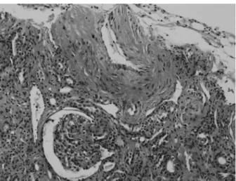
A renal biopsy and bronchoscopy were promptly per- formed. Bronchoscopic examination showed mild hyper- emia in both bronchi, with red bronchial alveolar lavage fluid. Light microscopic examination of the renal biopsy showed proliferative endarteritis in interlobular arteries and fibrinoid necrosis and intramural thrombi of small arterioles without crescent formation or necrotizing lesions, consis- tent with hypertensive nephrosclerosis (Fig. 3). Steroid therapy was rapidly decreased and stopped. A trans-bron- chial lung biopsy showed macrophage infiltration into alve- olar spaces and goblet cell metaplasia in bronchial mucosa generally seen in heavy smokers. Vanillyl-mandelic acid, metanephrine, epinephrine, and norepinephrine in 24-h urine samples were within normal levels. Tests for anti-glo- merular basement membrane antibody and anti-neutrophil
CASE REPORT
A 27-year-old man visited the emergency room with he- moptysis. His blood pressure was 180/100 mm Hg, and he had tachycardia (110/min) and tachypnea (24/min). He had been a heavy smoker, but had never been diagnosed with hypertension. Laboratory data was as follows: white blood cell count 13150/μL; hemoglobin level, 11.3 g/dL; platelet count, 110×103/μL; serum urea nitrogen level, 32 mg/dL;
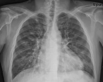
and serum creatinine level, 3.11 mg/dL. His serum electro- lyte levels were as follows: sodium, 120 mEq/L; potassium, 2.5 mEq/L; chloride, 80 mEq/L; calcium, 7.5 mg/dL; and phosphorus, 3.2 mg/dL. Urinalysis showed microscopic he- maturia and proteinuria (+++). Total protein to creatinine ratio in spot urine was 6.08 mg/g. Pro-brain natriuretic pep- tide level was markedly increased. Chest radiograph (Fig.
1) and computed tomography showed bilateral infiltration, suggesting alveolar hemorrhage. Renal ultrasound using a Doppler scan showed increased echogenicity in both nor- mal-sized kidneys without renal artery stenosis. Goodpas- ture’s syndrome, SLE, microscopic polyangiitis or PAN was strongly suspected.
On the second hospital day, his serum creatinine level in- creased to 4.65 mg/dL. Anti-nuclear antibody test was neg- ative, complement levels were normal, plasma renin activi- ty was 6.27 ng/L·s (reference, 0.28-0.69 ng/L·s), serum aldosterone level was 6.54 nmol/L (reference, 0.11-0.28 nmol/L), and serum parathyroid hormone level was 159.1 pg/mL (reference, 14-72 pg/mL). Peripheral blood smear (PBS) showed some helmet cells suggestive of micrcoangi- opathic hemolysis. Fundoscopic examination showed cot- ton wool spots and splinter hemorrhage with papillary ede-
ma, consistent with hypertensive changes (Fig. 2). Fig. 1. Chest radiograph showing infiltration in both lower lobes and cardio- megaly.
Fig. 2. Fundoscopic examination showing multiple cotton wool spots and a splinter hemorrhage.
Hoon Suk Park, et al.
Yonsei Med J http://www.eymj.org Volume 53 Number 6 November 2012 1226
therapy was rapidly decreased and stopped. The patient`s re- nal function improved as his blood pressure was controlled.
Malignant hypertension is characterized by elevated blood pressure accompanying encephalopathy or acute ne- phropathy as target organ damage.3 In malignant hyperten- sion, the damaged endothelium increases permeability and activates coagulation cascade, including platelet activation and fibrin deposition. Red blood cells are destroyed within vessels and helmet cells or schistocytes can be seen in PBS, resulting in end-organ ischemia.4 In contrast, in immune- mediated systemic diseases such as Goodpasture’s syn- drome, SLE, microscopic polyangiitis and PAN, immune complex or direct vasculitis related to autoantibodies de- stroys body organs, and the inflammatory cell infiltration or active necrotizing lesion is seen on histology.2
Few cases of malignant hypertension mimicking immune- mediated pulmonary renal syndrome have been reported.1,5,6 Hida, et al.1 showed that alveolar capillaries might be in- jured as the capillaries in systemic circulation were injured by malignant hypertension, resulting in alveolar hemor- rhage. However, Sato, et al.5 and Aithal, et al.6 concluded that left ventricular dysfunction resulting from systemic hy- pertension can cause pulmonary edema; thus leading to hemorrhage. Pulmonary circulation is somewhat free of systemic hypertension because it is separated from the sys- temic circulation by the heart; however, it can be affected by systemic hypertension with left ventricular dysfunction.
Renal ischemia from renal artery stenosis, malignant hyper- tension or renin producing tumor may cause hyponatremic- hypertensive syndrome. Secondary aldosteronism with the concomitant elevation of serum renin level and the subse- quent activation of ADH caused by volume depletion from pressure natriuresis manifest hyponatremia together with hypokalemia in this syndrome.7 Therefore, the initial hypo- natremia and hypokalemia in our patient can be one of the manifestations by malignant hypertension.
Rapid ELISA tests for anti-GBM antibody and ANCAs yielding results within 30 minutes may help for early differ- ential diagnoses in the cases where immune-mediated pul- monary renal syndrome are suspected, although these rapid tests cannot be substituted for renal biopsy for confirmative diagnosis because small numbers of false negative tests ex- ist and correct diagnosis in the cases with RPGN is essen- tial for improving the patients’ overall prognosis.8,9
Thus, we suggest that for patients with pulmonary renal syndrome, common illnesses like hypertension should not be overlooked.
cytoplasmic antibody (ANCA) were negative. An echocar- diogram showed eccentric left ventricular hypertrophy with decreased systolic and diastolic dysfunctions and a dilated left atrium. In the further reports of the patient’s renal biop- sy, immunofluorescent studies showed fine granular deposi- tion of C3 and C1q only within the vessel walls and elec- tron microscopic examination showed focal effacement of the epithelial cell foot processes, focally swollen endotheli- al cell with irregularly thickened capillary basement mem- brane and normal mesangial basement membrane without deposition.
His serum creatinine level increased to 5.15 mg/dL on the fourth day, but he did not require dialysis. Serum creati- nine level gradually decreased with strict blood pressure control and was 4.03 mg/dL at discharge. The patient last visited the outpatient clinic 10 months after discharge: his blood pressure was 120/80 mm Hg and well controlled with antihypertensive mediation, and his serum creatinine level was 1.98 mg/dL.
DISCUSSION
A patient presenting with malignant hypertension with pul- monary hemorrhage and rapidly deteriorating renal function presented at our hospital. This unusual case suggested im- mune-mediated pulmonary renal syndrome. Immune-medi- ated pulmonary renal syndrome has a poor prognosis in the absence of early appropriate immunosuppression.2 Steroid therapy was administered in our patient, however when the renal biopsy showed only thrombotic microangiopathy without crescent formation or necrotizing vasculitis, the
Fig. 3. Light microscopy examination showing intima fibrous hyperplasia, smooth muscle cell hyperplasia in the media of interlobular artery, and a collapsed glomerulus by ischemia (H&E ×200).
Alveolar Hemorrhage in Malignant Hypertension
Yonsei Med J http://www.eymj.org Volume 53 Number 6 November 2012 1227 3. Slama M, Modeliar SS. Hypertension in the intensive care unit.
Curr Opin Cardiol 2006;21:279-87.
4. Haas AR, Marik PE. Current diagnosis and management of hyper- tensive emergency. Semin Dial 2006;19:502-12.
5. Sato Y, Hara S, Yamada K, Fujimoto S. A rare case of alveolar haemorrhage due to malignant hypertension. Nephrol Dial Trans- plant 2005;20:2289-90.
6. Aithal S, Marley N, Venkat-Raman G. An unusual non-immuno- logical cause of renal pulmonary syndrome. Clin Nephrol 2009;
72:322-5.
7. Agarwal M, Lynn KL, Richards AM, Nicholls MG. Hypona- tremic-hypertensive syndrome with renal ischemia: an underrec- ognized disorder. Hypertension 1999;33:1020-4.
8. Saxena R, Isaksson B, Bygren P, Wieslander J. A rapid assay for circulating anti-glomerular basement membrane antibodies in Goodpasture syndrome. J Immunol Methods 1989;118:73-8.
9. Westman KW, Bygren PG, Eilert I, Wiik A, Wieslander J. Rapid screening assay for anti-GBM antibody and ANCAs; an important tool for the differential diagnosis of pulmonary renal syndromes.
Nephrol Dial Transplant 1997;12:1863-8.
ACKNOWLEDGEMENTS
This case report was supported by a grant of the Korea Health- care Technology R&D Project, Ministry of Health and Wel- fare, Republic of Korea (A102065).
REFERENCES
1. Hida K, Wada J, Odawara M, Kunitomi M, Hayakawa N, Kashi- hara N, et al. Malignant hypertension with a rare complication of pulmonary alveolar hemorrhage. Am J Nephrol 2000;20:64-7.
2. Hirayama K, Yamagata K, Kobayashi M, Koyama A. Anti-glo- merular basement membrane antibody disease in Japan: part of the nationwide rapidly progressive glomerulonephritis survey in Japan. Clin Exp Nephrol 2008;12:339-47.