• 검색 결과가 없습니다.

D Radiation Exposure to Premature Infantsin a Neonatal Intensive Care Unit inTurkey

N/A
N/A
Protected

Academic year: 2022

Share "D Radiation Exposure to Premature Infantsin a Neonatal Intensive Care Unit inTurkey"

Copied!
4
0
0

로드 중.... (전체 텍스트 보기)

전체 글

(1)

416 Korean J Radiol 9(5), October 2008

Radiation Exposure to Premature Infants in a Neonatal Intensive Care Unit in

Turkey

Objective: The aim of this work was to determine the radiation dose received by infants from radiographic exposure and the contribution from scatter radiation due to radiographic exposure of other infants in the same room.

Materials and Methods: We retrospectively evaluated the entrance skin doses (ESDs) and effective doses of 23 infants with a gestational age as low as 28 weeks. ESDs were determined from tube output measurements (ESD TO ) (n = 23) and from the use of thermoluminescent dosimetry (ESD TLD ) (n = 16). Scattered radiation was evaluated using a 5 cm Perspex phantom. Effective doses were estimated from ESD TO by Monte Carlo computed software and radiation risks were estimated from the effective dose. ESD TO and ESD TLD were correlated using linear regression analysis.

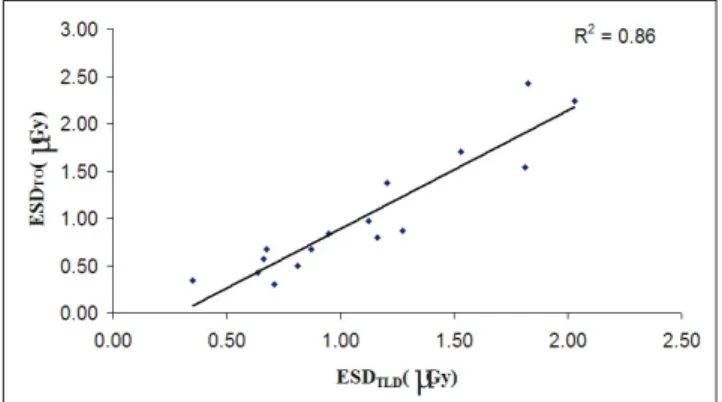
Results: The mean ESD TO for the chest and abdomen were 67 μ Gy and 65 μ Gy per procedure, respectively. The mean ESD TLD per radiograph was 70 μ Gy. The measured scattered radiation range at a 2 m distance from the neonatal intensive care unit (NICU) was (11-17 μ Gy) per radiograph. Mean effective doses were 16 and 27 μ Sv per procedure for the chest and abdomen, respectively. ESD TLD was well correlated with ESD TO obtained from the total chest and abdomen radiographs for each infant (R

2

= 0.86). The radiation risks for childhood cancer estimated from the effective dose were 0.4 × 10

-6

to 2 × 10

-6

and 0.6 × 10

-6

to 2.9 × 10

-6

for chest and abdomen radiographs, respectively.

Conclusion: The results of our study show that neonates received acceptable doses from common radiological examinations. Although the contribution of scat- ter radiation to the neonatal dose is low, considering the sensitivity of the

neonates to radiation, further protective action was performed by increasing the distance of the infants from each other.

iagnostic radiology is increasingly used in the assessment and treatment of neonates requiring intensive care. It is often necessary to perform a large number of radiographic examinations that depend on the birth weight of the infant, gestational age and medical problems (1). The age at which exposure takes place is critical in the determination of radiation risk. During fetal development and early childhood, intense tissue proliferation and differentiation takes place, and it is known that proliferating cells are more susceptible to the induction of cancer (2). Radiographic examinations of neonates are particularly critical because of delayed radiogenic cancers as a consequence of a relative longer life expectancy. The small size of neonates, especially of premature infants, places all organs within the useful beam, resulting in a higher effective dose per radiograph than may be the case with older children and adults. Therefore, radiation doses for neonatal X-ray examina- Turan Olgar, PhD

1

Esra Onal, MD

2

Dogan Bor, PhD

1

Nurullah Okumus, MD

2

Yildiz Atalay, MD

2

Canan Turkyilmaz, MD

2

Ebru Ergenekon, MD

2

Esin Koc, MD

2

Index terms : Radiation dose Entrance skin dose Effective dose Radiography

DOI:10.3348/kjr.2008.9.5.416

Korean J Radiol 2008 ; 9 : 416-419 Received October 17, 2007; accepted after revision March 24, 2008.

1

Ankara University, Institute of Nuclear Science, 06100 Tandogan, Ankara, Turkey;

2

Gazi University Faculty of Medicine, Division of Neonatology 06500 Besevler, Ankara, Turkey

Address reprint requests to : Turan OLGAR, PhD, Ankara University, Institute of Nuclear Science, 06100, Tandogan, Ankara, Turkey.

Tel. 90 (312) 212 03 84-154 Fax. 90 (312) 212 7343 e-mail: [email protected]

D

(2)

tions should be kept to a minimum. It is a common practice to keep infants in large rooms together in most neonatal intensive care units (NICU). Our NICU is designed with four incubators present in one room and initially no specific precaution is taken for other infants while a radiographic examination is performed on one of the patients. It is also important to ensure that radiation doses from repeated radiographic examinations carried out frequently in the same room of the NICU should be kept at a minimum. The aim of this work was to determine the radiation doses received by the infants from radiographic exposure and also the contribution from scatter radiation due to radiographic exposure of other infants in the same room.

MATERIALS AND METHODS

Population

A total of 23 neonates with a gestational age as low as 28 weeks and a mean body weight of 1,500 grams were included in the study. The mean duration of hospitalization of the infants was 50 days (range, 6-112 days). The most common requested radiographic examinations for the infant chest and abdomen were used in this study.

Radiographic Technique

All radiographic examinations were performed on one GE AMX-4 and two GE VMX+ mobile X-ray units (GE Medical Systems, Milwaukee, WI). The measured half value layers of these X-ray systems at 50 kVp were 2.24, 2.17 and 2.15 (mm Al), respectively. Performance tests of this equipment (such as accuracy kVp and collimation settings and tube output) were carried out according to the protocols given by the Institute of Physics and Engineering in Medicine (IPEM) (3). Radiographs were acquired using Kodak T-mat G/RA film and Kodak Lanex Regular Screen combination (Kodak Health Imaging, Eastman Kodak, Rochester, NY) with a 400 relative film-screen combina- tion speed. Most examinations were carried out with the baby in an incubator and placed directly on top of the cassette, with a focus-to-film distance of 100 cm. However, some examinations were performed with the baby in a radiant warmer and the distance between the focus-to-film

was measured and recorded. Radcal ion chambers (Model 9010 Radiation Monitor Controller, 90 × 6-6 and 90 × 6-180 ionization chambers, MDH Radcal Monrovia, CA) were used for tube output and scatter radiation measure- ments, respectively. The tube outputs were measured over a range of tube voltages (kVp). A focus to film distance (FFD) of 100 cm was chosen for these measurements.

In order to evaluate the contribution of the scatter radiation, a 5 cm Perspex phantom to simulate an infant was irradiated with the exposure parameters typical for clinical examinations. Since the distance between the NICUs was approximately 2 m, scattered radiation was measured at the NICU level at this distance.

Radiographic Dose Evaluation

Entrance skin doses (ESDs) were calculated from the tube output measurements of the X-ray units in accordance with the following formula:

where the ISL factor is an inverse-square law correction from the focus-to-chamber distance (100 cm) to the focus- to-skin distance (FSD), mAs is the product of tube current (mA) and exposure time(s) used in the infants studies. BSF is the backscatter factor and is taken as 1.1 for the 50-70 kVp range used for a neonate with a body thickness of 5 cm (4). The factor is an inverse-square law correction from the focus-to-chamber distance to FSD, and ( μ en / ρ ) Tis Air is the

mass energy absorption coefficient ratio of tissue to air.

The mass energy absorption coefficient ratio is equal to 1.05 for the kVp range used in this study (5).

Lithium fluoride thermoluminescent dosimeter (TLD) chips (3.7 × 3.7 × 0.9 mm) (Model 100: Harshaw Chemical, Solon, OH) in plastic handling pockets (3 for each) were used to measure the total entrance skin dose for both chest and abdomen radiographs for 16 infants, and a Model 3500 Reader (Harshaw Chemical) was used for TLD readout. These pockets were attached to a single point on the skin of a patient for each examination and a pediatrician for each examination determined these locations. In order to minimize batch-to-batch variability, a

ESDs = Output (μ GymAs

-1

) × mAs (1)

× BSF × ISL × ( μ en / ρ ) Tis Air

Radiation Exposure to Premature Infants in Neonatal Intensive Care Unit

Korean J Radiol 9(5), October 2008 417

Table 1. Summary of Exposure Parameters, ESD

TO

and ED Examination Number of Radiographs Tube Potential

mAs ESD

TO

per Radiograph ED per Radiograph

(kVp) (μ Gy) (μ Sv)

Chest 14 (2-31) 49 (46-51) 1.9 (1.6-3.5) 67 (44-136) 15 (10-31)

Abdomen 05 (1-24) 48 (45-51) 0.2 (1.6-2.5) 65 (25-197) 22 (7-63)0

Note.─ *Each value of the table is mean and range (in parentheses). ESD

TO

= entrance skin doses from tube output, ED = effective dose

(3)

preselected group of TLDs was calibrated initially and variations of the sensitivities were kept within ± 5%.

Calibration of TLDs was carried out for radiographic system using the same X-ray beam qualities as with the neonate studies. A Radcal ion chamber was taken as a reference chamber, and the manufacturer recommenda- tions were considered for these calibrations. A linear regression analysis with the least squares method was used to determine the correlation between ESD TO and ESD TLD . Background radiation was also measured by the use of TLD chips placed outside the radiographic room over the hospitalization time for each infant. Then these values were subtracted from each infant TLD that was used for infant ESD measurements. Effective doses (ED) for each examination were calculated from ESD TO and Monte Carlo conversion factors given from PCXMC software (STUK Helsinki, Finland) (6).

Estimates of radiation risk can be made from the ED.

The major risk of concern for an irradiated neonate is childhood cancer. Since most of the neonates in an NICU will be pre-term, fetal risk factors for estimating the risk of a childhood cancer following neonatal exposure were used for the neonate risk calculations (7).

RESULTS

The mean numbers of radiographs received by one infant were 14 and 5 for chest and abdomen examinations, respectively. The mean ESD TO per radiograph was calculated by dividing the sum of the ESD TO by the total

radiography numbers for each examination. The mean ESD TO per radiograph were 67 μ Gy and 65 μ Gy for the chest and abdomen, respectively. The mean ESD TLD was 70 μ Gy. ESD TLD was well correlated with ESD TO obtained from the total chest and abdomen radiographs for each infant (R

2

= 0.86) (Fig. 1).

The measured scattered radiation range at a 2 m distance from the NICU was 11-17 μ Gy per radiograph.

Considering the mean ESD as 65 μ Gy per radiograph, the contribution of the scatter radiation is negligible. The radiation dose of an infant received from the scatter radiation from the irradiation of other infants is approxi- mately 1/4,600 times the direct exposure of the infant.

Table 1 summarizes the radiographic data, ESD TO and ED for each examination accrued in this study.

Using the overall risk factors for inducing a fatal childhood cancer in the first decade of life following Olgar et al.

418 Korean J Radiol 9(5), October 2008

Fig. 1. Correlation of the ESD

TLD

with ESD

TO

Table 2. Comparison of Exposure Parameters, ESD

TO

, ED and Risk Factors for Childhood Cancer with Previously Published Data

ESD TO ED

( μ Gy) ( μ Sv) Risk (×10

-6

)

Reference Examination kVp/mAs

per per (ED × 13 × 10

-2

)

Radiograph Radiograph

This study Chest 49/1.9 67 15 2

Abdomen 48/2.0 65 22 2.9

Brindhaban and Al-Khalifah (11) Chest 57/1.6 60 26 -

Abdomen 57/1.6 60 32 -

Jones et al. (12) Chest 62/2.0 56.7 15.4 2

Abdomen 62/2.5 73.6 21.9 2.8

Armpilia et al. (1) Chest 53.1/2 36 7.8 1

Abdomen 53/2.0 39 10.2 1.3

Robinson and Dellagrammaticas (13) Chest 60/1.0 53 - -

Abdomen 60/1.0 57 - -

Schneider et al. (14) Chest - 78 - -

Lowe et al. (15) Chest - 16-160 - -

Chapple et al. (4) Chest - 55 - -

Note.─ ESD

TO

= entrance skin doses from tube output, ED = effective dose

μ

μ

(4)

Radiation Exposure to Premature Infants in Neonatal Intensive Care Unit

Korean J Radiol 9(5), October 2008 419

prenatal radiation exposure based on ICRP60 (2.8 × 10

-2

Sv

-1

to 13 × 10

-2

Sv

-1

) yields somatic risk estimates of 0.4

× 10

-6

to 2 × 10

-6

and 0.6 × 10

-6

to 2.9 × 10

-6

for chest and abdomen radiographs, respectively (7).

DISCUSSION

The ESDs were comparable to the mean values reported by the Commission of European Communities (EC) as 80 μ Gy (8) and by the National Radiological Protection Board (NRPB) as 50 μ Gy for chest radiographs (9).

Exposure of infants to radiation in the vicinity of the exposed infants in the same room is very low, practically for the range of the lowest detectable dose.

The difference between ESD TLD and ESD TO may be attributable to variations on the tube outputs and errors of the focus-to-skin distance measurements. The ESD values per radiograph were found to be higher than the values reported in the literature (1, 4). The use of low tube potential (kVp) values in this study probably is the reason for higher doses. According to Duggan et al. (10), increas- ing the tube potential from 50 kVp to 60 kVp reduced ESD by 9%. Table 2 shows a comparison between exposure parameters, ESD TO , ED and risk factors for childhood cancer per examination for this study and other studies.

This is the first dosimetry study performed for an NICU in Turkey and we measured radiation near the exposed infant in the same room to check safe distances in the NICU for scatter radiation. If the high kV technique had been used in this study, the doses delivered to patients could have been substantially reduced. There is a continu- ing need for assessment of radiation dose in the neonate healthcare centers along with a regular review of dose reduction procedures. The results obtained from this study show that the ESD TLD and ESD TO indicated no significant differences.

Because of their sizes, a relatively large area of the infants was irradiated and it was more difficult to shield radiosensitive organs. In addition, measurement of infant dose must be performed in sterile conditions as soon as possible because of the high sensitivity of the infants to infection.

The results of our study show that neonates received acceptable doses from common radiological examinations.

Although the contribution of scatter radiation to the

neonatal dose is low, considering the sensitivity of the neonates to radiation, further protective action should be performed by increasing the distance between patients until the effect of scatter radiation disappears.

References

1. Armpilia CI, Fife IA, Croasdale PL. Radiation dose quantities and risk in neonates in a special care baby unit. Br J Radiol 2002;75:590-595

2. Richardson RB. Past and revised risk estimates for cancer induction by irradiation and their influence on dose limits. Br J Radiol 1990;63:235-245

3. Institude of Physics and Engineering in Medicine.

Recommended standards for the routine performance testing of diagnostic X-ray imaging systems. Report No77, York: IPEM, 1997

4. Chapple CL, Faulkner K, Hunter EW. Energy imparted to neonates during X-ray examinations in a special care baby unit.

Br J Radiol 1994;67:366-370

5. Wall BF, Harrison RM, Spiers FW. Patient dosimetry techniques in diagnostic radiology. Institute of Physical Science in

Medicine. Report No. 53, York, UK: IPEM, 1988

6. Tapiovaara M, Lakkisto M, Servomaa A. PCXMC- A PC-based Monte Carlo Program for Calculating Patient Doses in Medical X-ray Examinations. Report STUK-A139. [Radiation and Nuclear Safety Authority (STUK), Helsinki], 1997 7. International Commission on Radiological Protection. 1990

Recommendations of the International Commission on Radiological Protection. Ann ICRP 1991;21:1-201

8. European commission. European guidelines on quality criteria for diagnostic radiographic images in paediatrics. Report EUR 16261 EN. Brussels: EC, 1996

9. Hart D, Wall BF, Schrimpton PC, Bungay DR, Dance DR.

Reference doses and patient size in paediatric radiology. NRPB- R318. Chilton: HMSO, 2000

10. Duggan L, Warren-Forward H, Smith T, Kron T. Investigation of dose reduction in neonatal radiography using specially designed phantoms and LiF: Mg, Cu, P TLDs. Br J Radiol 2003;76:232-237

11. Brindhaban A, Al-Khalifah K. Radiation dose to premature infants in neonatal intensive care units in Kuwait. Radiat Prot Dosimetry 2004;111:275-281

12. Jones NF, Palarm TW, Negus IS. Neonatal chest and abdominal radiation dosimetry: a comparison of two radiographic techniques. Br J Radiol 2001;74:920-925

13. Robinson A, Dellagrammaticas HD. Radiation doses to neonates requiring intensive care. Br J Radiol 1983;56:397-400

14. Schneider K, Kohn MM, Ernst G. The derivation of reference dose values to chest X-rays in radiography. Radiat Prot Dosim 1998;80:199-202

15. Lowe A, Finch A, Boniface D, Chaudhuri R, Shekhdar J.

Diagnotic image quality of mobile neonatal chest X-rays and the

radiation exposure incurred. Br J Radiol 1999;72:55-61

수치

Table 1 summarizes the radiographic data, ESD TO and ED for each examination accrued in this study.

참조

관련 문서