Acepromazine inhibits hERg potassium ion channels expressed in human embryonic kidney 293 cells
8
0
0
전체 글
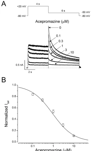
(2) 76 action potential [16]. Blocking of hERG channels leads severe cardiovascular adverse effects such as acquired long QT synd romes and then increases risk for torsade de pointes arrhythmias, and sudden death [17,18]. The mammalian ether-à-go-go gene families belonging to hERG are highly conserved and have common characters [19]. Mouse ether-à-go-go related gene (mERG) B is expressed selectively in the heart and has a similar electrophysiological feature with hERG B, the human homolog of mERG B [20]. The phenothiazine antipsychotics (thioridazine, chlorpromazine) inhibit hERG channels, which contribute a critical role in arrhythmogenesis [21-24]. Even though, acep romazine has been often used in veterinary medicine and few human intoxication of acepromazine has been reported, the toxicological mechanisms of acepromazine in animals as well as in human have not been studied yet. Thus, we investigated whether the hERG potassium channel, a major target of arrthy thmogenic drugs, was affected by acepromazine to evaluate the toxic potentials in human and animal. . Joo YS et al. Germany) was used for fast drug application. Solutions were rapidly switched around the cell using a piezoelectric translator displacing the q-tubing laterally to expose the cells to the drugcontaining solution for a defined period of time, and then rapidly return to the drug-free solution. Solutions were delivered under gravity from reservoirs placed above the preparation, and the application timing was controlled by a perfusion valve control system (VC-8, Warner Instruments, Hamden, CT, USA).. Electrophysiology For electrophysiological recording, the cover glasses containing adherent hERG-HEK293 recombinant cells were transferred to the recording chamber (RC-13, Warner Instruments) mounted on the stage of an inverted microscope (IX70, Olympus, Tokyo,. Methods Cell culture The hERG-HEK293 recombinant cell line (CYL3039, Millipore, Billerica, MA, USA) was used for electrophysiological recording, as previously reported in detail [25]. The cells were maintained in D-MEM/F-12 (Invitrogen, Grand Island, NY, USA) supplemented with 10% fetal bovine serum, 1% nonessential amino acid, and 400 μg/ml geneticin, according to the manufacturer’s directions. The cells were plated on cover glasses (12 mm diameter; Fisher Scientific, Pittsburgh, PA, USA) and placed in 35 mm culture dishes at least 24 hours prior to patch -clamp recordings.. Solutions and drugs The external bath solution contained 140 NaCl, 5 KCl, 1 CaCl2, 1 MgCl2, 10 HEPES, and 10 glucose in mM, and was adjusted to pH 7.3 using NaOH. Osmolarity of the solution measured using a vapor pressure osmometer (Vapro 5520, Wescor, Logan, UT, USA) was 300~310 mOsm. The internal pipette solution contained 140 KCl, 1 CaCl 2 , 1 MgCl 2 , 10 HEPES, 10 EGTA in mM and was adjusted to pH 7.3 using KOH. The average osmolarity of internal solution was 290 mOsm. Acepromazine (Santa Cruz Biotechnology, Dallas, Texas, USA) was dissolved in dimethylsulfoxide (DMSO, Sigma, St. Louis, MO, USA) as a stock solution of 100 mM, and the stock solution was then diluted with the external solution to obtain the desired concentration. The concentration of DMSO in the final dilution was <0.1%, and this DMSO concentration had no effect on the hERG currents [25]. The q-tubing glass pipette mounted on a piezoelectric translator (P-601 PiezoMove Z Acturator, Physik Instrumente, Karlsruhe, Korean J Physiol Pharmacol 2017;21(1):75-82. Fig. 1. Concentration-dependent inhibition of hERG currents by acepromazine. (A) hERG currents were elicited by a 4-sec depolarizing pulse to +20 mV from a holding potential of –80 mV and repolarization to –50 mV for 6 sec to measure the peak tail currents in the absence and presence of acepromazine. The dotted line marks zero current. (B) Normalized hERG tail currents (Itail) to the control were plotted as a function of acepromazine concentration. The experimental data were fitted with a Hill equation. Data were expressed as the means±S.E.M. https://doi.org/10.4196/kjpp.2017.21.1.75.
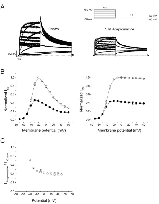
(3) 77. Inhibition of hERG potassium channel by acepromazine. Japan). Cells were continuously perfused with an external bath solution. hERG currents were recorded using a Multiclamp 700 B microelectrode amplifier and pClamp 10.1 software (Molecular Devices, Sunnyvale, CA, USA) in a whole-cell configuration of the patch-clamp technique at room temperature (22~24oC). Glass micropipettes were pulled from glass capillaries (PG101654, World Precision Instruments, Sarasota, FL, USA) using a programmable horizontal microelectrode puller (P-97, Sutter Instrument, Novato, CA, USA). The tip resistances of the patch pipettes were 2~4 MΩ when filled with the internal solution. The liquid junction potentials between pipette and bath solutions were in the range 3~5 mV, and zeroed before the gigaohm seal was formed. The current signals were filtered at 2 kHz, digitized at 10 kHz, and saved on a PC using DigiData 1322 and pClamp 10.1 software (Molecular Devices).. Statistical analysis Data analysis was performed using pClamp 10.1 software (Molecular Device) and Origin 8.0 software (Origin Lab Corp., Northampton, MA, USA). The averaged data are presented as the mean±S.E.M. A paired Student's t-test was used for the statistical analysis of paired data. A one-way analysis of variance with Bonferroni's test was applied for the comparison of multiple groups. p<0.05 was considered statistically significant.. Fig. 2. Effect of acepromazine on current-voltage (I -V ) relationships. (A) Whole-cell hERG currents were evoked by depolarizing pulses from –70 mV to +60 mV for 4 sec in steps of 10 mV every 15 sec from a holding potential of –80 mV and repolarization to –50 mV for 6 sec in the absence and presence of acepromazine (1 μΜ). (B) The I -V relationships of the steadystate (ISS) and peak tail (Itail) currents of the hERG channels under the control conditions (○) and after the application of acepromazine (●). The I SS and I tail at each voltage in the presence of acepromazine were normalized to those in the absence of acepromazine. I tail data were fitted to a Boltzmann function. (C) The voltage-dependent block of the I tail by acepromazine was expressed as a relative current (IAcepromazine/ IControl). * Significant difference from data obtained at –10 mV when compared to –40 mV (p<0.01). Data are expressed as the means±S.E.M. www.kjpp.net. Korean J Physiol Pharmacol 2017;21(1):75-82.
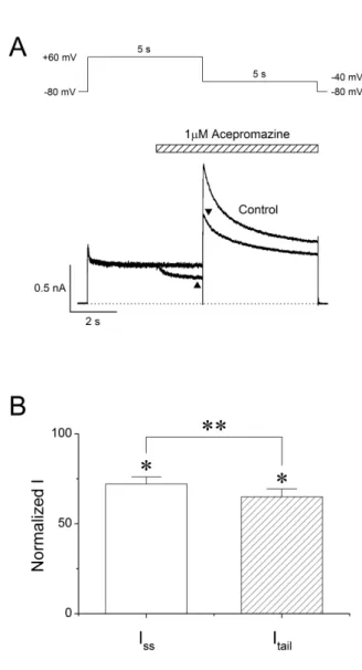
(4) 78. Results Concentration-response relationships for blocking of hERG currents by acepromazine Fig. 1A shows the representative hERG currents traces recorded in the absence and presence of acepromazine (0.1, 0.3, 1, 3, and 10 μΜ). The hERG currents were evoked with 4 sec depolarization to +20 mV from a holding potential of –80 mV followed by a repolarization to –50 mV for 6 sec. The voltage protocol was applied every 15 sec. The peak tail currents of the hERG at –50 mV were normalized to the control values and plotted for drug concentrations (Fig. 1B) to evaluate the effect of acpromazine on the activated states of hERG channels. Acepromazine inhibited the hERG tail currents at –50 mV in a concentration-dependent manner. At the concentrations of 0.1, 0.3, 1, 3, and 10 μ Μ acepromazine, the tail currents reduced by 16.3±1.7, 26.3±2.1, 46.4±2.8, 70.5±2.7, and 89.3±0.8% (n=10), respectively. The concentration-response curve was fitted by using Hill equation, yielding an IC50 value of 1.5±0.1 μΜ and Hill coefficient of 1.1±0.1 (n=10).. Joo YS et al. activation ranging from 28.6±4.5% at –40 mV to 56.2±1.6% at –10 mV (Fig. 2C, n=6, p<0.01). However, the inhibitions of Itail by acepromazine were not significantly different in the range of 0~+60 mV, and the voltage range after the channels were fully activated.. Voltage-dependent inhibition of hERG currents by acepromazine To identify the current-voltage (I -V ) relationships of the hERG currents, the channels were activated by applying 4 sec depolarizing step-pulses (Δ10 mV, –70 mV~+60 mV) from a holding potential of –80 mV in every 15 sec followed by a 6 sec repolarizing pulse to –50 mV in the absence and presence of 1 μΜ acepromazine, which was the near IC50 value for hERG currents based on the Fig. 1. The steady-state hERG currents (Iss) were measured at the end of the depolarizing step-pulses and the peak hERG tail currents (Itail) were measured during the repolarizing periods (Fig. 2A). Fig. 2B shows the average I-V relationships for Iss and Itail before and after the application of 1 μM acepromazine. Under the control conditions, Iss was activated at a membrane potential of approximately –50 mV and reached the maximum at –20 mV. Acepromazine significantly inhibited the Iss at the membrane potentials between –40 and +60 mV (Fig. 2B, left). To investigate the effects of acepromazine on the voltage dependency of hERG channel activation step, the Itail were analyzed and normalized data were plotted against the membrane potential with or without acepromazine (Fig. 2B, right). Acepromazine significantly inhibited the Itail at the membrane potentials between –40 and +60 mV. When the plots of normalized tail currents were fitted to a Boltzmann function, acepromazine shifted the halfactivation membrane potential (V1/2) of activation curves to the hyperpolarizing direction (i.e., from –38.6±0.8 mV to –45.0±0.8 mV, n=6, p<0.001), and decreased the slope factor (k) from 5.6±0.2 to 4.6±0.2 (p<0.001). The voltage-dependent inhibition of the Itail by acepromazine increased sharply for voltages of channel Korean J Physiol Pharmacol 2017;21(1):75-82. Fig. 3. Interaction of acepromazine with inactivated and open state of hERG channels. (A) The hERG currents were elicited by 5-sec depolarizing pulse to +60 mV from a holding potential of –80 mV, followed by a repolarizing pulse to –40 mV for 5 sec to induce peak tail currents. Acepromazine (1 μΜ) was rapidly applied during the depolarizing pulse and continued to the repolarizing pulse as indicated by the hatched bar. The currents were measured at the end of depolarization pulse (▲, ISS) and at the time of peak tail currents (▼, I tail), during the drug application. The dotted line marks zero current. (B) Averaged data showed that the inhibition of ISS and Itail by acepromazine. *p<0.001 when compared to control. **p<0.001 when compared between ISS and Itail. Data are expressed as the means±S. E.M. https://doi.org/10.4196/kjpp.2017.21.1.75.
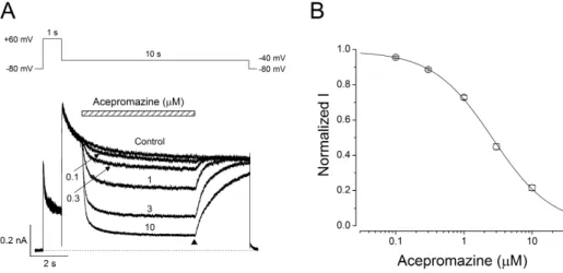
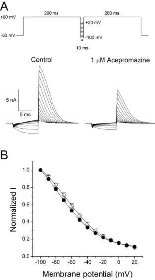
(5) 79. Inhibition of hERG potassium channel by acepromazine. Interaction of acepromazine with both open and inactivated state of hERG channels To investigate the effect of acepromazine with both the inactivated and open state of the hERG channels, a fast drug perfusion system was used [25,26]. The hERG currents were elicited by a 5 sec depolarizing pre-pulse to +60 mV from a holding potential of –80 mV, followed by 5 sec of repolarization to –40 mV (Fig. 3A, n=9). Acepromazine (1 μΜ) was applied from the middle of depolarizing pulse to the end of repolarizing pulse, inhibiting the steady-state hERG currents by 27.9±1.3% at the end of the depolarizing pre-pulses (p<0.001) and the peak hERG tail currents by 35.0±1.5% immediately after repolarization to –40 mV (Fig. 3B, p<0.001). Thus, acepromazine preferentially inhibited the hERG channels in the open states than the inactivation.. Open channel block of the hERG currents by acepromazine Fast drug application system was also used for further inves tigation of the acepromazine effect on the open states of hERG channels. The hERG currents were elicited by a 1sec depolarizing pre-pulse to +60 mV, rapidly inactivating the hERG channels followed by 10 sec of repolarization to –40 mV, and this pulse protocol induced a fast recovery of hERG channels from inac tivation and remained hERG channels to the open states. Acepromazine was applied and removed quickly during the peak hERG tail currents for 5 sec. Fig. 4A shows a rapid and reversible inhibition of hERG tail currents by fast application of acepromazine in a concentration-dependent manner. The hERG tail currents was reduced by 4.5±0.7, 11.5±0.6, 27.2±1.2, 55.2±1.7,. and 78.4±1.7% at 0.1, 0.3, 1, 3, and 10 μ Μ of acepromazine, respectively (Fig. 4B, n=12). Based on this experiment, the calculated IC50 value for the open states of hERG channels was 2.4±0.2 μ Μ with Hill coefficient of 1.2±0.1. This IC50 value was higher than the IC50 value for the activated states of hERG channels in the above experiment (Fig. 1B, IC50=1.5 μΜ) with almost the same value of Hill coefficient (1.1 vs. 1.2). This difference in the IC50 values was most likely explained by a higher affinity of acepromazine for the activated state of hERG channels than the open state.. Effect of acepromazine on steady-state inactivation of hERG channels To study the effect of acepromazine on the steady-state in activation of hERG channels, the double-pulse protocol with steps of inter-stimulus repolarizing pulse was used (Fig. 5A). The cells were depolarized to +60 mV for 200 msec to induce inactivation and hyperpolarized from –100 mV to +20 mV in 10-mV voltage steps for 10 msec. And then a second depolarizing pulse of +60 mV was applied for 200 msec. Peak current amplitudes during the second depolarizing pulses were analyzed and normalized data to the maximum values were plotted to evaluate the steadystate inactivation of hERG channels. The acquired data under the control condition and 1 μM acepromazine were fitted to Boltzmann equation (Fig. 5B). The half-inactivation voltage (V1/2) and slope factor (k) were –64.2±3.5 mV and 22.8±0.4 in controls (n=7), and –74.7±3.1 mV and 24.5±0.4 in the presence of 1 μM acepromazine (n=7). The steady-state inactivation curve shifted to the hyperpolarized direction by acepromazine (p<0.001). However, the slope factor did not change significantly in the. Fig. 4. Concentration-dependent effect of acepromazine on open state hERG channels. (A) The hERG currents were elicited by a 1-sec depolarizing pulse to +60 mV from a holding potential of –80 mV, followed by a repolarizing pulse to –40 mV for 10 sec to evoke peak tail currents. 1 sec after the start of repolarizing pulse, acepromazine was rapidly applied for 5 sec and then removed during the repolarizing pulse by fast drug application system. The bar indicates the time for the application of acepromazine. (B) Graph showed a concentration-response relationship for an open channel block of the hERG current by acepromazine. The normalized currents at the end of the drug application (▲) were plotted for the concentrations of acepromazine. The data were fitted using a Hill equation. Data were expressed as the means±S.E.M. www.kjpp.net. Korean J Physiol Pharmacol 2017;21(1):75-82.
(6) 80. Fig. 5. Effect of acepromazine on steady-state inactivation of hERG channels. (A) Representative current traces showed the steady-state inactivation under the control conditions and in the presence of 1 μM acepromazine, which were evoked by a double-pulse protocol with steps of inter-stimulus repolarizing pulses. After the first 200-msec depolarizing pulse of +60 mV inducing hERG channel inactivation, 10 msec inter-stimulus step pulses from –100 to +20 mV, with a 10 mV increment were applied, followed by a second depolarizing pulse of +60 mV. (B) Normalized steady-state inactivation curves under control conditions (○) and in the presence of 1 μM acepromazine (●). Solid lines represent fitting with Boltzmann function. Data are expressed as means±S.E.M.. presence of acepromazine.. Discussion The effect of acepromazine on the hERG potassium channels expressed in HEK293 cells was investigated using the patchclamp technique. The results of our study can be summarized as follows: 1) acepromazine inhibited the hERG channels in a concentration-dependent manner; 2) the drug also blocked hERG Korean J Physiol Pharmacol 2017;21(1):75-82. Joo YS et al. channels voltage dependently between –40 mV and +60 mV; 3) acepromazine inhibited the hERG currents by acting on both the inactivated (during the depolarization) and open (during the repolarization) states of the channels; 4) the result of fast drug application study was consistent with the open-channel block mechanism of hERG channel by acepromazine. These results suggest that acepromazine directly inhibits the hERG potassium channels, responsible for the repolarization of action potential in cardiac tissues, preferentially by binding to the open and inactivated states of ion channels. Even though, the primary pharmacological mechanisms of neurobehavioral effects of phenothiazine antipsychotics, including acepromazine could be attributed to their potent antagonism of dopamine receptors [27-29], these drugs also caused many central and peripheral adverse reactions by acting on the dopamine receptors (on-target adverse effects) and by nonspecific actions on other receptor types and ion channels (offtarget adverse effects) [30-32]. One of serious morbidity after the ingestion of the phenothiazine antipsychotics usually resulted from cardiotoxicity including hypotension, conduction delay and ventricular arrhythmia [33]. Especially, it has been reported that thioridazine and chlorpromazine directly inhibited hERG channels, through which these drugs could induce long QT syndrome [21-24]. Taken together with several reports showing that hERG currents inhibition is an important arrhythmogenic mechanism of acquired long QT syndrome and torsade de pointes [17,18], our results suggest that acepromazine can cause arrhythmia possibly through the inhibition of hERG potassium channels as an off-target adverse effect. cDNAs cloned from erg mRNA in cardiac tissue of the rat, guinea pig, rabbit, dog, and human showed a highly homologues. The dog and rabbit cDNAs were 99% identical to the human sequence and the rat and guinea pig’ were 96% identical to the human sequences [34]. Drugs reported to induce QT prolongation or torsade de pointes in human also prolonged QT intervals with 92% accuracy in canine Purkinje fiber model, an in vivo model for acquired long QT syndrome and drug-induced arrhythmogenesis [35]. Furthermore, canines have an important role in the preclinical assessment of QT interval prolongation in drug toxicity test [36]. A study assessing the potential neurobehavioral and cardiovascular toxicity of candidate drugs in a canine model observed that the dog’ heart rate was increased by acepromazine treatment [37]. Therefore, the drug-induced cardiac toxicity, especially related to long QT syndrome, seems to shares common molecular targets in both human and some other species such as dog, and rabbit. It has been also reported that heart rates were increased in the two human intoxication cases of acepromazine, even though one of them was an overdose of acepromazine and etorphine combination [10,14]. These results suggest that acepromazine overdoses can induce certain forms of adverse cardiovascular effects, including arrhythmias. However, there were still no https://doi.org/10.4196/kjpp.2017.21.1.75.
(7) 81. Inhibition of hERG potassium channel by acepromazine. reports showing that acepromazine induced the arrhythmias in human overdose cases and even veterinary uses. The pharmacokinetic or toxicokinetic data of acepromazine in human have been not well documented. For veterinary use, the potency of acepromazine is species dependent: from 0.5 mg/kg in dogs to 5 mg/kg in horse, and the maximum plasma concentration (Cmax) of acepromazine was ~0.19 μ Μ when administered at a dose of 0.15 mg/kg [38] and the Cmax of ~0.18 μΜ when administered at a dose of 0.1 mg/kg [39] in horses. In a human toxicology case, after 8 hr of 950 mg acepromazine ingestion, the plasma acepromazine level was 0.19 μΜ [10]. In our study, acepromazine inhibited hERG channel with an IC50 value of 1.5 μΜ, which was about ten times higher than the Cmax based on the sedative uses of acepromazine in the animals. These results suggest that acepromazine inhibits the hERG channels with relatively low potency. Chlorpromazine, a closely related structural analog of acepromazine, showed a higher IC50 value (21.6 μΜ) with a relative potential of long QT syndrome, even though its therapeutic plasma concentrations range (61.4~260 nM) were much lower than the IC50 [23,24]. Because phenothiazines have a high lipophilicity and high affinity to lipids in the tissues, the tissue concentration of acepromazine was expected to be higher than plasma level [40]. Together with these reports, our results suggest that if acepromazine accumulates in cardiac tissue with high affinity, it has an arrhythmogenic potential through the inhibition of hERG potassium channels in cardiac tissues even at lower plasma concentration level. Acepromazine has been widely used as a sedative in combi nation with various anesthetics or analgesics in veterinary medicine [5,41]. Moreover, many sedatives and anesthetics have been known to induce long QT syndrome though the inhibition of hERG potassium channels in human [42] and animals [43]. Even though acepromazine has a relatively higher IC50 for hERG channel inhibition, if it used in combination with drugs with potential of prolongation of QT intervals through the hERG channel inhibition, such as sedatives and anesthetics, it might be dangerous to human and animals through a synergistic drug interaction. This interpretation could be support by the reports that acepromazine significantly increased heart rate and blood pressure when it combined with halothane, ketamine, and butorphanol [44,45]. These synergistic effects of hERG channel inhibition through the drug interaction could be another explanation of acepromazine possibly inducing long QT syndrome even at higher IC50 in our study. This study shows that hERG currents were inhibited by acepromazine. In consistent with that hERG channels have high homology with other mammalian species, these results suggest that acepromazine have a potentially arrhythmogenic action through the inhibition of hERG potassium channels in cardiac tissues and the prolongation of QT intervals.. www.kjpp.net. Acknowledgements We thank Prof. Hahn (Department of Physiology, College of Medicine, The Catholic University of Korea, Korea) for helpful comments for the preparation of this manuscript. This research was supported by Basic Science Research Program through the National Research Foundation of Korea (NRF) funded by the Ministry of Science, ICT and future Planning (NRF2014R1A1A2056820).. Conflicts of Interest The authors declare no conflicts of interest.. References 1. Collard JF, Maggs R. Clinical trial of acepromazine maleate in chronic schizophrenia. Br Med J. 1958;1:1452-1454. 2. O'reilly PO, Michaud M, Wojcicki HM, Keogh RP. Acetylpromazine (acepromazine) in the treatment of chronic mental illness. Can Med Assoc J. 1958;79:381-383. 3. Simpson RW. The effects of notensil in chronic mental illness. J Ment Sci. 1958;104:179-181. 4. Urquhart R, Forrest AD. Clinical trial of promazine hydrochloride and acetylpromazine in chronic schizophrenic patients. J Ment Sci. 1959;105:260-264. 5. Rankin DC. Sedatives and tranquilizers. In: Grimm KA, Lamont LA, Tranquilli WJ, Greene SA, Robertson SA, editors. Veterinary anesthesia and analgesia. 5th ed. Oxford: John Wiley & Sons; 2015. p.196-206. 6. López-Sanromán FJ, Gómez Cisneros D, Varela del Arco M, Santiago Llorente I, Santos González M. The use of low doses of acepromazine as an aid for lameness diagnosis in horses: An accelerometric evaluation. Vet Comp Orthop Traumatol. 2015;28: 312-317. 7. Monteiro ER, Coelho K, Bressan TF, Simões CR, Monteiro BS. Effects of acepromazine-morphine and acepromazine-methadone premedication on the minimum alveolar concentration of isoflurane in dogs. Vet Anaesth Analg. 2016;43:27-34. 8. Perrin KL, Denwood MJ, Grøndahl C, Nissen P, Bertelsen MF. Comparison of etorphine-acepromazine and medetomidineketamine anesthesia in captive impala (aepyceros melampus). J Zoo Wildl Med. 2015;46:870-879. 9. Berns SD, Wright JL. Pediatric acepromazine poisoning: the importance of child-resistant packaging for veterinary drugs. Am J Emerg Med. 1993;11:247-248. 10. Algren DA, Ashworth A. Acute acepromazine overdose: clinical effects and toxicokinetic evaluation. J Med Toxicol. 2015;11:121-123. 11. Bryant SM, Mycyk MB. Human exposure to pet prescription medications. Vet Hum Toxicol. 2002;44:218-219. 12. Clutton RE. Attempted suicide with acepromazine maleate: a case report. Vet Hum Toxicol. 1985;27:391. 13. Gaulier JM, Sauvage FL, Pauthier H, Saint-Marcoux F, Marquet P, Korean J Physiol Pharmacol 2017;21(1):75-82.
(8) 82 Lachâtre G. Identification of acepromazine in hair: an illustration of the difficulties encountered in investigating drug-facilitated crimes. J Forensic Sci . 2008;53:755-759. 14. Sterken J, Troubleyn J, Gasthuys F, Maes V, Diltoer M, Verborgh C. Intentional overdose of Large Animal Immobilon. Eur J Emerg Med. 2004;11:298-301. 15. Stowell LI. Suicide with the veterinary drug acepromazine. J Anal Toxicol. 1998;22:166-168. 16. Thomas D, Karle CA, Kiehn J. The cardiac hERG/IKr potassium channel as pharmacological target: structure, function, regulation, and clinical applications. Curr Pharm Des. 2006;12:2271-2283. 17. Raschi E, Vasina V, Poluzzi E, De Ponti F. The hERG K+ channel: target and antitarget strategies in drug development. Pharmacol Res. 2008;57:181-195. 18. Sanguinetti MC, Tristani-Firouzi M. hERG potassium channels and cardiac arrhythmia. Nature. 2006;440:463-469. 19. Martinson AS, van Rossum DB, Diatta FH, Layden MJ, Rhodes SA, Martindale MQ, Jegla T. Functional evolution of Erg potassium channel gating reveals an ancient origin for IKr. Proc Natl Acad Sci U S A. 2014;111:5712-5717. 20. Lees-Miller JP, Kondo C, Wang L, Duff HJ. Electrophysiological characterization of an alternatively processed ERG K+ channel in mouse and human hearts. Circ Res. 1997;81:719-726. 21. Drolet B, Vincent F, Rail J, Chahine M, Deschênes D, Nadeau S, Khalifa M, Hamelin BA, Turgeon J. Thioridazine lengthens repola rization of cardiac ventricular myocytes by blocking the delayed rectifier potassium current. J Pharmacol Exp Ther. 1999;288:12611268. 22. Hoehns JD, Stanford RH, Geraets DR, Skelly KS, Lee HC, Gaul BL. Torsades de pointes associated with chlorpromazine: case report and review of associated ventricular arrhythmias. Pharmacotherapy. 2001;21:871-883. 23. Kim KS, Kim EJ. The phenothiazine drugs inhibit hERG potassium channels. Drug Chem Toxicol. 2005;28:303-313. 24. Thomas D, Wu K, Kathöfer S, Katus HA, Schoels W, Kiehn J, Karle CA. The antipsychotic drug chlorpromazine inhibits HERG potassium channels. Br J Pharmacol. 2003;139:567-574. 25. Chae YJ, Jeon JH, Lee HJ, Kim IB, Choi JS, Sung KW, Hahn SJ. Escitalopram block of hERG potassium channels. Naunyn Schmie debergs Arch Pharmacol. 2014;387:23-32. 26. Ganapathi SB, Kester M, Elmslie KS. State-dependent block of HERG potassium channels by R-roscovitine: implications for cancer therapy. Am J Physiol Cell Physiol. 2009;296:C701-710. 27. Dahl SG, Hough E, Hals PA. Phenothiazine drugs and metabolites: molecular conformation and dopaminergic, alpha adrenergic and muscarinic cholinergic receptor binding. Biochem Pharmacol. 1986; 35:1263-1269. 28. Hals PA, Hall H, Dahl SG. Phenothiazine drug metabolites: dopa mine D2 receptor, alpha 1- and alpha 2-adrenoceptor binding. Eur J Pharmacol . 1986;125:373-381. 29. Peroutka SJ, Synder SH. Relationship of neuroleptic drug effects at brain dopamine, serotonin, alpha-adrenergic, and histamine. Korean J Physiol Pharmacol 2017;21(1):75-82. Joo YS et al receptors to clinical potency. Am J Psychiatry. 1980;137:1518-1522. 30. Ashoor A, Lorke D, Nurulain SM, Kury LA, Petroianu G, Yang KH, Oz M. Effects of phenothiazine-class antipsychotics on the function of α7-nicotinic acetylcholine receptors. Eur J Pharmacol. 2011;673:25-32. 31. Hals PA, Hall H, Dahl SG. Muscarinic cholinergic and histamine H1 receptor binding of phenothiazine drug metabolites. Life Sci. 1988;43:405-412. 32. Lee IS, Park TJ, Suh BC, Kim YS, Rhee IJ, Kim KT. Chlorpromazineinduced inhibition of catecholamine secretion by a differential blockade of nicotinic receptors and L-type Ca 2+ channels in rat pheochromocytoma cells. Biochem Pharmacol. 1999;58:1017-1024. 33. Schmidt W, Lang K. Life-threatening dysrhythmias in severe thioridazine poisoning treated with physostigmine and transient atrial pacing. Crit Care Med. 1997;25:1925-1930. 34. Wymore RS, Gintant GA, Wymore RT, Dixon JE, McKinnon D, Cohen IS. Tissue and species distribution of mRNA for the IKr-like K+ channel, erg. Circ Res. 1997;80:261-268. 35. Gintant GA, Limberis JT, McDermott JS, Wegner CD, Cox BF. The canine Purkinje fiber: an in vitro model system for acquired long QT syndrome and drug-induced arrhythmogenesis. J Cardiovasc Pharmacol. 2001;37:607-618. 36. Gralinski MR. The dog's role in the preclinical assessment of QT interval prolongation. Toxicol Pathol. 2003;31 Suppl:11-16. 37. Tontodonati M, Fasdelli N, Moscardo E, Giarola A, Dorigatti R. A canine model used to simultaneously assess potential neurobe havioural and cardiovascular effects of candidate drugs. J Phar macol Toxicol Methods. 2007;56:265-275. 38. Wieder ME, Gray BP, Brown PR, Hudson S, Pearce CM, Paine SW, Hillyer L. Identification of acepromazine and its metabolites in horse plasma and urine by LC-MS/MS and accurate mass measure ment. Chromatographia. 2012;75:635-643. 39. Hashem A, Keller H. Disposition, bioavailability and clinical efficacy of orally administered acepromazine in the horse. J Vet Pharmacol Ther. 1993;16:359-368. 40. Daniel WA. Mechanisms of cellular distribution of psychotropic drugs. Significance for drug action and interactions. Prog Neuropsychopharmacol Biol Psychiatry. 2003;27:65-73. 41. Brock N. Acepromazine revisited. Can Vet J. 1994;35:458-459. 42. Darpö B. Spectrum of drugs prolonging QT interval and the inci dence of torsades de pointes. European Heart Journal Supplements. 2001;3:K70-80. 43. Finley MR, Lillich JD, Gilmour RF Jr, Freeman LC. Structural and functional basis for the long QT syndrome: relevance to veterinary patients. J Vet Intern Med. 2003;17:473-488. 44. Steffey EP, Kelly AB, Farver TB, Woliner MJ. Cardiovascular and respiratory effects of acetylpromazine and xylazine on halothaneanesthetized horses. J Vet Pharmacol Ther. 1985;8:290-302. 45. Ward JL, Schober KE, Fuentes VL, Bonagura JD. Effects of sedation on echocardiographic variables of left atrial and left ventricular function in healthy cats. J Feline Med Surg. 2012;14:678-685.. https://doi.org/10.4196/kjpp.2017.21.1.75.
(9)
수치
+2
관련 문서
다양한 번역 작품과 번역에 관한 책을 읽는 것은 단순히 다른 시대와 언어, 문화의 교류를 넘어 지구촌이 서로 이해하고 하나가
Zao Wou-Ki generated the best H1 result for the entire Asian continent in Hong Kong; 53% of Zhang Daqian’s turnover was hammered there, and lots of new
The index is calculated with the latest 5-year auction data of 400 selected Classic, Modern, and Contemporary Chinese painting artists from major auction houses..
1 John Owen, Justification by Faith Alone, in The Works of John Owen, ed. John Bolt, trans. Scott Clark, "Do This and Live: Christ's Active Obedience as the
faster reactions: stable carbocation & unstable reactants.. electron donating groups &
Currents in the Cheju Strait have the eastward barotrophic component of about 5cm/sec through the year and become stronger in summer by the addition
The solidification process of metal alloy was expressed by the change of solid fraction, and the solid fraction was controlled by varying size and
In gi ngi va,LCs are found i n oralepi thel i um ofnormalgi ngi va and i n smal l er amountsi nthesul cul arepi thel i um,buttheyareprobabl yabsentfrom thejuncti onal epi thel
