원 원 저저
of Clinical Toxicology pISSN: 1738-1320 / eISSN: 2508-6332
INTRODUCTION
Agent Orange (AO) was an herbicide and defoliant used by the United States and its military allies during the Vietnam War. From 1964 to 1973, about 320,000 Korean military participated in the Vietnam War and were exposed to AO. AO was a mixture of 2,4-
고엽제 노출이 폐렴의 치료 결과에 미치는 영향
중앙보훈병원 응급의학과1, 경희대학교병원 응급의학과2
김동성
1∙이정엽
1∙계유찬
1∙정의기
1∙정기영
2The Effects of Agent Orange in Patient with Pneumonia
Dong Sung Kim, M.D.
1, Jungyoup Lee, M.D.
1, Yu Chan Kye, M.D.
1, Euigi Jung, M.D.
1, Ki Young Jeong, M.D., Ph.D.
2Department of Emergency Medicine, Veterans Health Service Medical Center, Seoul
1, Department of Emergency Medicine, Kyung Hee University Medical Center, Seoul
2, Korea
Purpose: Agent Orange (AO) is a herbicide and defoliant used by the United States and its military allies during the Vietnam War. Pneumonia is a common cause of death among Vietnam veterans in our hospital. There have been no previous studies researching any association between AO exposure and the prognosis for pneumonia. The pri- mary objective of this study was to investigate associations between AO exposure and 30-day mortality due to pneumonia. The secondary objective was to examine the clinical factors associated with therapeutic outcomes in veterans with pneumonia, and to assess the prevalence of combined diseases in AO-exposed veterans.
Methods: This study retrospectively included veteran patients diagnosed with pneumonia in the emergency depart- ment and hospitalized between February 2014 and March 2018. The enrolled patients were grouped according to their defoliant exposure history, and the clinical information of defoliant-exposed and non-defoliant-exposed groups were compared. Patients were divided according to 30-day mortality, and significant factors influencing mortality were evaluated by using univariate analysis and multivariate analysis. The final multivariate model revealed the effect of AO exposure on therapeutic outcomes of pneumonia.
Results: A total of 1006 patients were analyzed. Of these, 276 patients had a history of AO exposure, whereas 730 patients had not been exposed. Factors positively associated with 30-day mortality were malignancy, respiratory rate, blood urea nitrogen, and albumin which was negatively associated with mortality.
Conclusion: Exposure to defoliant is not associated with 30-day mortality in patients with pneumonia. However, veterans with defoliant exposure are associated with a high prevalence of diabetes mellitus, hypertension, cere- brovascular accident, malignancy, and chronic kidney disease.
Key Words: Agent orange, Dioxin, Defoliant, Pneumonia, Veteran
책임저자: 이 정 엽
서울특별시 강동구 진황도로 61길 53 중앙보훈병원 응급의학과
Tel: 02) 2225-4184 Fax: 02) 2225-1480 E-mail: [email protected]
투고일: 2020년 2월 10일 1차 심사일: 2020년 3월 6일 게재 승인일: 2020년 4월 3일
dichlorophenoxyacetic acid and 2,4,5-trichlorophe- noxyacetic acid, which also contained dioxin conta- minants, including 2,3,7,8-tetrachlorodibenzo-p-diox- in
1). In Korea, about 142,448 Vietnam war veterans are alive and being treated for AO complications in veterans hospitals. Numerous research articles have been published regarding the health effects of AO
2-9). Previous studies have reported deaths due to cancers of the stomach, small intestine, liver, larynx, lung, blad- der, and thyroid gland; in addition, chronic myeloid leukemia and angina pectoris, chronic obstructive pul- monary disease, chronic kidney disease and chronic liver disease were all increased with AO exposure
10). Among defoliant related disease, following diseases (stroke, diabetes and chronic liver disease) were asso- ciated with mortality in patients with pneumonia
11). Pneumonia is a leading cause of morbidity and mor- tality in old age
12,13). There have been no previous studies investigating associations between AO expo- sure and the prognosis for pneumonia. The primary objective of this study was to investigate associations between AO exposure and 30-day mortality due to pneumonia. The secondary objective was to investi- gate the clinical factors associated with therapeutic outcomes in veterans with pneumonia and to assess the prevalence of combined disease in AO-exposed veterans.
METHODS
1. Hospital setting and study design
The study hospital was a 1,400-bed secondary acad- emic hospital with an annual Emergency Department (ED) census of 30,000. All patients presenting with pneumonia who were admitted to our ED have been registered in a pneumonia registry, since our ED was established in February 2014. We used the pneumonia registry and collected additional information by retro- spective electronic medical record (EMR) chart review.
Veteran patients who were diagnosed with pneumonia in the ED and hospitalized between February 2014 and March 2018 were included. We collected patient data Patients who were discharged from the ED or trans-
ferred to another hospital were excluded. Pneumonia severity index (PSI) or CURB-65 scoring system were used to evaluate disease severity and evaluate the need for hospital admission
14,15).
We divided enrolled patients according to defoliant exposure history and compared the clinical informa- tion for the defoliant-exposed and non-defoliant- exposed groups. We divided the patients according to 30-day mortality and investigated significant fac- tors influencing mortality by using univariate analy- ses and subsequent multivariate analyses. The final multivariate model showed the effect of AO exposure on therapeutic outcomes of pneumonia. This study was approved by our Institutional Review Board, and informed consent was waived due to the retrospec- tive nature of the study. (IRB number : 2019-04-008)
2. Data collection and classification
Based on the pneumonia registry, we established a standardized form that contained 80 variables, includ- ing demographic factors, clinical factors, laboratory data, and therapeutic results. Vital signs were record- ed at the triage stage in the ED. Initial laboratory data after ED admission were recorded. The demographic data included age, sex, diabetes mellitus (DM), hyper- tension (HTN), chronic lung disease, chronic liver dis- ease, congestive heart failure (CHF), chronic kidney disease (CKD), cerebrovascular accident (CVA), malig- nancy, tuberculosis (TB) history, nursing home resi- dency, and AO exposure history. The clinical parame- ters included systolic blood pressure (SBP), heart rate (HR), body temperature (BT), respiratory rate (RR), white blood cell (WBC) count, hemoglobin (Hb), platelet count, blood urea nitrogen (BUN), serum cre- atinine (Cr), blood glucose, serum albumin, serum C- reactive protein (CRP), serum sodium (Na), serum potas- sium (K), blood pH, arterial oxygen partial pressure (pO
2), and pleural effusion. The therapeutic results included 30-day mortality and in-hospital mortality.
We classified patients with respect to community-
acquired pneumonia (CAP), health care-associated
pneumonia (HACP) or hospital-acquired pneumonia
(HAP). HAP was defined as pneumonia that occurred
more than 48 hours after hospitalization. Patients who were hospitalized in an acute care hospital for more than two days within 90 days of the infection, resided in a nursing home or long-term care facility, attended a hemodialysis clinic, recently received intravenous antibiotics or chemotherapy, or sought wound care within 30 days of the infection were classified as having
HACP
12,16-18). In terms of cancer history, patients with
active disease were classified as patients with cancer, while those with no evidence of disease were classified as patients without cancer. Patients who were unable to move by themselves were defined as bedridden.
Defoliant exposure history was identified by EMR review, which was provided by the Ministry of National Defense. The defoliant exposure history is registered according to Vietnam war record and the period of defo- liant use. Exposure levels were recorded in three steps, but we did not use the data. We also classified patients with respect to sepsis and septic shock
19). In cases of missing data, our research team re-examined the EMR and obtained additional information from patients by phone. We excluded cases without therapeutic results.
3. Statistical analyses
The means and standard deviations (SD) of contin- uous variables were calculated and the means com-
pared using the Student’s T-test. The frequencies (per- centages) of binominal variables were analyzed using the χ
2or Fisher’s exact test. Enrolled patients were divided into two groups according to their 30-day mortality, and the frequencies compared by univari- ate analyses. We selected the clinically significant variables with reference to the results in the univariate analyses. Multivariate logistics regression analyses were performed to find independent risk factors using the candidate variables. Variables were removed from the multivariate model in a stepwise manner, and the final model with the best fit was determined using Bayesian information criterion: this criterion introduces a penalty term to the number of parameters in a model, such that one model is considered better than another if it has a smaller Bayesian information criterion value
20). The final model showed the effect of AO exposure and other significant factors associated with 30-day mor- tality due to pneumonia. A two-sided test was used with a 5% significance level. All calculations were conducted using Stata version 12.0 (StataCorp, College Station, TX).
RESULTS
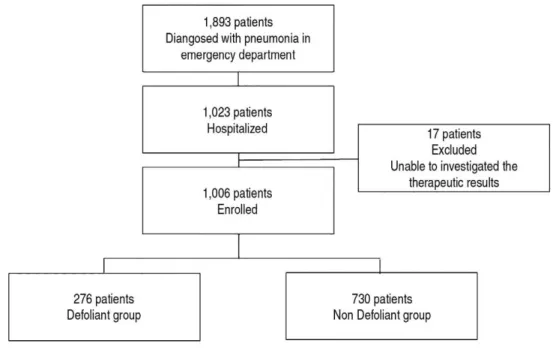
A total of 1,893 patients were diagnosed with pneu- monia in the ED and 1,023 patients were hospitalized
Fig. 1. Flow chart of study.
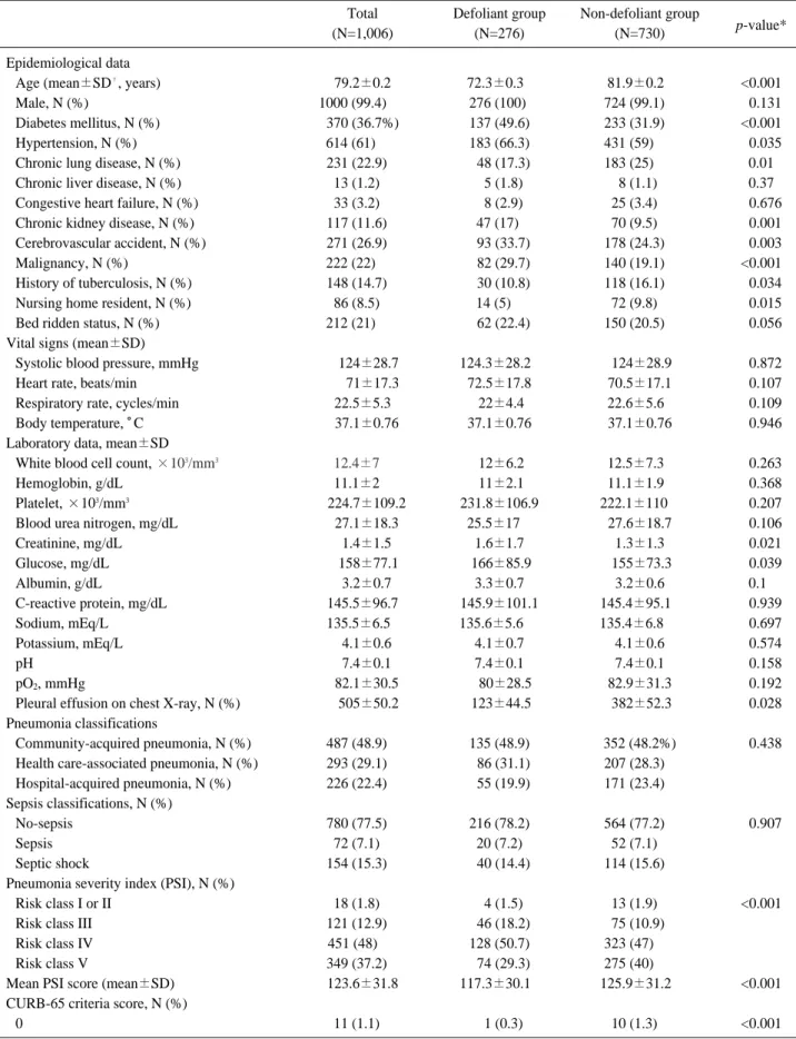
Table 1. Baseline characteristics of defoliant group and non-defoliant group
Total Defoliant group Non-defoliant group
p-value*
(N=1,006) (N=276) (N=730)
Epidemiological data
Age (mean±SD
�, years) 79.2±0.2 72.3±0.3< 81.9±0.2< <0.001
Male, N (%) 1000 (99.4)<<. 276 (100). 724 (99.1)0. <0.131
Diabetes mellitus, N (%) 370 (36.7%) 137 (49.6) 233 (31.9)0. <0.001
Hypertension, N (%) 614 (61)<<.. 183 (66.3) 431 (59)000 <0.035
Chronic lung disease, N (%) 231 (22.9)<. <48 (17.3) 183 (25)000 0.01
Chronic liver disease, N (%) 13 (1.2)<. <5 (1.8) 8 (1.1). 0.37
Congestive heart failure, N (%) 33 (3.2)<. <8 (2.9) 25 (3.4)0. <0.676
Chronic kidney disease, N (%) 117 (11.6)<. 47 (17). 70 (9.5)0. <0.001
Cerebrovascular accident, N (%) 271 (26.9)<. <93 (33.7) 178 (24.3)0. <0.003
Malignancy, N (%) 222 (22)<<.. <82 (29.7) 140 (19.1)0. <0.001
History of tuberculosis, N (%) 148 (14.7)<. <30 (10.8) 118 (16.1)0. <0.034
Nursing home resident, N (%) 86 (8.5)<. 14 (5)<. 72 (9.8)0. <0.015
Bed ridden status, N (%) 212 (21)<<.. <62 (22.4) 150 (20.5)0. <0.056 Vital signs (mean±SD)
Systolic blood pressure, mmHg <.124±28.7 124.3±28.2< .124±28.9 <0.872
Heart rate, beats/min <<.71±17.3 72.5±17.8 70.5±17.1 <0.107
Respiratory rate, cycles/min 22.5±5.3 .22±4.4 22.6±5.6< <0.109
Body temperature, 。C <37.1±0.76 37.1±0.76 37.1±0.76 <0.946
Laboratory data, mean±SD
White blood cell count, ×10
3/mm
312.4±7<. .12±6.2 12.5±7.3< <0.263
Hemoglobin, g/dL 11.1±2<. .11±2.1 11.1±1.9< <0.368
Platelet, ×10
3/mm
3<224.7±109.2 231.8±106.9 222.1±110<. <0.207
Blood urea nitrogen, mg/dL <27.1±18.3 25.5±17<. 27.6±18.7 <0.106
Creatinine, mg/dL <1.4±1.5 1.6±1.7 1.3±1.3 <0.021
Glucose, mg/dL <.158±77.1 .166±85.9 .155±73.3 <0.039
Albumin, g/dL <3.2±0.7 3.3±0.7 3.2±0.6 0.10
C-reactive protein, mg/dL 145.5±96.7 145.9±101.1 145.4±95.1< <0.939
Sodium, mEq/L 135.5±6.5< 135.6±5.6<< 135.4±6.8<< <0.697
Potassium, mEq/L <4.1±0.6 4.1±0.7 4.1±0.6 <0.574
pH <7.4±0.1 7.4±0.1 7.4±0.1 <0.158
pO
2, mmHg <82.1±30.5 <.80±28.5 82.9±31.3 <0.192
Pleural effusion on chest X-ray, N (%) <.505±50.2 .123±44.5 .382±52.3 <0.028 Pneumonia classifications
Community-acquired pneumonia, N (%) 487 (48.9)<. 135 (48.9) 352 (48.2%) <0.438 Health care-associated pneumonia, N (%) 293 (29.1)<. <86 (31.1) 207 (28.3)0.
Hospital-acquired pneumonia, N (%) 226 (22.4)<. <55 (19.9) 171 (23.4)0.
Sepsis classifications, N (%)
No-sepsis 780 (77.5)<. 216 (78.2) 564 (77.2)0. <0.907
Sepsis 72 (7.1)<. 20 (7.2) 52 (7.1)0.
Septic shock 154 (15.3)<. <40 (14.4) 114 (15.6)0.
Pneumonia severity index (PSI), N (%)
Risk class I or II 18 (1.8)<. <4 (1.5) 13 (1.9)0. <0.001
Risk class III 121 (12.9)<. <46 (18.2) 75 (10.9).
Risk class IV 451 (48)<<. 128 (50.7) 323 (47)000
Risk class V 349 (37.2)<. <74 (29.3) 275 (40)000
Mean PSI score (mean±SD) 123.6±31.8 117.3±30.1< 125.9±31.2< <0.001
CURB-65 criteria score, N (%)
0 11 (1.1)<. <1 (0.3) 10 (1.3)0. <0.001
(Continued to the next page)
during the study period. We were unable to investi- gate the therapeutic results in 17 patients, therefore 1,006 patients were included in the final study popu- lation (Fig. 1). Table 1 presents the baseline charac- teristics of the enrolled patients and compares the clinical information of the defoliant-exposed and non- defoliant-exposed groups using univariate analyses.
The mean age ± SD of the enrolled patients was 79.2
±0.2 years, and 99.4% were men. The crude 30-day mortality was 25.8% and mortality was not signifi- cantly different in the two groups. There were 276 patients who had been exposed to AO, while 730 had not been exposed. The prevalence of DM, HTN, CKD, CVA, and malignancy were higher in the defoliant- exposed group, although the defoliant-exposed group was significantly younger. The two groups were not significantly different on initial vital signs. With respect to the laboratory data, creatinine and glucose concen- trations were greater in the defoliant-exposed group and pleural effusion was more frequently diagnosed in the non-defoliant group. The number of patients who were classified as having sepsis or septic shock were not significantly different between the groups. In terms of pneumonia classification, the proportion of patients with CAP, HACP, or HAP were not different between the two groups. According to PSI and CURB65, dis- ease severity was higher in the non-defoliant group (p<0.001).
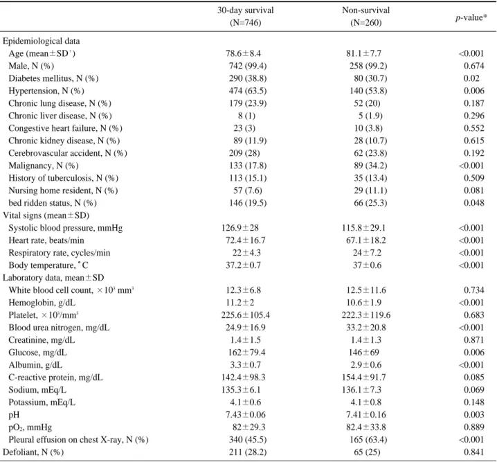
In univariate analyses comparing the survival and non-survival groups, the following variables were
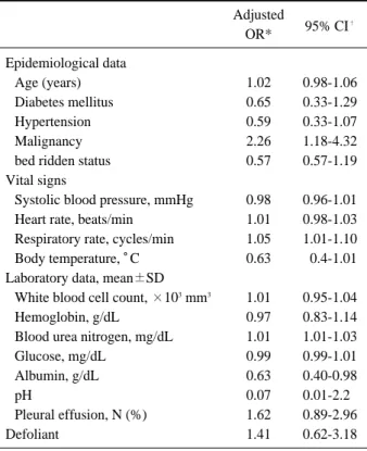
significantly associated with 30-day mortality: age, DM, HTN, malignancy, bedridden status, vital signs (SBP, HR, RR, and BT), laboratory data (WBC, Hb, BUN, glucose, albumin, and pH). These factors were candidate variables for the multivariate analyses (Table 2). We performed multivariate logistic regression analy- ses with candidate variables and defoliant exposure history. Factors positively associated with 30-day mor- tality were malignancy, RR, BUN, and albumin which was negatively associated with mortality (Table 3). As a primary outcome, defoliant exposure history was not associated with 30-day mortality in patients with pneumonia. Defoliant exposure was associated with greater prevalence of DM, HTN, CVA, malignancy, and CKD.
DISCUSSION
The AO used in Vietnam caused sequelae in numer- ous Vietnam veterans. In 2007, the government of the Republic of Korea enacted a law requiring compen- sation and treatment for defoliant complications.
Based on the provisions of this law, the Korean gov- ernment has supported veterans who participated in the military campaign in Vietnam and citizens who worked in areas contaminated by AO. In Korea, the following 20 diseases are recognized as sequelae of defoliant intoxication: non-Hodgkin lymphoma, soft tissue sarcoma, chloracne, peripheral neuropathy, por- phyria cutanea tarda, Hodgkin’s disease, lung cancer, Table 1. Baseline characteristics of defoliant group and non-defoliant group
Total Defoliant group Non-defoliant group
p-value*
(N=1,006) (N=276) (N=730)
1 285 (28.4)<. 101 (36.7) 184 (25.3)0.
2 433 (43.2)<. 117 (42.5) 316 (43.3)0.
3 203 (20.2)<. 44 (16). 159 (21.9)0.
4 60 (5.9)<. 10 (3.6) 50 (6.8)0.
5 9 (0.9). <2 (0.7) 7 (0.9).
Mean CURB-65 (mean±SD) <<.2±0.9 1.8±0.8 2.1±0.9 <00.0006
In hospital mortality N (%) 339 (33.8%) <83 (30.4) 256 (35)000 <0.164
30 day mortality, N (%) 260 (25.8)<. <65 (23.5) 195 (26.7)0. <0.307
* Univariate analysis of defoliant group and non-defoliant group.
�
Standard deviation.
Continuous variables are presented as the mean with standard deviations and compared with the student T test.
Categorical data are presented as number (%) of patients and analyzed using the χ
2or Fisher exact test.
laryngeal cancer, bronchial cancer, multiple myeloma, prostate cancer, Buerger’s disease, diabetes mellitus, B cell chronic lymphocytic leukemia, chronic myeloge- nous leukemia, Parkinson’s disease, ischemic heart dis- ease, amyloid light-chain amyloidosis, salivary gland cancer, and gall bladder cancer. The following diseases are recognized as suspected complications of defoliant exposure: photodermatitis, psoriasis, seborrheic der- matitis, chronic urticaria, xerotic eczema, central ner-
vous system diseases, cerebral infarction, multiple scle- rosis, amyotrophic lateral sclerosis, myopathy, malig- nant neoplasms, liver disease, hypothyroidism, hyper- tension, cerebral hemorrhage, arteriosclerosis, and hyper- lipidemia.
In our study, the defoliant-exposed group of veterans was younger than the non-exposed veterans. This result reflects the Vietnam War having been the last war in which the Korean army participated (1961-1971).
Table 2. Univariate analysis of 30-day survival and non-survival group
30-day survival Non-survival
p-value*
(N=746) (N=260)
Epidemiological data
Age (mean±SD
�) 78.6±8.40 81.1±7.70 <0.001
Male, N (%) 0742 (99.4) 0258 (99.2) <0.674
Diabetes mellitus, N (%) 0290 (38.8) 0080 (30.7) 0.02
Hypertension, N (%) 0474 (63.5) 0140 (53.8) <0.006
Chronic lung disease, N (%) 0179 (23.9) .52 (20) <0.187
Chronic liver disease, N (%) .8 (1) 005 (1.9) <0.296
Congestive heart failure, N (%) 23 (3). 010 (3.8) <0.552
Chronic kidney disease, N (%) 0089 (11.9) 0028 (10.7) <0.615
Cerebrovascular accident, N (%) 209 (28). 0062 (23.8) <0.192
Malignancy, N (%) 0133 (17.8) 0089 (34.2) <0.001
History of tuberculosis, N (%) 0113 (15.1) 0035 (13.4) <0.509
Nursing home resident, N (%) 057 (7.6) 0029 (11.1) <0.081
bed ridden status, N (%) 0146 (19.5) 0066 (25.3) <0.048
Vital signs (mean±SD)
Systolic blood pressure, mmHg 126.9±2800. 115.8±29.10 <0.001
Heart rate, beats/min 72.4±16.7 67.1±18.2 <0.001
Respiratory rate, cycles/min .22±4.3 .24±7.2 <0.001
Body temperature, 。C 37.2±0.70 .37±0.6 <0.001
Laboratory data, mean±SD
White blood cell count, ×10
3mm
312.3±6.80 12.5±11.6 <0.734
Hemoglobin, g/dL 11.2±200. 10.6±1.90 <0.001
Platelet, ×10
3/mm
3225.6±105.4 222.3±119.6 <0.683
Blood urea nitrogen, mg/dL 24.9±16.9 33.2±20.8 <0.001
Creatinine, mg/dL 1.4±1.5 1.4±1.3 <0.871
Glucose, mg/dL .162±79.4 146±690 <0.006
Albumin, g/dL 3.3±0.7 2.9±0.6 <0.001
C-reactive protein, mg/dL 142.4±98.30 154.4±91.70 <0.085
Sodium, mEq/L 135.3±6.100 136.1±7.300 <0.069
Potassium, mEq/L 4.1±0.6 4.1±0.8 <0.148
pH 7.43±0.06 7.41±0.16 <0.003
pO
2, mmHg 0.82±29.3 82.4±33.8 <0.889
Pleural effusion on chest X-ray, N (%) 0340 (45.5) 0165 (63.4) <0.001
Defoliant, N (%) 0211 (28.2) .65 (25) <0.841
* Univariate analysis of 30 day survival and non-survival group.
�
Standard deviation.
Continuous variables are presented as the mean with standard deviations and compared with the student T test.
Categorical data are presented as number (%) of patients and analyzed using the χ
2or Fisher exact test.
Although Vietnam veterans were younger, the preva- lence of DM, HTN, CKD, CVA, and malignancy were greater in Vietnam veterans. In Korea, all of these diseases, except CKD, are recognized as complica- tions of defoliant exposure. Further study of the asso- ciations between defoliant exposure and CKD is need- ed. In the defoliant-exposed group, serum creatinine, and blood glucose concentrations were greater than in the non-defoliant-exposed group. These results may have been related to the greater prevalence of CKD and DM in the defoliant-exposed group (Table 1)
10).
Most previous studies of the adverse effects of AO exposure have addressed chronic diseases, and stud- ies of associations with acute diseases requiring critical care are lacking. Given pneumonia has been the lead- ing cause of death in Vietnam veterans in our ED, we aimed to investigate whether defoliant exposure affected the course of pneumonia. This study was the first to investigate associations between historical defo- liant exposure and therapeutic outcomes of pneumonia.
Although we were unable to demonstrate clear asso- ciations between pneumonia and AO exposure, we will
continue to investigate the influence of historical AO exposure on current acute disease.
In our study, clinical factors associated with 30-day mortality were malignancy, high RR, high BUN, and low albumin. In previous studies, these factors were found to be significant predictors of a poor prognosis in pneumonia
21,22). Our study population consisted of elderly patients with an average age of 79 years and a 30-day mortality rate of 25.8%. The analyses indicated that malignancy, high RR, high BUN, and low albu- min could be good predictors for mortality in elderly patients.
This study has several limitations. First, because the degree of defoliant exposure was not investigated, we couldn’t investigate dose-response relationships for defoliant exposure. Second, the percentage of men among the enrolled patients was high, because almost all Korean veterans were male. Therefore, we could not determine if sex influenced the therapeutic results.
Third, potential biases may have been introduced because of the retrospective nature of the study.
CONCLUSION
In conclusion, historical defoliant exposure was not associated with 30-day mortality due to pneumonia.
Defoliant exposure was associated with high preva- lence of DM, HTN, CVA, malignancy, and CKD.
ORCID
Dong Sung Kim (https://orcid.org/0000-0002-9174-2178) Jungyoup Lee (https://orcid.org/0000-0003-2597-850X)
ACKNOWLEDGEMENTS
This work was supported by a VHS Medical Center Research Grant, Republic of Korea [grant number: 16002]
REFERENCES
01. Kang HK, Dalager NA, Needham LL, et al. Health status of Army Chemical Corps Vietnam veterans who sprayed defoliant in Vietnam. Am J Ind Med 2006;49:875-84.
Table 3. Multivariate logistic regression analysis Adjusted
95% CI
�OR*
Epidemiological data
Age (years) 1.02 0.98-1.06
Diabetes mellitus 0.65 0.33-1.29
Hypertension 0.59 0.33-1.07
Malignancy 2.26 1.18-4.32
bed ridden status 0.57 0.57-1.19
Vital signs
Systolic blood pressure, mmHg 0.98 0.96-1.01
Heart rate, beats/min 1.01 0.98-1.03
Respiratory rate, cycles/min 1.05 1.01-1.10
Body temperature, 。C 0.63 00.4-1.01
Laboratory data, mean±SD
White blood cell count, ×10
3mm
31.01 0.95-1.04
Hemoglobin, g/dL 0.97 0.83-1.14
Blood urea nitrogen, mg/dL 1.01 1.01-1.03
Glucose, mg/dL 0.99 0.99-1.01
Albumin, g/dL 0.63 0.40-0.98
pH 0.07 0.01-2.20
Pleural effusion, N (%) 1.62 0.89-2.96
Defoliant 1.41 0.62-3.18
* Odds ratio
�