Modulation of Adhesion Proteins Integrin β1 and FAK, and Cytoskeletal Protein Actin by Spermine in MCF-7 Cells
Hye Jin Jee†and Byeong Gee Kim*
Department of Biological Sciences, College of Natural Sciences Pusan National University, Busan 609-735, Korea
Received November 7, 2011 /Revised December 13, 2011 /Accepted December 19, 2011Polyamines are essential for cell growth and differentiation; however their precise roles are unclear yet. In the present study, the cytotoxic effect of spermine (spm) on MCF-7 cells was investigated. In the MTT assay of MCF-7 cells treated with spm, cell viability was significantly decreased in a time-and dose-dependent manner. Cell viability measurement was confirmed by trypan blue staining.
FACS analysis shows that sub-G1 was increased in a time-and dose-dependent manner too. When the cells were treated with spm, cells started to show morphological changes within 2 hrs. The expression of adhesion proteins (FAK and integrin β1), and cytoskeletal protein (actin) was checked by Western blotting analysis. Integrin β1 levels were slightly decreased, and FAK and actin levels were rapidly decreased with spm treatment. In confocal laser scanning microscopy, the distribution of actin did not change but the expression decreased in a dose-dependent manner with spm treatment. FAK was even- ly distributed under the plasma membrane in the untreated control. However, at 10 μM spm FAK seemed to move toward the cell nucleus. Integrin β1, which was mainly found in the focal point of the plasma membrane in the untreated control, dispersed through the entire plasma membrane in spm treatment. The present results indicate that cytotoxic effects of spm are triggered by the disruption of adhesion proteins and cytoskeletal protein.
Key words : Actin, ECM, FAK, Integrin, spermine
*Corresponding author
*Tel:+82-51-510-2286, Fax:+82-51-581-2962
*E-mail : [email protected]
†Current address
*Department of Biochemistry, College of Medicine, Dong-A University, Busan, Korea
Introduction
Polyamines such as putrescine (put), spermidine (spd), and spermine (spm) are small, flexible molecules, which bind to nucleic acids and proteins and thus affect their con- formation and biological activity [25]. Polyamines are im- portant multifunctional cellular components and are consid- ered mediators of cell growth and division. Recently, poly- amines have also been known to be implicated in cell death.
It appears that polyamines are bivalent regulators of cellular functions, promoting proliferation or cell death depending on the cell type and on environmental signals [12,18].
Among polyamines, spm was most effective in inhibiting cancer cell growth. The cytotoxicity of spm has been sug- gested by the production of H
2O
2and aldehyde(s) from spm in the presence of amine oxidase [1,2].
Polyamines are also essential for the normal attachment of cells to the extracellular matrix (ECM), which explains
at least some of the reliance of cell migration and the organ- ization of the cytoskeleton on normal polyamine levels [22].
The migration of cells depends on their ability to make and break attachments to the ECM. The ECM controls cells through signals via a family of cell surface receptors called integrins [10]. The malignancy of a tumor is associated with its ability to undergo migration and metastasis. Metastases form after a series of sequential, interlinked steps involving a variety of adhesive interactions between cancer cells and host tissues [7]. The mechanism that plays a role in the cyto- toxic properties of polyamine may be related to the down-regulation of integrin-associated signaling. Integrin family proteins are receptors that connect cells to the ECM.
Integrins function as heterodimeric transmembrane cell sur-
face receptors consisting of α- and β- subunits that bind sev-
eral ECM proteins such as collagen, fibronectin, and laminin
at the extracellular domain [11]. Integrin-associated signal-
ing factors have been demonstrated to directly regulate the
apoptotic machinery, as well as invasion. The apoptotic
mechanism associated with loss of attachment to the ECM
is called anoikis, which is also controlled by integrin medi-
ated signaling in cooperation with the cell cycle regulators
and pro-apoptotic proteins [9]. Among the integrin family
members, integrin β1 has been shown to play an important
role in the invasiveness, metastasis formation, and drug re- sistance of cancer cells [26]. As integrins bind to the ECM, they aggregate and direct the formation of a cytoskeletal and signaling complex that results in the assembly of actin fila- ments [19]. Integrin binding to ECM protein or integrin crosslinking increases tyrosine phosphorylation of focal ad- hesion kinase (FAK) [20].
FAK, a tyrosine kinase, is a peripheral membrane protein and interact with integrin. FAK is believed to role as a cen- tral molecule in integrin-mediated signaling. It is one of the core adhesion proteins involved in cellular motility and pro- tection against apoptosis [6,21]. FAK resides at sites of in- tegrin clustering, so called the focal adhesions, that are prominent in cells that are grown in tissue culture. FAK car- ries out protein-protein interaction adaptor functions at sites of cell attachment to the ECM, thereby contributing to fo- cal-adhesion ‘scaffolding’, and also transmits adhesion-de- pendent and growth-factor-dependent signals into the cell interior. FAK influences the dynamic regulation of in- tegrin-associated adhesions, and the actin cytoskeleton that is tethered there, through diverse molecular interactions [16].
Actin remodeling is another crucial element of the cell-motility process. Adhesion dynamics are tightly linked to control of actin assembly and disassembly and FAK con- tributes to both, the latter usually by influencing RhO-GTPase pathways [5]. Integrins transduce signals by as- sociating with adapter proteins that connect to the trans- membrane growth factor receptors, cytoplasmic kinases, and cytoskeleton. The binding of integrin-ECM protein initiates by integrin clustering in the plane of cell membrane, promot- ing the assembly of cytoskeletal actin [23]. Actin reorganiza- tion stimulates the further association of integrin to large multi-protein aggregates, stimulating cell-matrix adhesion that allows, through cytoskeletal actin anchoring, peculiar cell and nuclear shape changes. These bio-molecular effects are necessary during cell cycle regulation/progression, ulti- mately directing the cell to proliferate, differentiate and/or die [8,27]. Adhesion and cytoskeletal proteins provide the basic infrastructure in maintaining the cell shape and function. Alteration of actin remodeling not only plays an important role in regulating the morphologic and pheno- typic events of a malignant cell but also provides a potential target for anticancer drug development. In the present study, to investigate the cytotoxic effect spm the expression of ad- hesion and cytoskeletal proteins including the integrin β1, FAK, and actin was studied in MCF-7 cells by Western blot- ting and confocal laser scanning microscopy.
Materials and Methods Chemicals
3-[4,5-dimethylthiazol-2-yl]-2,5-diphenyl tetrazoliμM (MTT), spd (N-[3-aminopropyl]-1,4-butanediamine), spm (N,N’- bis[3-aminopropyl]-1,4-butanediamine), 2-{[3-(trifluoro meth- yl)phenyl]amino} benzoic acid (Fluofenamic acid), trypan blue, Fluo-3/AM, propidiμM iodide (PI), ribonuclease A, Dulbecco’s modified Eagle’s mediμM (DMEM, with L-gluta- mine and 1,000 mg/l Glucose) were purchased from Sigma Chemical Co (St. Louis, MO, USA). Fetal bovine serum (FBS) was purchased from GIBCO (New York, USA). All other chemicals were purchased from standard commercial sources.
Cell culture
The MCF-7 human breast carcinoma cell line was ob- tained from the American Type Culture Collection (ATCC, Rockville, MD), and maintained in DMEM containing phe- nol red with 10,000 units/ml penicillin G, 10 mg/ml strepto- mycin, and 10% heat-inactivation FBS in a humidified at- mosphere of 95% air; 5% CO
2at 37℃. Culture media were changed every 2 or 3 days. Cells were harvested using tryp- sin-EDTA and subcultured at weekly intervals.
Measurement of cell viability
Cell viability were determined by using microculture tet- razolium technique (MTT) and trypan blue. The MTT assay is a quantitative colorimetric assay for mammalian cell sur- vival and proliferation, based on the ability of live cells to utilize a pale yellow substrate, and its subsequent mod- ification into a dark blue formazan product by these cells.
MCF-7 cells were plated at 5x10
4cells/ml in 48 wells in phenol red-free DMEM (2% DCC-serum). Cells were al- lowed to adhere 24 hrs, then incubated for additional 1, 2, and 3 days in different drug concentrations. Details for MTT assay are described in elsewhere [14]. Trypan blue is a wa- ter-soluble dye used for the dye exclusion test for cell via- bility to distinguish between viable and nonviable cells by seeping into nonviable cells with damaged plasma mem- branes causing them to appear blue. MCF-7 cells were plated onto 6-well plates at approxi-mately 2×10
5cells per well and incubated for 24 hrs. After the cells were treated with spm at various concentrations, cells were harvested by trypsiniza- tion and resuspended in 0.4% trypan blue solution for 5 min.
An aliquot of the cell suspension was taken in a hemocy-
tometer, observed under phase contrast microscope to de- termine cell viability.
Assessment of morphology
The cells were either attached on slide glass by cytospin centrifugation for 5 min at 900 rpm using cellspin (Hanil).
The cells were then fixed with 4% paraformaldehyde (Sigma, MO, USA) at room temperature for 20 min. Fixed cells were washed three times with PBS for 10 min and incubated with 0.2% Triton X-100 for 15 min. After three washes with PBS, the cells were incubated with an appropriate primary anti- body in 1% bovine serum albumin (BSA) at room temper- ature for 2 hrs. For secondary reaction, the cells were in- cubated with an appropriate FITC -conjugated secondary an- tibody at room temperature for 2 hrs. The cells were in- cubated with Texas red phalloidin at room temperature for 2 hrs. Finally, the cells were mounted on glass slides and observed under Confocal microscope (LSM510, Carl Zeiss, Germany).
Flow cytometric analysis
Cells were harvested, fixed with 95% ethanol for 24 hrs, incubated with 0.05 mg/ml PI and 1 μg/ml RNase A at 37°C for 1 hr, and analyzed by flow cytometry, using an Epics XL and analysis software (EXPO32
TM; Beckman Coulter, MI, USA). The cells belonging to the sub-G1 population were considered to be apoptotic cells; the percentage of each phase of the cell cycle was determined.
Western blot analysis
The cells treated with spm were harvested and lysed in lysis buffer (10 mM Tris/HCl, pH 7.4, 150 mM sodium chlor- ide (NaCl), 5 mM EDTA, pH 8.0, with 1%Triton X-100, 0.01 mg/mL aprotinin and 0.04 mg/ml phenylmethyl-sulfonyl- fluoride (PMSF)). Equivalent amount of protein (20 μg) from each sample was subjected to electrophoresis using 10-12%
sodium dedocyl sulfate polyacrylamide gel electrophoresis (SDS-PAGE). Total proteins were estimated by the method of Bradford assay. After SDS-PAGE proteins were trans- ferred onto nitrocellulose paper (Bio-Rad) in a Bio-Rad elec- troblotting device. Ponseau S staining of transferred mem- branes was used to monitor loading equivalence and transfer efficiency. Nitrocellulose matrices were preblocked with 5%
skim milk in TBST (Tris buffer saline, pH 7.4 containing 0.1%
Tween-20) buffer for 1h at room temperature. Following TBST washing preblocked matrices was 4℃ overnight with
various primary antibodies, and then with secondary antibody. The immune-complexes were detected using the ECL (enhanced chemiluminescence) detection kit (Amersham, USA).
Statistical analysis
All experiments were carried out at least in triplicate.
Results were expressed as mean±SEM. Statistical sig- nificances between the control and the treatment group were determined by one-way analysis of variance and then fol- lowed by the Student T test. In all cases, a p value of less than 0.05 was considered statistically significant.
Results Effect of polyamine on cell viability
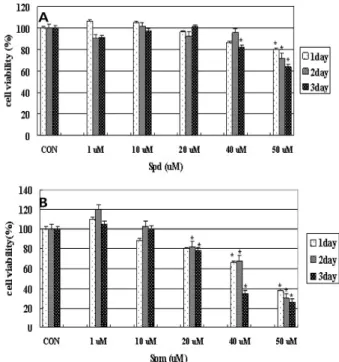
The effect of polyamine on the viability of MCF-7 cells was examined using the MTT staining method. In order to determine the effect of polyamines on MCF-7 cell viability, cells were treated for 1~3 days with spm and spd. As shown in Fig. 1, spd did not have any significant effect on cell viability. However, when cells were treated for 3 days with
Fig. 1. Effect of spd and spm on cell viability in MCF-7 cells.
Cells were plated in 48-wells at 5x10⁴ cells/1 ml in DMEM. Cells treated with (A) spd (1-50 μM) and (B) spm (1-50 μM) for 3 days, and then viability was meas- ured by the MTT assay. Statistical analysis was per- formed by the student’s test. *
p
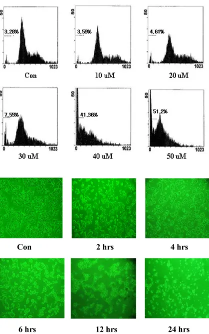
<0.05 vs. controlFig. 2. Cell viability after 24 hrs of spm treatment (%: percent of live cells). Cells were plated in 6-wells at 2x10⁵cells/2 ml in DMEM and incubated for 24 hrs. After the cells were treated with spm (10-50 μM) for 24 hrs, via- bility was measured by the trypan blue assay. Data shown are representative of three independent experiments.
1-50 μM of spm, cell viability decreased in a dose- and time-dependent manner as shown in Fig. 1B. At the lower concentration (under 10 μM), spm did not affect cell viability. But rather cell viability slightly increased to 120%
of the control at 1μM spm treated for 2 days. Cells treated with 20 μM spm showed about 80% viability after 3days incubation compared to the control. However, at 40 μM spm treated, cell viability dropped to 37% of the control on 3days incubation. At 50 μM spm cell viability dropped below 37%
of the control in 3 days incubation. Put, which is the pre- cursor of spd and spm, did not give any noticeable effect on cell viability (data not shown).
Effects of spm on MCF-7 viability assessed with trypan blue
MCF-7 viability was also assessed using trypan blue dye exclusion assay after 24 hrs of treatment with spm. It is re- ported that live healthy cells will exclude the dye and appear opaque, while the dead and very weak cells take the dye and stain blue when observed under a light microscope. The results clearly showed that cell viability started to decrease within 24 hrs treatment of spm in a dose-dependent manner as shown in Fig. 2. Cells treated with 50 μM spm showed about 38% viability for 1day incubation compared to the control in the MTT assay. But in the trypan blue assay, cells treated with 50 μM spm showed about 62% viability for 1 day incubation.
Effects of spm on MCF-7 viability assessed by flow cytometry
To explore more clearly the viability inhibitory effect of
spm in MCF-7 cells, the cell cycle was analyzed using propi- dium iodide staining and flow cytometry. The results showed a typical sub-G1 arrest pattern in spm treated cells.
As shown in Fig. 3, sub-G1 populations increased in a dose-dependent manner by spm treatment. At 50 μM spm, cell death reached 51.2%. The flow cytometric analysis dem- onstrated that cells appear to accumulate at sub-G1 phases following spm treatment, with a concomitant decrease in the percentage of cells in the S and G2-M phase when compared to the control. These results suggest that spm causes the in- duction and accumulation of MCF-7 cells at sub-G1phase, resulting cell death subsequently.
Morphological changes of spm treated cells
To study the cytotoxic effect of spm in detail, the early effect on cell morphology was observed using phase-contrast microscope. A high concentration of spm treatment rendered morphological changes in MCF-7 cells as shown in Fig. 4.
Cells started shrinking and rounding approximately 2 hrs after treatment. After 12 hrs, cells were considerably de- tached from the culture dish surface, and after 24 hrs, most of the cells were detached and also lost cell-cell attachment.
Spm induced morphological changes as well as inhibition of adhesion, migration and spreading.
Effect of spm on the expression of integrin β1, FAK, and actin proteins
The change in protein level of FAK, Integrin β1, and actin
were analyzed by Western blotting. As shown in Fig. 5,
when cells were treated with spm for 24 hrs, FAK expression
level decreased drastically. However, integrin β1 expression
Fig. 3. Identification of cell death by sub-G1 analy- sis (%: percent of dead cells). Cells were incubated for 24 hrs in 6-wells at 2x10⁵ cells/2 ml in DMEM. After cells were treat- ed with spm (10-50 μM) for 24 hrs, viability was measured by flow cytometry. Data shown are representative of three in- dependent experiments.
Fig. 4. Phase-contrast microscopy of MCF-7 cells treated spm 100 μM. Cells were plated in 6-wells at 2x10⁵cells per 2 ml in DMEM and incubated for 24 hrs. After the cells were treated with spm (100 μM) and ob- served at regular intervals (2, 4, 6, 12, and 24 hrs) using phase-contrast microscope.
Data shown are representative of three in- dependent experiments.
levels increased slightly in 10 μM spm, then slightly de- creased at higher concentrations. Actin levels also dimin- ished in a dose-dependent manner compared to the control.
Immunofluorescence analysis of FAK, integrin β1, and actin
The alteration in the cellular distribution of cytoskeletal proteins was checked by confocal microscopy. As shown in Fig. 6, actin and integrin β1 appeared evenly expressed in the plasma membrane of the untreated cells, mainly located at focal contacts homogeneously distributed along the mem- brane of spread cells. Actin, which appeared under the plas- ma membrane of the cell in control, was decreased in a dose-dependent manner with spm treatment. Integrin β1 al-
so decreased in spm treatment. Besides that level change, some alterations of integrin β1 distribution in the plasma membrane were noticed. Integrin β1, mainly found in the focal point of the plasma membrane in the untreated control, dispersed through the entire plasma membrane in spm treatment. FAK levels also decreased by spm treatment.
However, FAK intracellular distribution was different from
that of actin and integrin β1. It seemed like FAK was evenly
distributed under the plasma membrane in the untreated
control. However, at 10 μM spm FAK seemed to move to-
ward the cell nucleus. The expression of FAK was almost
completely knocked out at 40 μM spm even though actin
was still under the membrane focal point as shown in the
merged picture of Fig. 6.
Fig. 5. Western blotting showing the effects of spm on the FAK, integrin β1, actin protein levels. Cells were treated with spm (10-40 μM) for 24 hrs. Equal amounts of proteins were separated by SDS-PAGE, and western blot analysis was performed using FAK-, integrin β1-, and actin-specific antibodies. Data shown are representative of three independent experiments.
Fig. 6. Immunofluorescence analysis of actin, integrin β1, and FAK. Cells were treated with spm (10 μM and 40 μM) in complete medium on glass coverslips for 24 hrs, then fixed and permeabilized. Co-localization of actin and integrin β1 was analyzed by confocal microscopy of the cells subsequently incubated with anti-integrin β1 (1:100) and anti-mouse IgG FITC-conjugated secondary antibody (1:100), followed by incubation with the phalloidin (1:100). Co-localization of FAK and actin was analyzed by confocal microscopy of the cells subsequently incubated with anti-Fak (1:100) and anti-rabbit IgG FITC-conjugated secon- dary antibody (1:100), followed by incubation with the phalloidin (1:100). Data shown are representative of three independent experiments.
Discussion
Polyamines are known to be essential and play multifunc- tional roles in cell viability, growth, proliferation and differ- entiation [23]. Polyamines have also been known to be im- plicated in cell death. It appears that polyamines are bivalent regulators of cellular functions, promoting proliferation or cell death depending on the cell type and on environmental signals [12,18]. Among them, spm was most effective in in-
hibiting cancer cell growth The cytotoxicity of spm has been
recognized for many years. Also, the recent study examines
that polyamine depletion inhibited migration [15] and de-
layed cell attachment to ECM proteins by disrupting the cy-
toskeleton [22]. Previous studies have shown that increased
spm concentrations in tumor tissues are correlated with can-
cer progression and that restriction of polyamine availability
inhibits tumor growth [13]. However, the present results
show that polyamines, especially spm, suppressed MCF-7
cell viability and also decreased the expression of several cytoskeletal proteins. The results showed that spm caused a time- and dose-dependent cell viability decrease. In the MTT assay of MCF-7 cells treated with spm, cell viability decreased rapidly. These results suggest that disturbance in intracellular spm level is related to cell death. The above results were confirmed again by the trypan blue assay. The trypan blue assay suggested that spm induced cell detach- ment before cell death through the disturbance of cytoskele- tal proteins in MCF-7 cells. Flow cytometric analysis con- firmed the above results. Flow cytometric analysis showed that significant cell death occurs in the cells treated with spm. At 50 μM spm, cell death reached 51.2%. The results demonstrated that cells accumulated at sub-G1 phases fol- lowing spm treatment, with a concomitant decrease in the percentage of cells in the S and G2-M phase, compared to the control. It is known that the appearance of surface rounding and blebbing has been linked with alterations of cytoskeletal structure and function. Spm treatment rendered morphological changes of MCF-7 cells. Cell started shrinking and rounding approximately 2 hrs after the treatment. After 12 hrs of spm treatment, cells were considerably detached from the culture dish surface, and after 24 hrs, most of the cells were detached and lost cell-cell attachment. These re- sults indicate that the treatment of spm in MCF-7 cells led to morphological changes suggesting an aberrant function of cytoskeleton proteins. The shape change may be due to significant alterations in the levels of cytoskeletal proteins, suggesting that integrin β1 and other cytoskeleton-bound proteins may be early targets of spm.
Cytoskeletal linkages also enable integrins to regulate cell shape and gene expression, mediating cell adhesion, spread- ing and migration of cells on ECM; all these processes are dependent on cytoskeletal actin [3]. Integins are physically linked to actin cytoskeleton to mediate the focal adhesions, which comprise large protein complexes containing both cy- toskeleton and signaling through FAK. It has been proposed that FAK plays a critical role in cell migration as well as in the regulation of cell survival and cell cycle progression [17]. FAK carries out protein–protein-interaction adaptor functions at sites of cell attachment to the ECM [16]. Both in Western blotting and confocal microscopy, FAK was af- fected most severely by spm treatment as shown in the pres- ent work. FAK seemed to move toward the cell nucleus.
Actin also showed level decrease, but without any cellular distribution in spm treatment. The membrane distribution
pattern of integrin was mainly in the focal adhesion point in the control, but the spm treatment induced big change to even membrane distribution. Compared to the level change of actin and FAK, integrin level did not show a dras- tic decrease.
According to the results of the present work, spm affects especially FAK, which influence the actin and integrin modulation. FAK rapidly disappeared with spm treatment in MCF-7 cells and cell adhesion capacity was decreased.
Therefore, the cytotoxic effect of spm seemed to be carried out by inducing the loss of cell attachment to the ECM, which is anoikis. Anoikis, a subset of apoptosis, is triggered by inadequate or inappropriate cell-matrix contacts [9].
Many kinase/phosphatase signaling molecules, including raf and phosphoinositide-3-kinase are known to be involved in the regulation of anoikis. Since bcl-2 expression levels are regulated by the above signaling molecules, the modulation of cytoskeletal proteins induced by spm might be correlated to the upstream event of apoptosis.
The level change of anti-apoptotic Bcl-2 and pro-apoptotic Bax proteins by spm treatment in MCF-7 cell was already reported in previous paper. Instead of caspase-3, caspase-7 played a key role in the downstream event of apotosis [14].
The control of FAK could be to the clinical application for the prevention of metastasis. FAK is often over expressed in cancer cells [24]. FAK is a multifunctional protein that plays a critical role in the integrin signaling [21] as well as a key molecule in tumor cell metastasis [16]. Therefore, un- derstanding of the adhesion and cytoskeletal protein modu- lation can be critical for the control of cancer.
Acknowledgement
This work was supported for two years by Pusan National University Research Grant.
References
1. Arancia, G., A. Calcabrini, M. Marra, P. Crateri, M. Artico, A. Martone, F. Martelli, and E. Agostinelli. 2004.
Mitochondrial alterations induced by serum amine oxidase and spermine on human multidrug resistant tumor cells.
Amino Acids
26, 273-282.2. Averill-Bates, D. A., Q. Ke, A. Tanel, J. Roy, G. Fortier, and E. Agostinelli. 2008. Mechanism of cell death induced by spermine and amine oxidase in mouse melanoma cells.
Int.
J. Oncol
. 32, 79-88.3. Calderwood, D. A., S. J. Shattil, and M. H. Ginsberg. 2000.
Integrins and actin filaments: Reciprocal regulation of cell adhesion and signalling
. J. Biol. Chem.
275, 22607-22610.4. Carragher, N. O. and M. C. Frame. 2004. Focal adhesion and actin dynamics: a place where kinases and proteases meet to promote invasion.
Trends Cell Biol.
14, 241-249 5. Chen, B. H., J. T. Tzen, A. R. Bresnickand, and H. C. Chen.2002. Roles of Rho-associated kinase and myosin light chain kinase in morphological and migratory defects of focal ad- hesion kinase-null cells.
J. Biol. Chem.
277, 33857-33863.6. Crowe, D. L and A. Ohannessian. 2004. Recruitment of focal adhesion kinase and paxillin to β 1 integrin promotes cancer cell migration via mitogen activated protein kinase activation.
BMC. Cancer
4, 18-23.7. Fidler, I. J. 1991. Cancer metastasis.
Br. Med. Bull.
47, 157-177.8. Frisch, S. M. and E. Ruoslahti. 1997. Integrins and anoikis.
Curr. Opin. Cell Biol.
9, 701-706.9. Frisch, S. M. and R. A. Screaton. 2001. Anoikis mechanisms.
Curr. Opin. Cell Biol.
13, 555-562.10. Garcia, A. J. and D. Boettiger. 1999. Integrin-fibronectin inter- actions at the cell-material interface: initial integrin binding and signaling.
Biomaterials
20, 2427-2433.11. Giancotti, F. G. and E. Ruoslahti.1999. Integrin signaling.
Science
285, 1028-1032.12. Han, L. C., C. Jiang, H. Li, W. Zhang, Y. Zhao, L. Zhang, Y. Zhang, W. Zhao, and B. Yang. 2007. Effects of polyamines on apoptosis induced by simulated ischemia/reperfusion injury in cultured neonatal rat cardiomyocytes.
Cell Biol. Int.
31, 1345-1352.
13. Holst, C. M., B. Frydman, L. J. Marton, and S. M. Oredsson.
2006. Differential polyamine analogue effects in four human breast cancer cell lines.
Toxicology
223, 71-81.14. Jang, E. S. and B. G. Kim. 2008. Mechanism of Apoptosis induced by spemine in MCF-7 breast cancer cells.
Korean J. Life Sci
. 18, 1177-1185.15. McCormack, S. A., J. Y. Wang, and L. R. Johnson. 1994.
Polyamine deficiency causes reorganization of F-actin and tropomyosin in IEC-6 cells.
Am. J. Physiol. Cell Physiol.
267, C715-C722.16. McLean, G. W., N. O. Carragher, E. Avizienyte, E. J. Evans, V. G. Brunton, and M. C. Frame. 2005. The role of focal adhesion kinase in cancer-a new therapeutic opportunity.
Nat. Rev. Cancer
7, 505-515.17. Mitra, S. K. and D. D. Schlaepfer. 2006. Integrin-regulated FAK-Src signaling in normal and cancer cells.
Curr. Opin.
Cell Biol.
18, 516-523.18. Pignatti, C., B. Tantini, C. Stefanelli, and F. Flamigni. 2004.
Signal transduction pathways linking polyamines to apoptosis.
Amino Acids
27, 359-365.19. Ray, R. M., M. J. Viar, A. Shirley, A. McCormack, and L.
R. Johnson. 2001. Focal adhesion kinase signaling is de- creased in polyamine-depleted IEC-6 cells.
Am. J. Physiol.
Cell Physiol.
281, C475-478.20. Richardson, A., R. K. Malik, J. H. Hildebrand, and J. T.
Parsons. 1997. Inhibition of cell spreading by expression of the C-terminal domain of focal adhesion kinase (FAK) is rescued by coexpression of src or catalytically inactive FAK:
a role for paxillin tyrosine phosphorylation.
Mol. Cell Biol.
17 6906-6914.
21. Sanders, M. A. and M. D. Basson. 2000. Collagen IV-depend- ent ERK activation in hμMan Caco-2 intestinal epithelial cells requires focal adhesion kinase.
J. Biol. Chem.
2000; 275, 38040-38047.22. Santos, M. F., M. J. Viar, S. A. McCormack, and L. R.
Johnson. 1997. Polyamines are important for attachment of IEC-6 cells to extracellular matrix
. Am. J. Physiol. Gastrointest Liver Physiol.
273, G175-G183.23. Schlaepfer, D. D., C. R. Hauch, and D. J. Sieg. 1999. Signaling through focal adhesion kinase.
Prog. Biophys. Mol. Biol.
71, 435-478.24. Thamilselvan, V. and M. D. Basson. 2004. Pressure activates colon cancer cell adhesion by inside-out focal adhesion com- plex and actin cytoskeletal signaling.
Gastroenterology
126, 8-18.25. Thomas, T. and T. J. Thomas. 2001. Polyamines in cell growth and cell death: molecular mechanisms and ther- apeutic applications.
Cell Mol. Life Sci
. 58, 244-258.26. van Golen, C. M., M. E. Soules, A. R. GraμMan, and E. L.
Feldman. 2003. N-Myc overexpression leads to decreased β1 integrin expression and increased apoptosis in hum an neuroblastoma cells.
Oncogene
22, 2664-2673.27. Warry, K. K., A. Mariotti, C. Zurzolo, and F. G. Giancotti.
1998. A requirement for caveolin-1 and associated kinase Fyn in integrin signaling and anchorage-dependent cell growth.