• 검색 결과가 없습니다.

Usefulness of Pulsatile Flow Aortic Aneurysm Phantoms for Stent-graft Placement

N/A
N/A
Protected

Academic year: 2021

Share "Usefulness of Pulsatile Flow Aortic Aneurysm Phantoms for Stent-graft Placement"

Copied!
8
0
0

로드 중.... (전체 텍스트 보기)

전체 글

(1)

* 이 논문은 2007년 5월 8일 접수되어 2007년 8월 27일 채택 됨.

책임저자: Gi-Young Ko, M.D., Department of Radiology, Asan Medical Center, College of Medicine, University of Ulsan, Seoul, Republic of Korea

E-mail : [email protected]

Usefulness of Pulsatile Flow Aortic Aneurysm Phantoms for Stent-graft Placement

― 스텐트그라프트 장치술을 위한 대동맥류 혈류 팬텀의 유용성 ―

Tae-Hyung Kim

1,2)

․ Gi-Young Ko

1)

․ Ho-Young Song

1)

․ In Kook Park

2)

Ji Hoon Shin

1)

․ Jin-Oh Lim

1)

․ Jin Hyoung Kim

1)

․ Eugene K. Choi

3)

1)

Department of Radiology, Asan Medical Center, College of Medicine, University of Ulsan

2)

Department of Biology, Graduate School of Dongguk University

3)

Weill Medical College of Cornell University, 1300 York Avenue, New York, NY 10021, USA.

― Abstract ―

To evaluate the feasibility and efficacy of a pulsatile aortic aneurysm phantoms for in-vitro study.

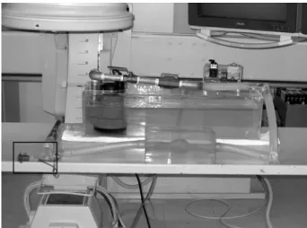
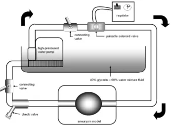
The phantoms consisted of a pulsating motor part(heart part) and an aortic aneurysm part, which mimicked true physiologic conditions. The heart part was created from a high-pressured water pump and a pulsatile flow solenoid valve for the simulation of aortic flow. The aortic aneurysm part was manufactured from paper clay, which was placed inside a acrylic plastic square box, where liquid silicone was poured.

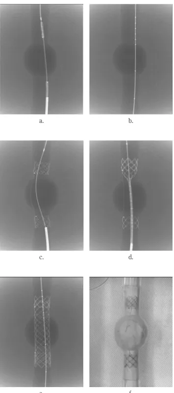
After the silicone was formed, the clay was removed, and a silicone tube was used to connect the heart and aneurysm part. We measured the change in pressure as related to the opening time(pulse rate, Kruskal-Wallis method) and pressure before and after the stent-graft implantation(n = 5, Wilcoxon's signed ranks test).

The changes in blood pressures according to pulse rate were all statistically significant( p <0.05). The systolic/diastolic pressures at the proximal aorta, the aortic aneurysm, and the distal aorta of the model were 157.80±1.92/130.20±1.92, 159.40±1.14/134.00±2.92, and 147.20±1.480/129.60±2.70mmHg, respectively, when the pulse rate was 0.5 beat/second. The pressures changed to 161.40±1.34/90.20±1.64, 175.00±1.58/93.00±1.58, and 176.80±1.48/90.80±1.92 mmHg, respectively, when the pulse rate was 1.0 beat/second, and 159.40±1.82/127.20±1.48, 166.60±1.67/138.00±1.87, and 161.00±1.22/135.40±1.67 mmHg, respectively, when it was 1.5 beat/second. When pulse rate was set at 1.0 beat/second, the pressures were 143.60±1.67/90.20±1.64, 147.20±1.92/84.60±1.82, and 137.40±1.52/88.80±1.64 mmHg after stent-graft implantation. The changes of pressure before and after stent-graft implantation were statistically significant( p <0.05) except the diastolic pressures at the proximal( p =1.00) and distal aorta( p = 0.157).

The aortic aneurysm phantoms seems to be useful for the evaluation of the efficacy of stent-graft before animal or clinical studies because of its easy reproducibility and ability to display a wide range of pressures.

Key Words : Experimental investigations, Hemodynamics/Flow dynamics, Aneurysm, Angiography

(2)

Ⅰ. INTRODUCTION

Aortic aneurysm is a pathological development of atherosclerosis that involves local enlargement of the aortic diameter and reduction in wall thickness

1)

. Surgical repair has been the standard treatment for abdominal aortic aneurysm(AAA), but it still carries a 4-6.5% mortality risk

2)

, even at experienced centers. Since Parodi et al. reported the first stent-graft repair of an AAA in 1991

3)

, investigators have become increasingly interested in stent-graft placement

4)

. Endovascular stent-graft implantation has been evaluated under various protocols, and investigators have improved the technique to offer a safer and less invasive solution for the treatment of aortic aneurysm

5,6)

.

Recently, various types of endovascular stent- grafts have been developed and used in clinical practice for the treatment of the aortic aneurysm.

Before endovascular stent-graft implantation was implanted into humans, numerous feasibility studies were conducted in animals and in in-vitro models

7-10)

.

However, most animals and in-vitro models to date have not accurately reflected human aortic size or physiologic conditions. To evaluate the applicability, safety, and biocompatibility of various types of stent-graft, an ideal flow model reproducing actual human aortic size and conditions has been necessary to reduce trial and error in animal and clinical studies.

We have recently developed an aortic aneurysm phantom, which could produce a wide range of aortic blood pressures and pulse rates. The purpose of this study was to evaluate the feasibility and efficacy of the aortic aneurysm model for in-vitro study of stent-graft placement.

Ⅱ. MATERIALS AND METHODS

1. The Pulsatile Flow Aortic Aneurysm Model The diameter of a normal aortic segment in the

phantom was derived from the mean cross diameter of the proximal descending thoracic aorta and the upper level of the iliac artery bifurcation in 10 normal aortograms and computed tomographic(CT) images.

The phantoms consists of a pulsating motor part(heart part) and an aortic aneurysm part, which mimicked real physiological conditions(Fig. 1). The heart part consisted of high-pressured water pumps(Hyupshin Water Design, Seoul, Korea) and a pulsatile flow solenoid valve(Hanwha Company, Seoul, Korea) for the simulation of aortic blood flow. The high-pressured water pump was used to produce a physiological pulsatile flow, and it was located inside an acrylic water bath. The pump was connected to a pulsatile flow solenoid valve.

Simulation of pulse rate was controlled by using a regulator(COTRON Cooperation, Seoul, Korea) of the solenoid valve. To closely mimic the real pumping action of the heart, the waveform of the pulsatile flow in the model was biphasic with a strong forward flow phase in systole(opened state of valve) followed by a slower forward flow phase in diastole (closed state of valve). As for the circulating fluid, a 40% glycerin – 60% water mixture fluid, maintained at 35℃, was used to simulate the hydrodynamic properties of blood(Fig. 2).

Fig. 1. Photograph of the pulsatile flow aortic aneurysm

model. The square box indicates the check valve system

for insertion of the guide wire and stent-graft

(3)

Fig. 2. Schematic diagram of pulsatile flow aortic aneurysm model. The large arrows indicate the direction of the circulating fluid

The aortic aneurysm part was created by a cast- reducing technique, which utilized a casting mold developed from paper clay(TODAN Instruction, Kyung-ki, Korea). The aneurysm phantom included segments representing the proximal and distal normal aorta and a segment of widened diameter representing an aortic aneurysm. The normal aortic segment was 20 mm in diameter and 9 cm in length from the upper and lower ends of the aneurysm, whereas, the segment representing the aneurysm was 45 mm in diameter and 50 mm in length. The clay mold was placed in a square box where liquid silicone(KE-1300, Shin-Etsu Chemical Co, Ltd., Tokyo, Japan) was subsequently poured. The hardening process of the silicone took approximately two days. After the silicone was formed, the clay was completely removed from the formed silicone model.

The total square dimension of the aortic aneurysm part was 19 cm in width, 23 cm in length, and 12 cm in height. A silicone tube was then used to connect the heart part and the aortic aneurysm part. For introduction of a guide wire and a stent-graft device, a check valve was connected to the distal normal aortic segments of the aortic aneurysm part.

2. A Stent-graft and Implantation Technique

A stent-graft(S&G Biotech Inc., Seoul, Korea) consisted of two parts : a graft stent and an inner bare stent(Fig. 3). The graft stent consisted of proximal and distal bare stents and a Dacron graft.

The stent was knitted from a single thread of 0.3 mm nitinol wire in an inter locked tubular configuration.

The stent was 22 mm in diameter and 3 cm in length. The Dacron graft was 20 mm in diameter and 6 cm in length. Two parts of the graft stents were connected to the Dacron graft using a 4-0 prolene blue monofilament on a tapered needle. The inner bare stent was knitted from a single thread of 0.3 mm nitinol wire in a non-interlocked tubular configuration. The stent was 22 mm in diameter and 12 cm in length. The inner bare stent was longer than graft stent for minimizing stent shortening.

Each of the graft stent and the inner bare stent were preloaded in a 12 Fr. introducer, separately.

Stent-graft introducing system consisted of two parts : a 12 Fr. outer Teflon sheath and a coil pusher(3.85 mm in outer diameter). Graft stents were preloaded in the introducing system. A preloaded graft stent set was advanced into the proximal aorta over a stiff guide wire. After deployment, the graft stent was centrally supported by a coaxial inner bare stent(Fig. 4). More details of the stent-graft and the implantation technique were previously described

10)

.

Fig. 3. Photograph of the expandable stent-graft

(4)

a. b.

c. d.

e. f.

Fig. 4. Radiographs and a photograph show the technical steps of separate stent-graft implantation. (a) A exchange guide wire was inserted through the check valve under fluoroscopic guidance, (b, c) Preloaded graft stent set was deployed across the aneurysm. (d, e, f) The graft stent was centrally supported by a coaxial inner bare stent

3. Evaluation of the Pulsatile Flow Aortic Aneurysm Model

To evaluate the feasibility and efficacy of the functional flow of the aortic aneurysm phantom, we performed two examinations.

First, pressures at the site of aneurysm and proximal and distal normal aortic segments of the aortic aneurysm phantom were measured 10 times with various opening rates of the valve(pulse rates) to evaluate whether the phantom could reproduce various blood pressures. The heart rate was divided into three study groups. Group I measurements were performed at a pulse rate of 0.5 beat per second, the pulse rate of group II at 1 beat per second, and the pulse rate of group III at 1.5 beats per second.

Second, in vitro stent graft placement was performed five times to evaluate the efficacy of the stent graft and hemodynamic changes of the aneurysm and proximal and distal aortic segments of the phantom following stent graft placement.

This examination was performed under group II conditions.

A 5 Fr. cobra catheter(Cook, Bloomington, IN, U.S.A.) was introduced into the proximal aortic segment over a 260-cm-long, 0.035-inch exchange guide wire(Terumo, Tokyo, Japan) through the check valve under fluoroscopic guidance. Digital subtraction angiography(DSA) was obtained using contrast medium to verify the location of the aortic aneurysm. Then, a stent graft introducing system was introduced beyond the aneurysm part over a stiff guide wire(Cook, Bloomington, IN, U.S.A.).

After the graft stent deployment, the coaxial inner bare stent was deployed at the same location of the graft stent to expand and support an interposed Dacron graft and the graft stent.

Pressures at the aneurysm part and proximal and

distal normal aortic segments were also measured

before and after stent graft placement at a pulse

rate at 1 beat per second.

(5)

4. Statistical Analyses

The results of the pressures are expressed as mean ± standard deviation. The Kruskal-Wallis method was used to test for differences in blood pressures within each subset of pulse rates. The Wilcoxon's signed rank test was used to evaluate the change of blood pressure before and after stent-graft implantation. The SPSS version 11.5 statistical package(SPSS, Chicago, Ill) was used to perform the analyses. A p -values of less than 0.05 were considered to indicate a statistically significant difference.

Ⅲ. RESULTS

The systolic and diastolic pressures at the proximal aorta, aortic aneurysm, and distal aorta were all significantly different according to the three groups( p < 0.05)(Table 1). The pressures (systolic/diastolic) of the model were 157.80±1.92/

130.20±1.92, 159.40±1.14/134.00±2.92, 147.20±

1.480/129.60±2.70 mmHg, at the proximal aorta, the aortic aneurysm, the distal aorta, respectively, under group I conditions. The pressures were 161.40±1.34/90.20±1.64, 175.00±1.58/93.00±1.58, 176.80±1.48/90.80±1.92 for the three respective aortic segments in group II, and 159.40±1.82/

127.20±1.48, 166.60±1.67/138.00±1.87, 161.00±1.22/

135.40±1.67 mmHg in group III.

We performed stent-graft implantation under group II conditions. The stent-graft deployment in the pulsatile flow aortic aneurysm phantom was successful and free of problems in all cases. During deployment of a separating stent-graft, no graft deformity or stent migration was noted. The changes of pressure before and after stent-graft implantation were statistically significant(Wilcoxon's signed ranks test, p < 0.05) except diastolic pressure at proximal ( p = 1.000) and distal aorta( p = 0.157)(Table 2). The pressures after stent-graft implantation were obtained at 143.60±1.67/90.20±1.64, 147.20±1.92/

84.60±1.82, 137.40±1.52/88.80±1.64 mmHg, in the proximal aorta, the aortic aneurysm, and the distal aorta, respectively. The aortic pressures decreased after stent-graft implantation(Fig. 5).

We confirmed the resolution of aortic aneurysm and exclusion of the aortic aneurysm by DSA imaging(Fig. 6), performed after the deployment of graft stent through the catheter.

Fig. 5. Pressure measurement of the model before and after stent-graft implantation at a pulse rate of 1.0 beat/

second condition

a. b.

Fig. 6. Digital subtraction angiograms(DSA) before and after stent-graft implantation

(a) DSA before stent-graft implantation show the aneurysm part(arrows) in two different aneurysm models. (b) DSA obtained after implantation of a stent-graft(curved arrows).

There is no evidence of endoleak

(6)

Ⅳ. DISCUSSION

In-vitro examinations and experimental studies are critical in the development of the new design and function of medical devices or materials, as it allows detection of any problems or defects prior to the initiation of clinical studies. The newly developed stent-graft has been evaluated under various experi󰠀

mental protocols, and investigators are improving the technique to offer a safer and less invasive solution for the treatment of aortic aneurysm

6-8)

. Some investigators have been using canine or ovine animal models to represent human aortic valves for the evaluation of newly designed stent-grafts

11,12)

. However, most experimental studies of stent-graft implantation did not accurately simulate actual human aortic size and shape. The ideal model will closely simulate the actual size of the aorta in humans for appropriate evaluation of stent-grafts under comparable situations. The choice of animal is also an important factor in being to able to overcome limitations in experimental studies focused on the study of aortic aneurysms.

The primary purpose of in-vitro examinations was to investigate the effectiveness, feasibility, and potential complications of newly developed devices prior to experimental and clinical study. Our phantom model of aortic aneurysm closely mimics the actual size and shape and physiologic conditions of true human aortic aneurysms. Some investigators have published reports on the use of actual human aortic phantom models for research of aortic aneurysms

12-16)

. Chong et al., for instance, investi󰠀

gated the hemodynamic flow patterns using flow visualization and laser Doppler aneurysm in a commercial bifurcated stent-graft for AAA repair

15)

. In this study, the AAA model was developed from clear silicone rubber with pulsatile flow being generated by a servo-controlled piston and a gear pump, which provided the unsteady and steady flow component, respectively. Rudin et al. reported the effectiveness of a coil and asymmetric stent using a elastomer vessel aneurysm models with blood flow

16)

.

In this phantom examination, dye-dilution imaging sequences were performed and time-density curves were constructed in disrupting standard vortex flow.

These results indicated that potentially favorable flow modification features can be created by using the described asymmetric stent design.

In some reported articles, phantom models developed from silicone materials have been widely used in implant materials and human phantom models in various areas in medicine

14-17)

. A model of an aortic aneurysm in our phantom can be easily created using silicone materials and paper clay. It is possible to perform stent-graft implantation mimicking similar clinical conditions under fluoroscopic guidance due to the similar thickness of the human phantom model with respect to that of the human body. Actual human aortic size and shape of the model can also easily be created by using paper clay and completely removed from the formed model after drying of the liquid silicone.

The in-vitro phantom model has been tried with various methods to simulate actual pulsatile flow of human aorta. We used the 40% glycerin - 60%

water mixed solution that has traditionally been used as circulating fluid to simulate the hydrodynamic properties of blood

14-16)

. To simulate the actual heart rate and blood pressure, we used a high- pressured water and a pulsatile flow solenoid valve.

The high-pressured water pump produced physiologic pulsatile flow, and the pump produced various pulse rates via control of the regulator of the solenoid valve. This allowed users to be capable of producing various pulse rates and blood pressures in our phantom model. The heart rate ranged from 30 beats/minute to 90 beats/minute, systolic pressure from 137.40±1.52 to 175.00±1.58, and diastolic pressure ranged from 84.60±1.82 to 130.20±1.92.

Therefore, we could simulate various clinical conditions by regulating the pulse rate and pulsatile pressure by adjustment of the opening rate of the solenoid valve.

Our phantom model has a side check valve

system for introduction of a guide wire, a catheter,

(7)

and a stent-graft introducing system, thereby allowing one to perform stent-graft placement under similar conditions of animal and clinical studies.

The side check value system also allows performance of stent-graft implantation and DSA on the phantom models. Investigators can thereby closely experience actual clinical conditions with our phantom model, facilitating the development of future experimental study designs using a functional flow model.

Our experiences have some limitation. First, the constant shape, diameter, length of aneurysm part used throughout our experiments did not reflect the true clinical condition of aortic aneurysms with their variable size and shape

18)

. In future studies, we will evaluate a newly designed aneurysm model that allows such a variability in aortic aneurysm morphology. Second, we did not perform CT and ultrasonographic evaluation of our phantom model, whereas, in clinical, practice, aortic aneurysms or dissection are evaluated by methods of sonography and CT before and after implantation of stent- grafts

18)

. In the next study, we will investigate the physiologic changes of the model according to blood pressure and pulse rate under ultrasonography and CT.

In conclusion, the pulsatile aortic aneurysm model of the actual human conditional flow seems to be useful in the evaluation of feasibility and efficacy of the newly designed stent-graft before animal or clinical studies because of its facile reproducibility and ability to apply a wide range of pressures.

ACKNOWLEDGMENTS

The study was supported by a grant(01-253) from the Asan Institute for Life Science, Seoul, Republic of Korea.

Reference

1. Sladen JG, Gilmour JL, Wong RW : Cumulative

patency and actual palliative in patients with claudication after aortofemoral bypass : prospective long-term follow-up of 100 patients, Am J Surg, 152, 190-195, 1996

2. Ernst CB : Abdominal aortic aneurysm. N Engl J Med, 328, 1167-1172, 1993

3. Parodi JC, Palmaz JC, Barone HD : Transfemoral intraluminal graft implantation for abdominal aortic aneurysms. Ann Vasc Surg, 5, 491-499, 1991

4. Han MC, Park JH : Interventional Radiology, ILCHOKAK, 319-328, 1999

5. Blum U, Langer M, Spillner G, et al. : Abdomi󰠀

nal aortic aneurysms : preliminary technical and clinical results with transfemoral placement of endovascular self-expanding stent-grafts, Radi󰠀

ology, 198, 25-31, 1996

6. Traul DK, Clair DG, Gray B, O'Hara PJ, Ouriel K : Percutaneous endovascular repair of infrarenal abdominal aortic aneurysm : a feasibility study, J Vasc Surg, 32, 770-776, 2000

7. Soula P, d'Othee BJ, Otal P, et al. : Macro󰠀

porous polyester-covered stent in an experimental abdominal aortic aneurysm model, J ENDOVASC THER, 8, 390-400, 2001

8. Maynar M, Qian Z, Hernandez J, et al. : An animal model of abdominal aortic aneurysm created with peritoneal patch : technique and initial results, Cardiovasc Intervent Radiol, 26, 168–176, 2003

9. Chuter TAM, Buck DG, Schneider DB, et al. : Development of a branched stent-graft for endovascular repair of aortic arch aneurysms, J ENDOVASC THER, 10, 940–945, 2003

10. Kang SG, Lee DY, Maeda M, et al. : Aortic dissection : percutaneous management with a separating stent-graft—preliminary results, Radiology, 220, 533–539, 2001

11. Beygui RE, Kinney EV, Pelc LR, et al. : Prevention

of spinal cord ischemia in an ovine model of

abdominal aortic aneurysm treated with a self-

expandable stent-graft, J Endovasc Surg, 6,

278-284, 1999

(8)

12. Sakaguchi S, Uchida BT, Timmermans HA, et al. : Twin-tube endografts for aortic aneurysms : an experimental feasibility study, J Vasc Interv Radiol, 10, 1092-1098, 1999

13. Abbott WM, Callow A, Moore W, et al. : Evalu󰠀

ation and perfomation standards for arterial prosthesis, J Vasc Surg, 17, 746-756, 1993 14. Chong CK, How TV, Black RA, et al. : Devel󰠀

opment of a simulator for endovascular repair of abdominal aortic aneurysm, Ann Biomed Eng, 26, 798-802, 1998

15. Chong CK, How TV : Flow pattern in an endovascular stent-graft for abdominal aortic aneurysm repair, Journal of Biomechanics, 37, 89-97, 2004

16. Ruden S, Wang Z, Kyprianou L, et al. : Measuremnet of flow modification in phantom aneurysm model : comparison of coils and a longgitudinally and axially asymmetric stent- initial findings, Radiology, 231, 272-276, 2004 17. Silver FH, Christiansen DL. : Biomaterials Science

and Biocompatibility, Springer, 279-304, 1999 18. Toshifumi Mita, Taseshi Arita, Naofumi Matsunaga,

et al. : Complications of endovascular repair for thoracic and abdominal aortic aneurysm : an imaging spectrum, RadioGraphics, 20, 1263- 1278, 2000

󰋯국문초록

스텐트그라프트 장치술을 위한 대동맥류 혈류 팬텀의 유용성

김태형

1,2)

․ 고기영

1)

․ 송호영

1)

․ 박인국

2)

․ 신지훈

1)

․ 임진오

1)

․ 김진형

1)

․ Eugene K. Choi

3)

울산대학교 의과대학 서울아산병원 영상의학과

1)

동국대학교 대학원 생물학과

2)

․코넬대학교 의과대학

3)

대동맥류 질환에 사용되는 스텐트그라프트의 체외실험을 위한 대동맥류 혈류 팬텀의 유효성과 실현가능성에 대해 평가하고자 한다. 팬텀은 인체의 혈류 조건과 유사한 상황을 재현할 수 있도록 심장부분과 대동맥류 부 분으로 구성되었다. 심장부분은 고압력 수중펌프와 솔레노이드 밸브를 사용하여 심장의 대동맥 혈류를 재현하 였고, 대동맥류 부분은 지점토를 사용하여 동맥류 모양을 재현하고 그 틀을 투명 실리콘으로 틀을 떠내는 방법 으로 제작하였다. 두부분은 실리콘 관으로 연결하였다. 제작된 팬텀에서 밸브의 개폐 시간에 따른 압력(수축기 /이완기) 변화를 측정하였으며, 스텐트그라프삽입술 전, 후의 압력변화를 측정하였으며, 통계적 유의성을 알아 보았다. 밸브의 개폐 시간에 따른 압력 변화는 통계적으로 유의한 결과를 보였다(P < 0.05). 0.5회/초의 개폐 조건에서는 팬텀의 대동맥 근위부, 대동맥류, 원위부의 압력은 각각 157.80±1.92/130.20±1.92, 159.40±

1.14/134.00±2.92, 147.20±1.480/129.60±2.70 mmHg이었으며, 1.0회/초의 개폐 조건에서는 161.40±1.34/

90.20±1.64, 175.00±1.58/93.00±1.58, 176.80±1.48/90.80±1.92 mmHg이었고, 1.5회/초의 개폐 조건에서는 159.40±1.82/127.20±1.48, 166.60±1.67/138.00±1.87, and 161.00±1.22/135.40± 1.67 mmHg이었다. 스텐트 그라프삽입술 전, 후의 팬텀의 압력변화는 대동맥부분에서 측정하였으며, 각각 143.60±1.67/90.20±1.64, 147.20±1.92/84.60±1.82, and 137.40±1.52/88.80±1.64 mmHg이었다. 결론적으로, 대동맥류 팬텀은 압력의 범위를 다양하게 적용할 수 있고, 팬텀 내에서 시술의 재현이 가능하여 동물실험 전 스텐트그라프트의 유용성 을 평가하기 위한 체외실험 기구로 유용할 것으로 기대된다.

중심 단어 : 실험적 조사, 혈류역학, 동맥류, 혈관조영술

수치

Fig. 1. Photograph of the pulsatile flow aortic aneurysm  model. The square box indicates the check valve system  for  insertion  of  the  guide  wire  and  stent-graft
Fig. 2. Schematic diagram of pulsatile flow aortic aneurysm  model.  The  large  arrows  indicate  the  direction  of  the  circulating  fluid
Fig. 4. Radiographs and a photograph show the technical  steps of separate stent-graft implantation
Fig. 6. Digital subtraction angiograms(DSA) before and after  stent-graft  implantation

참조

관련 문서