266
Although occasional patients with chronic myeloid leukemia (CML) have chromosomal changes other than Philadelphia chromosome early in the disease, in typical cases the 9;22 translocation remains the sole abnormality throughout the disease course in chronic phase. When disease pro- gression occurs, however, 75-80% develop additional chromosome aberrations. These secondary changes sometimes precede the more aggressive manifestations hematologically and clinically and thus may serve as valuable prognostic indicators. ider(9)(q10)t(9;22)(q34;q11.2) is very rare and a recurrent chromosomal abnormality associated with acute lymphoblastic leukemias (ALL) and lymphoblastic crisis of CML. And ider(9)(q10)t(9;22)(q34;q11.2) is a lymphoid-specific rear- rangement and the patients with this abnormality are of older age on average. They commonly show pre-B cell lineage immunophenotype and L2 morphology. We report a case of ider(9) (q10)t(9;22)(q34;q11.2) as secondary aberration in a patient with lymphoblastic crisis of CML.
(Korean J Clin Pathol 1999; 19: 266-70)
Key words :ider(9)(q10)t(9;22)(q34;q11.2), Blast crisis, Chronic myeloid leukemia
266
만성골수성백혈병의 이차적 핵형변화로서 ider(9)(q10)t(9;22)(q34;q11.2) 동반 1예
ider(9)(q10)t(9;22)(q34;q11.2) as Secondary Karyotypic Aberration of Chronic Myeloid Leukemia
Gui Jeon Choi, M.D., Dong Seok Jeon, M.D., Hyo Jin Chun, M.D., Jae Ryong Kim, M.D., Hong Suk Song, M.D.*, and Joong Won Lee, M.D.**
Departments of Clinical Pathology and Internal Medicine*, College of Medicine, Keimyung University;
Department of Clinical Pathology, College of Medicine, Kyungpook University**, Taegu, Korea.
최귀전∙전동석∙전효진∙김재룡∙송홍석*∙이중원**
계명대학교 의과대학 임상병리학교실, 내과학교실*, 경북대학교 의과대학 임상병리학교실**
서 론
Philadelphia 염색체(이하 Ph) 양성 만성골수성백혈병 환자에 서 동반되는 이차성 염색체 이상은 초진 당시에는 불과 9% 정도 이지만 모세포 발증기로 이행될 경우는 75 - 80%에 이른다고 하 며, 이러한 이차성 염색체 이상은 모세포 발증기의 전환과정 및 병인에 매우 중요한 역할을 하며 예후인자가 되고 있다[1]. 흔히 관찰되는 이차적인 염색체 이상으로 trisomy 8, i(17q), extra
Ph[+Ph, +der(22)t(9;22)], trisomy 19 및 이들 염색체 이상 의 상호조합형이 대략 70%를 차지하며 나머지 약 30%에서 trisomy 21(7%), Y염색체의 결손(5%), monosomy 7(3%), monosomy 17(3%) 및 trisomy 17(4%)의 빈도로 관찰되며, 약 1%에서 t(3;21)(q26;q22)이 관찰된다[2-3]. 이러한 이차적인 염색체 이 상은 때때로 혈액학적, 임상적 양상보다 선행되어 나타나므로 중 요한 예후 인자로 생각될 뿐만 아니라 감별진단에도 보조적인 도 움이 되고 있다[1-3]. 또한 t(9;22)의 소견은 급성골수성백혈병 의 매우 낮은 빈도에서, 소아의 급성림프구성백혈병의 2-6%에서, 성인의 급성림프구성백혈병에서는 17-30%의 빈도로 나타나며 불량한 예후를 나타내는 소견이라고 간주되고 있다[4-9].
ider(9)(q10)t(9;22)(q34;q11.2)는 매우 드문 핵형으로서 만 성골수성백혈병의 모세포 발증기에서 3예, 급성림프구성백혈병에
266266
접 수
:
1998년 10월 7일 접수번호:
KJCP1224 수정본접수:
1998년 11월 25일교 신저 자
:
최 귀 전우 700-310 대구시 중구 동산동 194 계명대학교 동산의료원 임상병리과 전화 : 053-250-7950, Fax : 053-250-7275
서 3예 등 6예의 문헌보고[10]가 확인 되었으며, 국내 보고는 아 직 없다. ider(9)(q10)t(9;22)는 i(9q)의 소견과 함께 림프구계 특이적인 핵형 변화로서 노령층에서, 그리고 pre-B 세포성 면역 표현형 및 FAB 분류상 L2형태를 특징으로 나타내는 핵형이다 [1, 10]. 저자들은 만성골수성백혈병에서 모세포 발증기로 이행 된 환자에서 Ph에 동반되는 이차적 핵형변화로서 ider(9)(q10)t (9;22)(q34;q11.2)의 소견을 나타낸 1예를 경험하였다.
증 례
환자 : 서○○, 남자, 42세
주소 : 약 6개월간의 만성피로감 및 관절동통
현병력 : 내원 6개월 전부터 피로감, 요통, 슬관절, 완관절 및 대퇴부 동통, 발한, 체중감소(6개월동안 약 8 kg 감소) 등의 증 상이 나타나 점차 악화되어 내원하였다.
과거력 : 10년전 경미한 뇌출혈의 병력이 있었다.
이학적 소견 : 외견상 만성병색이였고 결막 창백, 간종대, 늑골 연하 15 cm까지 감지되는 심한 비장종대 및 우측서혜부에 1.5 cm 정도의 단단하고 유동성의 림프절 종대도 관찰되었다.
검사소견 : 내원당시 말초혈액으로 시행한 일반혈액 검사상, 백혈구수는 278,000/ L, 혈색소는 10.8 g/dL, 헤마토클릿 37.4%, 혈소판 285,000/ L이었고 경미한 대소부동증 및 변형적
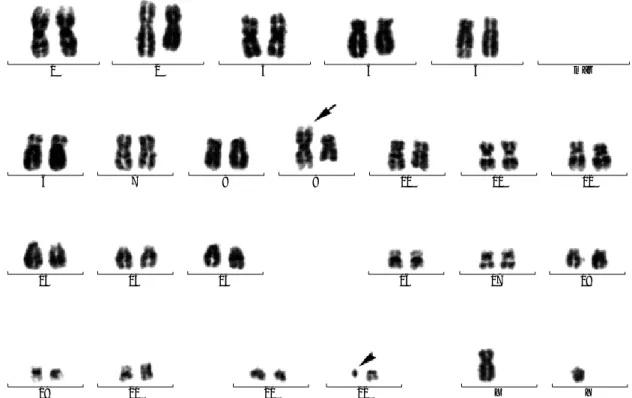
혈구증의 소견이 관찰되었다. 적혈구침강속도는 29 mm/hr, LDH 380.5 U/L, 요산은 8.5 mg/dL이었고, 혈청 철은 21 g/dL로 감소되어 있었다. 말초혈액의 백혈구 감별계산상 모세포는 11%, 전골수구 5%, 골수구 18%, 후골수구 4%, 간상호중구 27%, 분 엽호중구 21%, 림프구 11%, 호산구 3%이었다. 골수생검상 세 포충실도는 증가되었으며, 골수흡인 도말검사에서 유핵세포의 감 별계산상 모세포 33.7%, 전골수구 1.4%, 골수구 12.2%, 후골수 구 6.0%, 간상호중구 5.6%, 분엽호중구 16.4%, 호산구 3%, 림 프구 16.2%이었다. 면역표현형 검사상 CD10, CD19, CD20, CD22 및 HLA-DR은 양성이었고 CD3, CD5, CD7, CD13, CD14, CD33 및 CD41은 음성이었다. 직접법 및 단기배양법으로 시행한 골수 염색체 핵형 분석은 46,XY,t(9;22)(q34;q11.2) [12]/46,XY,ider(9)(q10)t(9;22)(q34;q11.2), der(22)t (9;22)(q34;q11.2)[10]/45,XY,-9, der(22)t(9;22) (q34;q11.2) [3]로 나타났다(Fig. 1, 2). 혈청 IgG, IgM 및 IgE는 각각 2,370 mg/dL, 335.5 mg/dL, 198.8 IU/mL이었다. 초진시 시행한 골 수검체의 major
BCR-ABL유전자 재배열을 역전사 중합효소 연쇄반응으로 검사한 결과 양성으로 분석되었다(Fig. 3).
치료 및 경과 : 환자는 접촉성 피부염과 기타 환자사정으로 집 중적인 항암요법을 즉시 시행하지 못하였고, allopurinol로 항암요 법을 시행 6일후 말초혈액의 혈소판은 72,000/ L, 총백혈구수는 480,000/ L, 모세포수도 37.3%로 증가되었다. 이후 prednisone, daunorubicin, vincristine, methotrexate, L-asparaginase 등의
Fig. 1. G-banded karyotype demonstrating 46,XY,ider(9)(q10)t(9;22)(q34;q11.2), der(22)t(9;22)(q34;q11.2). The arrow indicates
the ider(9)(q10)t(9;22)(q34;q11.2), and the arrowhead indicates the Philadelphia chromosome.
1
6 7 8 9 10 11 12
13
19 20 21 22 X Y
14 15 16 17 18
2 3 4 5 mar
집중적인 항암요법을 받았고, 요법 9일째 백혈구수는 700/ L (모세포는 계수되지 않음), 혈색소 9.0 g/dL, 혈소판 14,000/ L 이었고 요법 14일째 백혈구수는 327/ L (모세포는 계수되지 않 음), 혈색소 6.0 g/dL, 혈소판 8,000/ L이었다. 항암요법 28일 째 시행한 골수천자도말 검사에서 모세포가 0.3%였고, 말초혈액 검사상 백혈구 2,630/ L, 혈색소 8.5 g/dL, 혈소판 559,000/ L 이었다. 이후 환자는 패혈증 및 폐농양의 소견도 나타내어 항생 제 투여가 추가되었고, 60일후 시행한 추적 골수검사상 완전관해 의 판정을 받았으며 핵형분석상 Ph 및 ider(9)(q10)t(9;22)의 소견은 관찰되지 않았다.
고 찰
만성골수성백혈병은 Ph 및
BCR-ABL융합유전자를 특징으 로하는 조혈간세포 질환[11]으로서 약 80% (60-90%) 정도에서 만성기를 거쳐 가속기와 모세포발증기로 진행되는 경과를 보인다 [12, 13]. 모세포발증기는 골수 및 말초에 미성숙세포가 과다증 식하며, 점진적인 빈혈과 혈소판감소증을 보이고, 가끔 모세포가
골수외부에 축적되기도 하며, 항암치료제에 반응이 격감되는 것 이 특징이다[1, 14].
t(9;22)의 소견은 세포유전학적인 수준에서는 급성림프구성백 혈병에서 나타나는 t(9;22)(q34;q11.2)와 동일하지만, 분자생물 학적인 수준에서 보면 급성림프구성백혈병의 50-80%에서 나타 나는 minor
BCR은 만성골수성백혈병에서 나타나는 major
BCR보다 근위부에 절단점이 위치하므로 만성골수성백혈병의 림프구 성 모세포발증기와는 감별이 될 수 있다[1, 14]. 본 증례에서도 환자는 초진시 이미 만성골수성백혈병의 림프구성 모세포발증기 상태였고
de novo급성림프구성백혈병과 감별이 어려웠는데 환 자의 이학적소견(간, 비종대 및 림프절 종대)과 염색체소견 및 major
BCR-ABL유전자에 대한 역전사 중합효소 연쇄반응의 결과를 종합하여 만성골수성백혈병의 림프구성 모세포 발증기로 진단하였다.
Diez-Martin 등[15]은 만성골수성백혈병에서 모세포발증기로 진행한 25예에서 이차성 염색체이상 유형, 면역표현형, 림프구 및 골수성 모세포발증기와의 상관관계를 조사하였는데, 골수성 모세 포발증기로 진행된 15예(86.6%)에서 extra Ph(+Ph), i(17q), +8, +19 및 이들의 조합형이 나타났고, 림프구성 발증기로 이행 된 8예에서 1예만 +Ph를 보여주었을 뿐 +8, +19, i(17q)의 소 견을 볼 수 없었다고 하였다. t(3;21)(q26;q22) 소견은 골수성 모세포발증기에 나타나는 소견이며[2, 3], i(17q) 및 inv(3) (q21q26)을 동반한 경우는 거핵모세포성 발증기로 이행하였다고 한다[15]. Ph와 동반된 이차적인 염색체이상의 유형이 림프구성 및 비림프구성백혈병의 감별에 도움을 줄 수 있는데, 비림프구성 백혈병으로의 진행을 예측하게 하는 염색체이상으로는 이미 언급 된 extra Ph(+Ph), i(17q), +8, +19 이외에도 t(8;21), t(15;17), inv(16), t(3;3) 및 inv(3)과 같은 3q26 이상 등이 있다[1, 2, 15].
ider(9)(q10)t(9;22)(q34;q11.2)는 매우 드문 핵형으로서 외 국의 경우 6예의 문헌보고가 있었다[10]. ider(9)(q10)t (9;22)(q34;q11.2)의 형성으로 9번 염색체의 단완은 결손되고 t(9;22) 전좌결과 생기는 9번 염색체의 장완 및 22번 염색체의 장완은 중복상태가 된다. 9번 염색체 단완의 결손[del(9)(p21- 22)]이나 불균형 전좌는 급성림프구성백혈병의 약 7-13%에서 관찰된다[16-20]고 하는데 i(9q)의 소견은 그중 1-2% 정도를 차지한다[21-22]. 흔히 9번 염색체 9p21-22부위가 결손되는데 여기에 위치하는 interferon 유전자와 purine대사에 중요한 효소 인 methylthioadenosine phosphorylase (MTAP)를 coding하는 유전자 결손이 병인발생기전과 질환의 진행에 관여한다고 한다 [17, 20, 23, 24].
9q에 위치하는 유전자의 과도발현 및
BCR-ABL융합유전자 등 ider(9q)가 질환의 발생기전에 중요한 역할을 할 것으로 관심 이 고조되어 있으나, ider(9q)의 보고가 극히 드물고 질환과 연루 된 치료적 접근이나 추적조사 기간의 부족으로 ider(9q)와 관련 된 예후적인 연구 및 접근에 어려움이 있다. 다만 전형적인 t(9;22)(q34;q11.2)결과 생긴 der(9)t(9;22)로부터 ider(9q)가
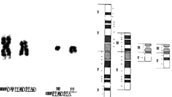
Fig. 2.Partial karyotype and ideogram demonstrating ider(9)
(q10)t(9;22)(q34;q11.2).
ider(9)(q10)t(9;22)
der(22)t(9;22)
9 ph 22
q
p
p p
q q q q
q
q
Fig. 3. RT-PCR for detecting major BCR-ABL gene rearrange-
ment. The band of 363-bp indicates the segments of BCR-ABL fusion transcripts. Lane M; size marker (100bp DNA ladder, GIBCO BRL); lane 1; K562 cells as positive control; lane 2; Bone marrow specimen from patient; lane 3; D/W as negative control.
bp M 2,072 1,500 600 300 200 100
363 bp
�
1 2 3
유래하였다고 생각되어지고 있다[10]. 본 증례의 환자는 비교적 예후가 양호하며 항암요법에 잘 치유되어 현재 완전관해상태로 외래로 치료 및 계속 추적조사중이지만, ider(9)(q10)t(9;22) 보 고 및 연구가 극히 드물기 때문에 아직 예후에 관한 판정을 내리 기에는 부족하다고 생각된다. 향후 ider(9)(q10)t(9;22)에 관한 검사실적 및 임상적 자료의 축적과 분석이 필요하다고 사료된다.
요 약
ider(9)(q10)t(9;22)는 매우 드문 핵형으로서 림프구계 특이 적인 핵형 변화이며 주로 노령층에서, 그리고 pre-B 세포성 면역 표현형 및 FAB 분류상 L2형태를 특징으로 나타내는 핵형이다.
아직 국내 보고된 바 없었으며, 저자들은 만성골수성백혈병에서 림프구성 모세포발증기로 이행된 환자의 골수 염색체 분석상 t(9;22)(q34;q11.2) 및 이차적인 핵형으로 ider(9)(q10)t(9;22) 를 경험하였다. 환자의 핵형은 46,XY,t(9;22)(q34;q11.2) [12]/46,XY,ider(9)(q10)t(9;22)(q34;q11.2), der(22)t(9;22) (q34;q11.2)[10]/45,XY,-9,der(22)t(9;22)(q34;q11.2)[3]로 분 석되었다.
참고문헌
1. Heim S and Miltelman F, ed. Cancer cytogenetics. 2nd ed. New York:
Wiley-Liss, 1995: 33-68.
2. Rubin CM, Larson RA, Bitter MA, Carrino JJ, Le Beau MM, Diaz MO, et al. Association of a chromosomal 3; 21 translocation with the blast
phase of chronic myelogenous leukemia. Blood 1987; 70: 1338-42.3. 최귀전 , 전효진 , 전동석 , 김재룡 , 권기영 . t(3;21)(q26;q22) 를 동반한 Ph 양성 만성골수성백혈병의 골수성 모세포발증기 1 예 . 대한임상 병리학회지
1997; 17: 21-7.4. Tuszynski A, Shut S, Young BD, Lister TA, Rohatiner AZ, Amess JAL et al. Detection and significance of bcr-abl mRNA transcripts and
fusion proteins in Philadelphia positive adult acute lymphoblastic leukemia. Leukemia 1993; 7: 1504-8.5. Bloomfield CD, Wurster-Hill D, Peng G, Le Beau M, Tantravahi R, Testa J, et al. Prognostic significance of the Philadelphia chromosome in
adult acute lymphoblastic leukemia. In: Gale RP and Heelzer D, ed. Acute lymphoblastic leukemia. 1st ed. New York: Wiley-Liss, 1990: 101-9.6. Gotz G, Weh H-J, Walter TA, Kuse R. Kolbe K, Dolken G, et al.
Clinical and prognostic significance of the Philadelphia chromosome in adult patients with acute lymphoblastic leukemia. Ann Hematol 1992; 64:
97-100.
7. Priest JR, Robison LL, McKenna RW, Lindquist LL, Warkentin PI, LeBein TW, et al. Philadelphia chromosome positive childhood acute lym-
phoblastic leukemia. Blood 1980; 56: 15-8.
8. Champlin RE and Golde DW. Chronic myelogenous leukemia: recent
advances. Blood 1985; 65: 1039-47.9. Berger R, Chen SJ, Chen S. Ph-positive acute leukemia. Cytogenetic and
molecular aspects. Cancer Genet Cytogenet 1990; 44: 143-52.10. Dierlamm J, Michaux L, Kroger N, Wlodarska I, Martiat P, Zeller W, et al. ider(9)(q10)t(9;22)(q34;q11) is a recurrent chromosomal abnor-
mality in acute lymphoblastic leukemia and lymphatic blastic phase of chronic myelogeous leukemia. Cancer Genet Cytogenet 1996; 89: 109-13.11. Heisterkamp N, Stephenson JR, Groffen J, Hansen PF, de Klein A, Bartram CR et al. Localization of the c-abl oncogene adjacent to a translo-
cation break point in chronic myelocytic leukemia. Nature 1983; 306: 239-42.12. Boggs DR. The pathogenesis and clinical patterns of blastic crisis of
chronic myeloid leukemia. Semin Oncol 1976; 3: 289-96.13. Vardiman JW. Chronic myelogenous leukemia and the myeloprolifera-
tive disorders. In : Knowles D. ed. Neoplastic hematopathology. 1st ed.Baltimore:Williams and Wilkins, 1992: 1405-38.
14. Kantarjian HM, Deisseroth A, Kurzrock R, Estrov Z, Talpaz M.
Chronic myelogenous leukemia : a concise update. Blood 1993; 82: 691- 703.
15. Diez-Martin J, Dewald GW, Pierre RV. Possible cytogenetic distinc-
tion between lymphoid and myeloid blast crisis in chronic granulocytic leukemia. A J Hematol 1988; 27: 194-203.16. Kowalczyk J and Sandberg AA. A possible subgroup of ALL with 9p-.
Cancer Genet Cytogenet 1983; 9: 383-5.
17. Chilcote RR, Brown E, Rowley JD. Lymphoblastic leukemia with lym-
phomatous features associated with abnormalities of the short arm of chro- mosome 9. N Engl J Med 1985; 313: 286-91.18. Carroll AJ, Castelberry RP, Crist WM. Lack of association between
abnormalities of the chromosome 9 short arm and either“
lymphomatous”
features of T-cell phenotype in childhood acute lymphocytic leukemia.Blood 1987; 69: 735-8.
19. Pollak C and Hagemeijer A. Abnormalities of the short arm of chromo-
some 9 with partial loss of material in hematological disorders. Leukemia 1987; 1: 541-8.20. Diaz MO, Rubin CM, Harden A, Ziemin S, Larson RA, Le Beau M,
et al. Deletions of inteferon genes in acute lymphoblastic leukemia. N Engl J Med 1990; 322: 77-82.21. Mertens F, Johansson B, Mitelman F. Isochromosomes in neoplasia.
Genes Chrom Cancer 1994; 10: 221-30.
22. Pui C-H, Carroll AJ, Raimondi SC, Schell MJ, Head DR, Shuster JJ,
et al. Isochromosomes in childhood acute lymphoblastic leukemia : A col- laborative study of 83 cases. Blood 1992; 79: 2384-91.23. Carrera CJ, Eddy RL, Shows TB, Carson DA. Assignment of the gene
for methylthioadenosine phosphorylase to human chromosome 9 by mouse-human somatic cell hybridization. Proc Natl Acad Sci USA 1984;. .
. .
81: 2665-8.
24. Olopade OI, Bohlander SK, Pomykala H, Maltepe E, Van Melle E, Le Beau MM, et al. Mapping of the shortest regeion of overlap of dele-
tions of the short arm of chromosome 9 associated with human neoplasia.
Genomics 1992; 14: 437-43.