INTRODUCTION
Acute kidney injury (AKI) is characterized by rapid disruption of renal function that occurs in diverse insults, including sepsis, nephrotoxins and ischemia/reperfusion injury that may occur during organ transplantation and cardiothoracic, vascular and general surgery.
1) AKI occurs in approximately 30% of all patients admitted to intensive care units and is associated with high mortality and mor- bidity.
2,3) Further, a number of recent experimental and clinical studies indicate that AKI is an independent risk factor for onset and deterioration of chronic kidney dis- ease.
4-6) Currently, AKI remains a significant health burden with its undefined pathogenesis and is a major unmet med- ical need without any pharmacological treatments.
7,8)
Kidney is an organ with high metabolic demand due to active transport of glucose, ions and nutrients. Thus, re- nal tubular mitochondrial dysfunction and ATP depletion are critical factors inducing AKI.
9,10) Proximal tubules re-
quire more active transport mechanisms than other tubule types because they reabsorb 70% of the glomerular fil- trate. Proximal tubule, particularly the S3 segment resid- ing in the outer medullary region, is highly vulnerable to hypoxic condition like ischemia/reperfusion injury (IRI), since this region is exposed to the lowest oxygen pressure (tissue oxygen tension 10~20 mmHg) with only 5~10%
of total renal blood flow.
11) Therefore, sufficient energy supply from fatty acid β-oxidation (FAO) is critical to the normal function of proximal tubule.
12) Regardless of the etiology, AKI induces mitochondrial dysfunction in prox- imal tubule.
13,14) Pathological signals like elevated levels of intracellular and mitochondrial Ca
2+, increased pro- duction of reactive oxygen species (ROS), mitochondrial membrane permeabilization and depolarization, and loss of ATP are hallmarks of mitochondrial dysfunction.
13)
Given that proximal tubule is highly sensitive to deple- tion of ATP, mitochondria is a potential therapeutic tar- get for preventing renal tubular cell death.
12,15) Here, we review the current findings regarding molecular mecha- nism of mitochondrial dysfunction, primarily focused on impaired fatty acid metabolism and ATP depletion, and recent developments in mitochondria-targeted therapeutic strategies in AKI.
http://wcms.jejunu.ac.kr
Copyright The Journal of Medicine and Life Science
Received: September 1, 2018; Revised: October 5, 2018; Accepted: October 7, 2018
* Correspondence to : Babu J. Padanilam
Department of Cellular and Integrative Physiology, University of Nebraska Medical Center, Omaha, NE 68198-5850
Tel: 1-402-559-3575, FAX: 1-402-559-4438 E-mail: bpadanilam@unmc.edu
https://doi.org/10.22730/jmls.2018.15.2.37 ISSN: 1738-1010
Mitochondrial fatty acid metabolism in acute kidney injury
By Hee-Seong Jang 1 , Babu J. Padanilam 1,2, *
1
Department of Cellular and Integrative Physiology, University of Nebraska Medical Center, Omaha, NE
2
Department of Internal Medicine, Section of Nephrology, University of Nebraska Medical Center, Omaha, NE
Abstract Mitochondrial injury in renal tubule has been recognized as a major contributor in acute kidney injury (AKI) pathogenesis. Ischemic insult, nephrotoxin, endotoxin and contrast medium destroy mitochondrial structure and function as well as their biogenesis and dynamics, especially in renal proximal tubule, to elicit ATP depletion.
Mitochondrial fatty acid β-oxidation (FAO) is the preferred source of ATP in the kidney, and its impairment is a critical factor in AKI pathogenesis. This review explores current knowledge of mitochondrial dysfunction and energy depletion in AKI and prospective views on developing therapeutic strategies targeting mitochondrial dysfunction in AKI.
Key words: Mitochondria, Energy metabolism, Fatty acid oxidation, ATP, Mitochondrial dysfunction, Ischemia/
reperfusion injury, Nephrotoxin, Sepsis, Acute kidney injury
Mitochondrial energetics
Mitochondria are highly dynamic intracellular organ- elles and have a network with other organelles, including endoplasmic reticulum and nucleus. Mitochondria can undergo physical changes like fusion, fission, and move- ment, and serve as a power plant generating cellular ener- gy.
16) The magnitude of generation of ATP is dependent on energy demand of the cell, based on their requirement for passive or active transport of ion, glucose, and nutrients.
The principle role of mitochondria is to generate cellular energy via electron transport and oxidative phosphoryla- tion, as well as calcium homeostasis and ROS generation.
Kidney uses 95~99% of energy derived from mitochon- drial oxidative metabolism.
17) Oxidative phosphorylation is more efficient by generation of 36 ATP per glucose, compared to 2 ATP by glycolysis. On the other hand, one fatty acid (FA) is more effective as it can generate 106 ATP via FAO. The proximal tubule has low glycolytic ca- pacity, and prefers FAO for generation of its high ATP de- mand.
18,19) Although FAO is the most efficient mechanism for producing ATP in proximal tubules, it is important to note that due to the high consumption of oxygen by prox- imal tubules, they are more susceptible than other cell types to changes in oxygen levels.
19) The distal tubule, on the other hand, has higher glycolytic metabolism and could switch to anaerobic metabolism in AKI, and may explain why it is relatively resistant to AKI.
19)
Mitochondrial fatty acid metabolism in AKI
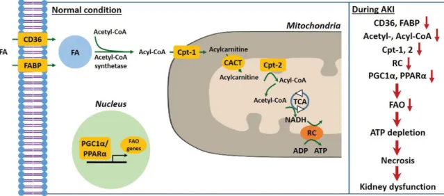
FA uptake, oxidation and synthesis are tightly balanced to achieve lipid homeostasis and to prevent lipid accumu- lation in various disease conditions. In the ischemic kid- ney, defects in the transport and oxidation of fatty acids can result in accumulation of fatty acids in the cytoplasm which may contribute to the decrease in ATP production and mitochondrial energetics.
12) FA are taken up in prox- imal tubule cells via specialized transport proteins on the plasma membrane, such as CD36, and also by retrieval from the glomerular filtrate by receptor-mediated albumin endocytosis.
18,20) Since FA is impermeable to outer mito- chondrial membrane (OMM), FAs are activated to acyl- CoA by acyl-CoA synthetases in the cytosol. Carnitine palmitoyltransferase-1 (CPT-1), the rate-limiting enzyme of carnitine shuttle on OMM, catalyzes the conversion of acyl-CoA to acylcarnitine, thus facilitating its passage through inner mitochondrial membrane (IMM) by carni- tine-acylcarnitine translocase (CPT-2).
18,20,21) CPT-2 on the IMM reconverts the acylcarnitine into an acyl-CoA.
The acyl-CoAs undergo FAO to generate acetyl-CoA, to fuel the tricarboxylic acid cycle and production of NADH and FADH
2
, that serve as electron donors to the electron transport chain for ATP production.
20) Deficiency of CPT- 1 results in energy failure and diverse kidney diseases including diabetic nephropathy and chronic kidney dis- ease.
18,20) In the ischemic kidney, the activity of CPT1 is
Figure 1. Mitochondrial fatty acid metabolism in AKI. FA enters into cytosol of renal proximal tubule cell (PTC) via FABP or CD36. In the cyto-
sol, FA are transformed from acetyl-CoA to acyl-CoA by addition of acetyl-CoA through acetyl-CoA synthetase and then transferred to mitochon-
drial matrix by carnitine shuttle, Cpt-1, CACT and Cpt-2, step by step. Acyl-CoA undergo β-oxidation to produce acetyl-CoA for TCA. NADH
generated by TCA is used as an electron donor for RC. During AKI, inhibition of genes related to fatty acid oxidation occurs to impair fatty acid
oxidation and that in turn deplete ATP, resulting in PTC necrosis and kidney dysfunction. FA, fatty acid; FAO, fatty acid β-oxidation; Cpt, carnitine
Opalmitoyltransferase; CACT, carnitine-acylcarnitine translocase; TCA, tricarboxylic acid cycle; RC, respiratory chain.
12,13,18,20,21,25,26,29,31,32)