• 검색 결과가 없습니다.

Inherited retinal degeneration in a Bengal cat

N/A
N/A
Protected

Academic year: 2021

Share "Inherited retinal degeneration in a Bengal cat"

Copied!
3
0
0

로드 중.... (전체 텍스트 보기)

전체 글

(1)

pISSN 2466-1384 eISSN 2466-1392 大韓獸醫學會誌 (2018) 第 58 卷 第 4 號 Korean J Vet Res(2018) 58(4) : 219~221 https://doi.org/10.14405/kjvr.2018.58.4.219

219

<Case Report>

Inherited retinal degeneration in a Bengal cat

Manbok Jeong

1,

*, Kristina Narfström

2

1

I Animal Medical Center, Bucheon 14427, Korea

2

Department of Veterinary Medicine and Surgery, College of Veterinary Medicine, University of Missouri-Columbia, Columbia, MO 65211, USA

(Received: July 9, 2018; Revised: August 3, 2018; Accepted: August 16, 2018)

Abstract: A 2-year-old intact female Bengal cat was presented with a 6-month history of visual impairment. The cat manifested bilateral negative menace responses and dazzle reflexes and sluggish pupillary light reflexes. Bilateral fundus changes included generalized tapetal hyperreflectivity, advanced retinal vascular attenuation, and increased pallor of the optic disc. A diagnosis of bilateral retinal degeneration was made. The clinical findings suggest that the investigated Bengal cat was most likely to have an inherited retinal degeneration. Further studies of the Bengal cat breed are needed to determine the prevalence of inherited retinal degeneration in this breed in Korea.

Keywords: Bengal cat, inherited retinal degeneration, pupillary light reflex, vision disorders

Inherited feline retinal degeneration, progressive retinal atrophy, evolves through a primary successive deterioration of photoreceptor cells in the retina [5]. It is a bilateral, sym- metrical, progressive retinal degeneration of the rods and cones, ultimately resulting in visual impairment. Generally, clinical signs of retinal degeneration include blindness, mydriasis, and incomplete pupillary light reflexes (PLRs).

Funduscopic examination reveals diffuse tapetal hyperreflec- tivity, retinal vascular attenuation, and pallor of optic nerve head [8].

This type of retinal degeneration was described in the Abyssinian, Siamese, and Persian cat breeds [1, 6, 9, 10, 15].

Of these, systematic studies have been performed using a breeding colony of Abyssinian cats and another colony of Persian cats. Most recently, the Bengal cat breed was found to have an early-onset, autosomal recessive progressive inherited retinal degeneration [13]. The purpose of the present paper was to report the observation of a presumed inherited retinal degeneration in a young Bengal cat in Korea.

A 2-year-old intact female Bengal cat was presented with loss of vision over the past 6 months. The owner had noticed the visual problem since she had moved to another house 6 months ago. There was no history of drug administration or surgical events at the time of the initial presentation. The cat was fed only commercial cat food.

A complete physical examination was unremarkable and the cat was unwilling to move around the examination room under photopic and scotopic conditions. On presentation,

there were no upper respiratory signs of infection including cough and sneezing. Menace responses and dazzle reflexes were absent in both eyes (OU) and PLRs were incomplete and sluggish OU. Intraocular pressures (IOPs) measured by rebound tonometry (Icare TonoVet; Icare Finland Oy, Fin- land) were 24 mmHg OU. Slit-lamp biomicroscopy (HS- 7000; Huvitz, Korea) showed anterior capsular cataract due to persistent pupillary membranes (iris to lens) in the right eye (Fig. 1) and posterior capsular cataract OU (Fig. 2). Flu- orescein staining showed no corneal defects. The fundus- copic changes (Vantage Plus; Keeler, UK) included severe diffuse tapetal hyperreflectivity, severe retinal vascular atten- uation, and optic nerve pallor OU (Fig. 3). Based on the information available, a diagnosis of bilateral retinal degener- ation was made.

Retinal degenerations appear to be observed more seldom

*Corresponding author

Tel: +82-32-677-5262, Fax: +82-32-677-5070 E-mail: 9757044@hanmail.net

Fig. 1. Anterior capsular cataract due to persistent pupillary

membranes (iris to lens) seen with diffuse illumination by slit-

lamp biomicroscopy in the right eye (A, before mydriasis; B,

after mydriasis).

(2)

220 Manbok Jeong, Kristina Narfström

in cats in comparison to findings in dogs [12]. The most common causes for a bilateral, symmetrical retinal degenera- tion are inherited degenerations/dystrophies, nutritional defi- ciencies and drug-related retinal degeneration. The ophthalmos- copic appearance of these different forms of retinopathies is more or less similar and includes tapetal hyperreflectivity, retinal vascular attenuation and depigmentation of the non- tapetal fundus [5].

Taurine is an essential amino acid for cats found in high concentrations in the heart muscle and in the retina, espe- cially photoreceptor cells [2, 5]. It has been considered to act as a neurotransmitter and to have a protective influence on cell membranes of the heart muscle and retina. Because the cat has limited ability to synthesize it from its precursor amino acid, dietary deficiency of taurine results in both hypertrophic cardiomyopathy and bilateral, symmetrical pro- gressive retinal degeneration. The earliest ophthalmoscopic finding of taurine deficiency retinopathy is increased granu- larity of the area centralis region followed by grayish discol- oration of the visual streak [2, 5]. In the advanced stage, there is a generalized retinal degeneration with retinal vascu- lar attenuation. The cat presented in this case report, although plasma taurine concentration was not measured, had been fed commercial cat food ever since birth. In addition, there were no ocular problems observed in a housemate fed the same commercial cat food.

Some studies were reported regarding adverse reaction to medications in the feline species [5]. More recently, feline retinal degeneration can be also generated from enrofloxacin toxicity [4, 6]. The enrofloxacin-associated retinal toxicity

produced acute and severe retinal degeneration and gener- ally permanent blindness in cats receiving parenteral or enteral enrofloxacin in a flexible dosing regimen (5 to 20 mg/kg, once a day) [6]. The cat in the present report has not received any medications and had not undergone anesthesia since birth.

Feline hereditary retinal degeneration has not been studied as systematically in cats as in dogs with the exception of studies in the Abyssinian and Persian cat breeds [10-12, 15].

Two different types of retinal diseases have been described in the Abyssinian cat with different modes of inheritance: A cone–rod dysplasia with an autosomal dominant mode of inheritance and rod–cone degeneration, which is recessively inherited [8, 11]. In the former, abnormalities of the PLRs and nystagmus were shown in affected kittens as young as at age 4 weeks. The earliest ophthalmoscopic findings included tapetal dullness and loss of detail, present by 8–12 weeks of age, with a fairly rapid progression, leading to eventual reti- nal vascular attenuation, tapetal hyperreflectivity, and depig- mentation in the nontapetal fundus by 1 year of age [3]. In the latter disease, funduscopic changes typically begin between one and two years of age. The retina of affected cats progress slowly to complete retinal degeneration in 3–4 years and affected cats become visually impaired by middle age or older. However, in a recent study, the progression of disease was found to be quite variable [10].

From the breeding colony of Persian cats, an early-onset, autosomal recessive, rapidly progressive retinalatrophy was reported [15]. Reduced PLRs could be noted as early as 2–3 weeks of age, and retinal degeneration was complete by 16 weeks [7]. Recently, presumed hereditary retinal degenera- tion was reported in a Siamese cat in Korea [14]. Ofri et al.

[13] characterized the pattern of progressive retinal degener- ation in Bengal cats as follows: an early-onset, autosomal recessive, and progressive retinal degeneration. According to the previous report, ophthalmoscopic appearance of retinal degeneration was observed at 9 weeks of age and became more obvious over the next 4 months. Visual impairment was behaviorally manifested by 1 year of age. Among the various breeds with inherited retinal degenerations described in cats to date, the cat in the present report is most likely to be con- sistent with the type of disease recently described in Bengal cats as evidenced by breed and onset of behavioral vision loss.

Aside from the bilateral, symmetrical retinal degenerations mentioned above, typical for hereditary retinal disease, reti- nal degeneration secondary to inflammatory disease and glaucoma can also be found in cats [7]. However, the fundu- scopic appearances of these latter forms are generally unilat- eral and asymmetrical. Retinal degeneration secondary to inflammatory disease usually results in multifocal areas of hyperreflectivity and/or focal pigmentation at the tapetal area and evidence of antecedent intraocular inflammation. The cat in the present report showed severe diffuse tapetal hyperre- flectivity, severe retinal vascular attenuation, and optic nerve pallor OU without evidence of prior and concurrent uveitis.

Fig. 2. Posterior capsular cataracts seen with retro-illumination by slit-lamp biomicroscopy in both eyes (A, right eye; B, left eye).

Fig. 3. Fundus photographs from a 2-year-old Bengal cat with

inherited retinal degeneration (A, right eye; B, left eye). There

is marked hyperreflectivity and grayish discoloration to the

tapetal fundus, and retinal vasculature is severely attenuated.

(3)

Inherited retinal degeneration in a Bengal cat 221

Also, the bilateral IOPs measured were within normal limits.

Furthermore, there were no signs of previous or concurrent glaucoma including optic disc cupping.

As dietary or drug-related factors were not identified as a cause of the retinal degeneration in the Bengal cat of this report, we concluded that the described hereditary retinal degeneration is most likely a hereditary type of retinal degen- eration even though no pedigree was available to prove our conclusion. This case report opens the possibility of pres- ence of an inherited retinal degeneration of Bengal popula- tion in Korea. Further work is thus needed to evaluate the prevalence of retinal disease in Korean Bengal cats. This is done through regular ophthalmic examinations of individual cats in the country, where after affected cats are removed from the breeding population.

References

1. Barnett KC. Retinal atrophy. Vet Rec 1965, 77, 1543-1560.

2. Barnett KC, Burger IH. Taurine deficiency retinopathy in the cat. J Small Anim Pract 1980, 21, 521-534.

3. Curtis R, Barnett KC, Leon A. An early-onset retinal dystrophy with dominant inheritance in the Abyssinian cat.

Clinical and pathological findings. Invest Ophthalmol Vis Sci 1987, 28, 131-139.

4. Ford MM, Dubielzig RR, Giuliano EA, Moore CP, Narfström KL. Ocular and systemic manifestations after oral administration of a high dose of enrofloxacin in cats. Am J Vet Res 2007, 68, 190-202.

5. Stile J. Feline ophthalmology. In: Gelatt KN, Gilger BC, Kern TJ (eds.). Veterinary Ophthalmology. 5th ed. pp. 1522- 1536, Wiley-Blackwell, Ames, 2013.

6. Gelatt KN, van der Woerdt A, Ketring KL, Andrew SE, Brooks DE, Biros DJ, Denis HM, Cutler TJ. Enrofloxacin-

associated retinal degeneration in cats. Vet Ophthalmol 2001, 4, 99-106.

7. Giuliano EA, van der Woerdt A. Feline retinal degeneration:

clinical experience and new findings (1994-1997). J Am Anim Hosp Assoc 1999, 35, 511-514.

8. Millichamp NJ. Retinal degeneration in the dog and cat. Vet Clin North Am Small Anim Pract 1990, 20, 799-835.

9. Menotti-Raymond M, David VA, Pflueger S, Roelke ME, Kehler J, O’Brien SJ, Narfström K. Widespread retinal degenerative disease mutation (rdAc) discovered among a large number of popular cat breeds. Vet J 2010, 186, 32-38.

10. Narfström K, David V, Jarret O, Beatty J, Barrs V, Wilkie D, O’Brien S, Menotti-Raymond M. Retinal degeneration in the Abyssinian and Somali cat (rdAc):

correlation between genotype and phenotype and rdAc allele frequency in two continents. Vet Ophthalmol 2009, 12, 285- 291.

11. Narfström K, Menotti Raymond M, Seeliger M.

Characterization of feline hereditary retinal dystrophies using clinical, functional, structural and molecular genetic studies.

Vet Ophthalmol 2011, 14 (Suppl 1), 30-36.

12. Narfström K. Hereditary and congenital ocular disease in the cat. J Feline Med Surg 1999, 1, 135-141.

13. Ofri R, Reilly CM, Maggs DJ, Fitzgerald PG, Shilo- Benjamini Y, Good KL, Grahn RA, Splawski DD, Lyons LA. Characterization of an early-onset, autosomal recessive, progressive retinal degeneration in Bengal cats. Invest Ophthalmol Vis Sci 2015, 56, 5299-5308.

14. Park S, Kang S, Lee E, Kim S, Noh H, Kwak J, Seo K.

Hereditary retinal degeneration in a Siamese cat. J Vet Clin 2015, 32, 209-211.

15. Rah H, Maggs DJ, Blankenship TN, Narfstrom K, Lyons

LA. Early-onset, autosomal recessive, progressive retinal

atrophy in Persian cats. Invest Ophthalmol Vis Sci 2005, 46,

1742-1747.

수치

Fig. 1. Anterior capsular cataract due to persistent pupillary membranes (iris to lens) seen with diffuse illumination by  slit-lamp biomicroscopy in the right eye (A, before mydriasis; B, after mydriasis).
Fig. 2. Posterior capsular cataracts seen with retro-illumination by slit-lamp biomicroscopy in both eyes (A, right eye; B, left eye).

참조

관련 문서

Of these loci, we identified previously unreported independent second effects in TNFAIP3 and TNFSF4 as well as differences in the association for a putative causal variant in

 As a affiliated company of Green Cross in the field of genome analysis, we provide disease diagnosis services, such as prenatal genetic test, oncogenomic analysis and

The index is calculated with the latest 5-year auction data of 400 selected Classic, Modern, and Contemporary Chinese painting artists from major auction houses..

Early efficacy signals in the mid dose cohort suggest positive biological activity and potential early clinical benefits. High dose adult cohort is currently ongoing with no

1 John Owen, Justification by Faith Alone, in The Works of John Owen, ed. John Bolt, trans. Scott Clark, &#34;Do This and Live: Christ's Active Obedience as the

As a result of the simple analysis, we found that variables related to drinking problems of college students were religion, family, residence, parents'

Second, as a result of the IPA analysis of the sub-factors of audience perception, the factors of entry route were located in the first quadrant, the

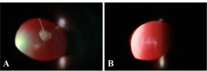
The Incomplete diffuse type PVD found primarily in patients with diabetic retinopathy and retinal detachment and complete diifuse type PVD found primarily in patients