좌측 유방암 방사선치료에서
CPAP(Continuous Positive Airway Pressure)의 유용성 평가
강남세브란스병원
정일훈·하진숙·장원석·전미진·김세준·정진욱·박별님·신동봉·이익재
목 적: 본 연구는 좌측 유방암 방사선치료에서 환자 호흡방법에 따른 영향에 대하여 치료계획을 통해 비교 분석하고, 깊은 들숨 호흡 정지법(deep inspiration breath hold, DIBH)에서 지속적 기도 양압(continuous positive airway, CPAP)의 유용성을 평가하고자 하였다.
대상 및 방법: 좌측 유방암 환자 총 10명을 대상으로 하였다. 모의치료에서 총 세 가지 호흡법을 적용하여 영상을 획득 하였다: 자유호흡법(free breathing, FB); 깊은 들숨 호흡 정지법(DIBH); 들숨 호흡 정지법(inspiration breath hold, IBH); 여기서 DIBH에서는 자가호흡확인 기구인 abches(APEX, Japan)와 IBH에서는 CPAP가 적용되었다. 치료계 획은 Ray Station(5.0.2.35, Sweden)을 이용하였고, 용적 강도변조 회전방사선치료법(volumetric modulated arc therapy, VMAT) one partial Arc을 환자당 동일 각도로 적용하였다. 치료계획용적(planning target volume, PTV) 의 총 처방선량은 50 Gy(2 Gy/fx)로 설정하였다. 치료계획분석은 PTV의 95 % dose(D95), 처방선량지수(conformity index, CI)와 선량균질지수(homogeneity index, HI)를 비교 분석하였다. 손상위험장기(Organ At Risk, OAR)인 폐, 심장, 왼내림 앞가지동맥(left anterior descending artery, LAD)을 분석하였다.
결 과: 동축의 폐(ipsilateral lung)의 평균용적 FB, DIBH(Abches), IBH(CPAP)에서 각각 1245.58±301.31 cm3, 1790.09±362.43 cm3, 1775.44±476.71 cm3였다. PTV의 평균 D95는 각각 46.67±1.89 Gy, 46.85±1.72 Gy, 46.97
±2.34 Gy였다. 평균 CI(HI)는 각각 0.95±0.02(0.91±0.01), 0.96±0.02(0.90±0.01), 0.95±0.02(0.92±0.02)였다. 폐 전 체의 V20(동측 폐)은 각각 10.74±4.50 %(20.45±8.65 %), 8.29±3.14 %(17.18±7.04 %), 9.12±3.29 %(18.85±7.85 %) 였다. 심장의 평균 선량(Dmean)은 각각 7.82±1.27 Gy, 6.10±1.27 Gy, 5.67±1.56 Gy였다. LAD의 최대선량(Dmax)은 각각 20.41±7.56 Gy, 14.88±3.57 Gy, 14.96±2.81 Gy였다. LAD에서 흉벽(Chest wall)까지 평균거리는 각각 11.33±
4.70 mm, 22.40±6.01 mm, 20.14±6.23 mm였다.
결 론: DIBH와 CPAP에서 FB보다 폐 용적이 커짐으로써, 방사선치료영역인 흉벽과 심장의 거리가 멀어지는 효과 때 문에 LAD의 선량을 감소시킬 수 있다. 좌측 유방암 방사선치료 시 DIBH방법의 대체방안으로 CPAP은 임상적 활용도 가 높을 것으로 사료된다.
▶ 핵심용어 : DIBH, CPAP, Left breast, Left Anterior Descending artery
서 론
우리나라 여성암 발병률 2위에 해당하는 암은 관상피 내암(ductal carcinoma in situ, DCIS) 이나 침윤성 암 (invasive cancer)이 대표적이다. 일반적으로 유방부분 절제(breast conserving surgery, BCS) 또는 전절제술 (modified radical mastectomy, MRM)을 시행 받은 경 우, 모든 환자에게 방사선치료가 시행된다.(1)
책임저자: 이익재, 강남세브란스병원 서울특별시 강남구 언주로 211 Tel: 02)2019-3148 E-mail: [email protected]
방사선치료는 국소제어(local control)와 생존을 향상시 키기 위하여 부작용을 고려해야 한다.(2-4) 좌측유방암 치료 에서 폐와 심장에 들어가는 방사선량으로 인하여 발생되 는 Lung toxicity와 Late cardiac toxicity가 중요하다.(8-11) 또한 오목가슴과 같은 흉벽 기형 환자나 유선내의 임파선 을 포함하여 방사선치료를 진행해야 하는 환자들은 심장 과 폐에 대한 방사선선량이 증가하는 위험성을 가지고 있
다.(5-7) 유방암 방사선치료에서 생기는 이러한 문제점을 보
완하기 위해 현재 다양한 방사선 치료방법이 개발되었다.
폐와 심장의 선량을 감소시키는 방법으로는 세기변조 방사선치료(intensity modulated radiation therapy, IMRT)와 환자의 호흡을 조절할 수 있는 abches를 사용하 는 방법이 있다. abches를 사용하여 호흡을 조절하는 원 리는 흉부를 지속적으로 부풀리게 하여 심장을 흉벽으로 부터 멀어지게 하는 것이다. 결국 호흡을 제어함으로써 흉 부 운동을 감소시켜 치료조사야 내에 포함되는 폐와 심장 의 방사선량을 감소시키는 것이다. 임상에서는 깊은 들숨 호흡 정지(deep inspiration breath hold, DIBH)기법이 치료에 적용되기 시작했다.(12,13)
DIBH는 깊은 숨을 들이마신 후 방사선치료 중에 숨을 참아야하기 때문에 환자의 어려움이 존재하고, 이로 인하 여 치료간 오차(intra-fraction error)에 대한 문제점이 발 생할 수 있다. 이러한 단점을 보완하기 위해 대체방안으로
본원에서는 지속적 기도 양압(continuous positive air- way, CPAP)를 이용한다. CPAP은 수면성 무호흡증(sleep apnea)환자에게 사용하는 장비이다. CPAP의 원리는 코와 연결된 마스크를 통해 연속적으로 낮은 압력으로 적당한 양의 공기압력(6-15 cmHg delay 15 min.)을 제공하여 기 도를 개방시켜주기에 폐가 확장된 상태에서 환자가 편안한 호흡을 유지하면서 치료를 진행할 수 있다(Fig. 1).
본 연구는 좌측 유방암 방사선치료에 환자 호흡방법에 따른 영향을 치료계획을 통해 비교 분석하고, DIBH 기법 에서 CPAP의 유용성을 평가하고자 하였다.
대상 및 방법
1. 실험재료
- Raystation(Elekta, 5.0.2.35, Sweden) - MIM software(6.6.14, United States) - VERSA HD(Elekta, Sweden)
- SOMATOM Definition AS(SIEMENS, Germany) - Dream station CPAP(PHILIPS, Netherlands) - abches(APEX, Japan)
- 10 cases of Left breast patients CT
2. 실험 방법
본원에서 시행한 좌측 유방암 환자 총 10명을 대상으로 하였다. 각 환자에 대한 치료계획은 Ray Station(5.0.2.35, Sweden)을 이용하였고, 용적 강도변조 회전방사선치료법 (volumetric modulated arc therapy, VMAT) one par- tial Arc을 환자당 동일 각도로 적용하였다. 치료계획 용적 (planning target volume, PTV) 처방선량은 50 Gy(2 Gy/
fx) 하였고, Raystation을 사용하여 치료계획을 수립하였 다. 모든 환자는 세 가지 호흡법이 적용되었다: 자유호흡법 (free breathing, FB); 깊은 들숨 호흡 정지법(DIBH); 들 숨 호흡 정지법(inspiration breath hold, IBH); 여기서 DIBH에서는 자가호흡확인 기구인 abches과 IBH에서는 CPAP가 적용되었다(Fig. 2).
세 가지 치료계획에서 PTV의 D95는 선량용적히스토그 램(dose volume histogram, DVH)을 이용하여 비교 분 Fig. 1. Principle of the CPAP device
석하였다. PTV에 대한 선량포함과 균질성을 평가하기 위 하여 처방선량지수(conformity index, CI), 선량균질지수 (homogeneity index, HI)를 구하였다.
•CI = BV95 %/PTV
(BV95 % = the body volume of the isodose of 95 % of the prescribed dose)
•HI = D5 %/D95 %
(D5 % = minimum dose to 5 % of the PTV, D95 %
= minimum dose to 95 % of the PTV)
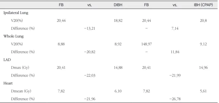
손상위험장기(Organ At Risk, OAR)인 폐, 심장, 왼내림 앞가지동맥(left anterior descending artery, LAD)을 선 택적으로 분석하였다. 호흡방법에 따른 폐의 변화는 동축 폐(ipsilateral lung)의 체적과 V20, 전체 폐(whole lung) 의 V20을 통해 확인하였다. 심장의 평균선량(Dmean)과 LAD의 최대선량(Dmax)을 구했다. 또한, LAD와 흉곽 (chest wall)까지 거리를 측정했다(Fig. 3).
(a) FB (b) DIBH (c) IBH(CPAP) Fig. 2. Comparison of isodose distribution in (a) FB, (b) DIBH and (c) IBH(CPAP)
결 과
FB, DIBH와 IBH(CPAP)의 치료계획용적(PTV) 평균 D95는 46.67±1.89 Gy, 46.85±1.72 Gy, 46.97±2.34 Gy이 고, CI(HI)는 각각 0.95±0.02(0.91±0.01), 0.96±0.02(0.90
±0.01), 0.95±0.02(0.92±0.02)였다.
동축의 폐(전체 폐)의 평균 FB, DIBH와 IBH(CPAP) 에서 각각 V20은 20.45±8.65 %(10.74±4.50 %), 17.18
±7.04 %(8.29±3.14 %), 18.85±7.85 %(9.12±3.29 %)였 다. 동축의 폐의 평균 부피는 각각 1245.58±301.31 cm3, 1790.09±362.43 cm3, 1775.44±476.71 cm3였다.
IBH(CPAP)에서 FB보다 크기가 43.11±27.05 % 증가했다.
심장의 평균 Dmean은 각각 7.82±1.27 Gy, 6.10±1.27 Gy, 5.67±1.56 Gy였다. LAD의 최대선량(Dmax)은 각각 20.41
±7.56 Gy, 14.88±3.57 Gy, 14.96±2.81 Gy였다(Table 1).
LAD에서 흉벽(chest wall)까지 평균거리는 각각 11.33±
4.70 mm, 22.40±6.01 mm, 20.14±6.23 mm였다(Table 2).
고안 및 결론
본 연구는 좌측 유방암 방사선치료에서 세 가지 호흡방 법(FB, DIBH(Abches), IBH(CPAP))에 따른 PTV와 OAR 인 폐, 심장, LAD의 방사선량을 비교분석하였다. PTV는 세 가지 호흡방법에 따른 치료계획 차이는 없었다.
LAD에 대한 Dmax는 DIBH와 IBH(CPAP)에서 FB보다 각각 22.03 %, 21.99 % 감소하였다. 이러한 결과는 폐 용적 이 커짐으로써 방사선 치료영역인 흉벽과 심장의 거리가 멀 어지는 효과 때문에 LAD의 선량을 감소시키는 현상으로 해석된다. 실제 본 연구에서 동축의 폐 용적은 IBH(CPAP) 에서 FB보다 43.11 % 증가함을 확인하였다. 또한, CPAP 를 이용한 IBH에서 폐 용적이 DIBH와 비슷하게 증가되었 고, 흉벽과 LAD 사이의 거리도 동일한 결과를 가졌다. 따 라서 좌측 유방 방사선치료에서 CPAP를 이용한 IBH는 LAD의 선량 감소과 폐 용적 증가로 이어질 수 있고, 이는 방사선치료에 의한 심장질환(heart disease)과 방사선폐렴 (radiation pneumonitis)과 같은 부작용을 줄일 수 있을
(a) FB (b) DIBH (c) IBH (CPAP) Fig. 3. Measurement distance of the heart to chest wall in (a) FB, (b) DIBH and (c) IBH(CPAP)
것으로 예상된다.
마지막으로 abches를 사용한 DIBH 치료에서는 의사소 통(communication)과 숨 참기(breath holding)가 치료 간 오차(intra-fraction error)를 발생할 수 있는 중요한 요소들이다. 이러한 단점을 보완하기 위해, 좌측 유방암 방 사선치료에 대한 IBH에서 CPAP는 지속적으로 기도를 개 방시켜주기에 폐가 확장된 상태에서 환자가 편안한 호흡을 유지하면서 치료를 진행할 수 있기에 임상적으로 활용도가 높을 것으로 사료된다.
참고문헌
1. 홍채선, 주상규, 박주영: Large Pendulous Breast 환자의 방사선 치료에 있어서 엎드린 자세의 유용성 평가. 대한방 사선치료기술학회지 2008; 37-43.
2. Kurtz J. The curative role of radiotherapy in thet- reatment of operable breast cancer. Eur J Cancer 2002;38:1961-74. (Breathing adapted radiotherapy of breast cancer: reduction of cardiac and pulmonary doses using voluntary inspiration breath-hold).
3. Hooning MJ, Botma A, Aleman BM, et al. Long-term Table 1. OARs(ipsilateral and whole lung, LAD and heart) dose depend on three different breathing techniques; FB, DIBH (Abches) and IBH(CPAP)
FB vs. DIBH FB vs. IBH (CPAP)
Ipsilateral Lung
V20(%) 20.44 18.82 20.44 20.8
Difference (%) -13.21 - 7.14
Whole Lung
V20(%) 8.88 8.92 148.97 9.12
Difference (%) -20.82 - 11.84
LAD
Dmax (Gy) 20.41 14.88 20.41 14.96
Difference (%) -22.03 -21.99
Heart
Dmean (Gy) 7.82 6.10 7.82 5.61
Difference (%) -21.96 -26.78
Table 2. Change of ipsilateral lung volume(cm3), and distance (mm) of the heart to chest wall depend on three different breathing techniques; FB, DIBH (Abches) and IBH(CPAP)
FB vs. DIBH FB vs. IBH (CPAP)
Ipsilateral Lung
Volume (cm3) 1245.58 1790.09 1245.58 1775.44
Difference (%) +46.24 +43.11
LAD
Distance (mm) 11.33 22.40 11.33 20.14
Difference (%) +59.67 +51.96
risk of cardiovascular disease in 10-year survivors of breast cancer. J Natl Cancer Inst 2007;99:365- 75. (The deep inspThe deep inspiration breath hold technique using Abches reduces cardiac dose in pa- tients undergoing left-sided breast irradiation).
4. Offersen B, Hojris I, Overgaard M. Radiation-in- duced heart morbidity after adjuvant radiotherapy of early breast cancer: Is it still an issue Radiother Oncol 2011;100:157-9. (The deep inspThe deep in- spiration breath hold technique using Abches re- duces cardiac dose in patients undergoing left-sided breast irradiation).
5. McGale P, Taylor C, Correa C, et al. Effect of ra- diotherapy after mastectomy and axillary surgery on 10-year recurrence and 20-year breast cancer mortality: meta-analysis of individual patient data for 8135 women in 22 randomized trials. Lancet.
2014;21:2127–2135.
6. Darby SC, Ewertz M, Hall P. Ischemic heart dis- ease after breast cancer radiotherapy. N Engl J Med.
2013;368:2527.
7. Taylor C, Correa C, Duane F, et al. Estimated risks of breast cancer radiotherapy: evidence from modern radiation doses to the lung and heart and from pre- vious randomized trials. J Clin Oncol. 2017;35:1641–
1649.
8. Early Breast Cancer Trialists’ Collaborative Group (EBCTCG). Effects of radiotherapy and of differenc- es in the extent of surgery for early breast cancer on local recurrence and 15-year survival: an overview of the randomised trials. Lancet 2005; 366: 2087- 06. (Deep inspiration breath hold technique reduces heart dose from radiotherapy for left-sided breast cancer, Cardiac dose estimates from Danish and Swedish breast cancer radiotherapy during 1977- 2001).
9. Van de SJ, Soete G, Storme G. Adjuvant radio- therapy for breast cancer significantly improves overall survival: the missing link. Radiother Oncol 2000;55:263-72. (Breathing adapted radiotherapy for breast cancer: Comparison of free breathing gating with the breath-hold technique).
10. Correa CR, Litt HI, Hwang WT, Ferrari VA, Solin LJ, Harris EE. Coronary artery findings after left- sidedcompared with right-sided radiation treat- ment for early-stage breast cancer. J Clin Oncol 2007;25:3031-7. (The deep inspThe deep inspiration breath hold technique using Abches reduces cardiac dose in patients undergoing left-sided breast irra- diation).
11. Darby SC, Ewertz M, McGale P, et al. Risk of isch- emic heart disease in women after radiotherapy for breast cancer. N Engl J Med 2013;368:987-98. (The deep inspiration breath hold technique using Abch- es reduces cardiac dose in patients undergoing left- sided breast irradiation).
12. Pedersen AN, Korreman S, Nystrom H, Specht L.
Breathing adapted radiotherapy of breast cancer:
reduction of cardiac and pulmonary doses using voluntary inspiration breath-hold. Radiother Oncol 2004;72:53-0.(Estimation of heart-position variabil- ity in 3D-surface-image-guided deep-inspiration breathhold radiation therapy for left-sided breast cancer).
13. Whoon Jong Kil, Tabitha Pham & Kyubo Kim (2019) Heart sparing breast cancer radiotherapy us- ing continuous positive airway pressure (CPAP) and conventional supine tangential fields: an alternative method for patients with limited accessibility to ad- vanced radiotherapy techniques, Acta Oncologica, 58:1, 105-109.
The efficacy of continuous positive airway pressure (CPAP) for patient with left breast cancer
Department of Radiation Oncology, Gangnam Severance Hospital
Jung Il Hun, Ha Jin Sook, Chang Won Suk, Jeon Mi Jin, Kim Sei Joon, Jung Jin Wook, Park Byul Nim, Shin Dong Bong, Lee Ik Jae
Purpose: This study examined changes in the position of the heat and lungs depending on the patient’s breath- ing method during left breast cancer radiotherapy and used treatment plans to compare the resulting radiation dose.
Materials and methods: The participants consisted of 10 patients with left breast cancer. A CT simulator(SIMENS SOMATOM AS, Germany) was used to obtain images when using three different breathing methods: free breathing(FB), deep inspiration breath hold(DIBH with Abches, DIBH), inspiration breath hold(IBH with CPAP, CPAP). A Ray Station(5.0.2.35, Sweden) was used for treatment planning, the treatment method was volumetric modulated arc therapy (VMAT) with one partial arc of the same angle, and the prescribed dose to the planning target volume (PTV) was a total dose of 50 Gy(2 Gy/day). In treatment plan analysis, the 95 % dose (D95) to the PTV, the conformity index(CI), and the homogeneity index (HI) were compared. The lungs, heart, and left anterior descending artery (LAD) were selected as the organs at risk(OARs).
Results: The mean volume of the ipsilateral lung for FB, DIBH, and CPAP was 1245.58±301.31cm3, 1790.09±362.43 cm3, 1775.44±476.71 cm3. The mean D95 for the PTV was 46.67±1.89 Gy, 46.85±1.72 Gy, 46.97±23.4 Gy, and the mean CI and HI were 0.95±0.02, 0.96±0.02, 0.95±0.02 and 0.91±0.01, 0.90±0.01, 0.92±0.02.
The V20 of Whole Lung was 10.74±4.50 %, 8.29±3.14 %, 9.12±3.29 % and The V20 of the ipsilateral lung was 20.45±8.65 %, 17.18±7.04 %, 18.85±7.85 %, the Dmean of the heart was 7.82±1.27 Gy, 6.10±1.27 Gy, 5.67±1.56 Gy, and the Dmax of the LAD was 20.41±7.56 Gy, 14.88±3.57 Gy, 14.96±2.81 Gy. The distance from the thoracic wall to the LAD was measured to be 11.33±4.70 mm, 22.40±6.01 mm, 20.14±6.23 mm.
Conclusion: During left breast cancer radiotherapy, the lung volume was 46.24 % larger for DIBH than for FB, and 43.11 % larger for CPAP than FB. The larger lung volume increases the distance between the thoracic wall and the heart. In this way, the LAD, which is one of the nearby OARs, can be more effectively protected while still satisfying the treatment plan. The lung volume was largest for DIBH, and the distance between the LAD and thoracic wall was also the greatest. However, when performing treatment with DIBH, the intra-fraction error cannot be ignored. Moreover, communication between the patient and the radiotherapist is also an important factor in DIBH treatment. When communication is problematic, or if the patient has difficulty holding their breath, we believe that CPAP could be used as an alternative to DIBH. In order to verify the clinical efficacy of CPAP, it will be necessary to perform long-term follow-up of a greater number of patients.