R E S E A R C H Open Access
Association of maxillary dental developmental abnormality with
precocious puberty: a case-control study
Yesel Kim 1 , Nam-Ki Lee 2 , Jae Hyun Kim 3 , Jeong-Kui Ku 4* , Bu-Kyu Lee 5 , Hoi-In Jung 1 and Sun-Kyu Choi 6
Abstract
Background: Dental studies of precocious puberty have focused on examination of jaw and dentition growth. The aim of the study was to analyze the relationship between precocious puberty and maxillary dental developmental abnormalities (DDAs).
Methods: This retrospective study was conducted on the Korean patients in whom dental panoramic and hand-wrist radiographs had been taken before they were 15 years of age. The maxillary DDAs were assessed as mesiodens, congenital missing teeth, peg-shape lateral incisors, or impacted teeth. The chronological ages of the control group members were within the normal range of the hand-wrist bone age. Others with a peak luteinizing hormone of ≥ 5 and < 5 IU/L were allocated to central precocious puberty (CPP) and peripheral precocious puberty (PPP), respectively.
Results: Of the enrolled 270 patients, 195, 52, and 23 were allocated to the control, CPP, and PPP groups, respectively.
The maxillary DDAs were significantly more prevalent in the CPP group than in the other groups. Among those with maxillary DDA, the mesiodens predominated. Age- and sex-adjusted multivariate analysis revealed maxillary DDA (odds ratio, 3.36; 95% CI, 1.60-7.05) and especially mesiodens (odds ratio, 5.52; CI, 2.29-13.28) to be significantly associated with CPP.
Conclusions: Maxillary DDAs were significantly more prevalent in the CPP group than in the PPP or control groups.
Among the many types of maxillary DDAs, mesiodens was significantly associated with CPP and may be considered a predictor of the development of CPP.
Keywords: Dental developmental abnormality, Gonadotropin-releasing hormone, Mesiodens, Precocious puberty, Supernumerary tooth
Background
Precocious puberty (PP) has recently become a topic of social focus. PP can be identified by signs of pubertal de- velopment in girls aged < 8 and in boys aged < 9 [1].
When the bone age —as determined using hand-wrist radiography by the Tanner-White or Greulich-Pyle atlas method [2] —is advanced compared to the chronological age, PP can be diagnosed differentially as central
precocious puberty (CPP) or peripheral precocious pu- berty (PPP) using a gonadotropin-releasing hormone stimulation test (GnRHST) [3 – 6].
Treatment of PPP is focused on the originated diseases such as congenital adrenal hyperplasia, McCune- Albright syndrome, severe hypothyroidism, disorders of the adrenal gland, tumors of the ovary or testis, and rare genetic syndromes [4]. On the other hand, the goal of treatment for CPP patients can be considered to match pubertal development with their peers to reduce the psy- chosocial problems and minimize the loss of growth po- tential. In patients with CPP, delayed treatment may result in growth loss and socio-psychological problems,
© The Author(s). 2020 Open Access This article is licensed under a Creative Commons Attribution 4.0 International License, which permits use, sharing, adaptation, distribution and reproduction in any medium or format, as long as you give appropriate credit to the original author(s) and the source, provide a link to the Creative Commons licence, and indicate if changes were made. The images or other third party material in this article are included in the article's Creative Commons licence, unless indicated otherwise in a credit line to the material. If material is not included in the article's Creative Commons licence and your intended use is not permitted by statutory regulation or exceeds the permitted use, you will need to obtain permission directly from the copyright holder. To view a copy of this licence, visit http://creativecommons.org/licenses/by/4.0/.
* Correspondence: [email protected]
4
Department of Oral and Maxillofacial Surgery, Section of Dentistry, Armed Forces Medical Command, Armed Forces Capital Dental Hospital, Seongnam-si 13634, Korea
Full list of author information is available at the end of the article
such as emotional distress and problem behavior, be- cause hormonally caused behavioral changes (e.g., ag- gression) may break out earlier in patients with CPP [7, 8]. Therefore, many studies have been conducted to identify the predictive factors of CPP which has been at- tributed to a dysfunction of the hypothalamic-pituitary- gonadal axis. As a result, endocrine-disrupting BMI [6], chemicals [7], central nervous (CNS) problems, or head trauma [8] have been suggested to be predictors of the future development of CPP.
At the chronologic age of six to seven, before puberty begins, mixed dentition starts as the deciduous teeth which are replaced with permanent teeth. Since the tim- ing of PP diagnosis is an important dental turning point, many researches have been conducted on the relation- ship between PP and dental development such as tooth eruption, tooth growth, and jaw growth [9–12]. How- ever, the relationship between PP and the dental param- eter is controversial because the above parameters vary, even in individuals without PP.
Dental developmental abnormalities (DDAs) are evi- denced by an abnormal tooth shape or number such as peg-shaped maxillary lateral incisors (peg-lateralis), con- genital missing tooth, impacted maxillary permanent teeth, germinated tooth, fused tooth, twinned tooth, taurodontism, or supernumerary teeth. The DDAs are more common in the maxillae than mandibles [13, 14].
Of these DDAs, supernumerary teeth are usually en- countered in the anterior maxillae and are called mesio- dens [14]. No association between mesiodens and other DDAs has been reported whereas peg-lateralis, congeni- tal missing lateral incisors, and impacted canines are in- terrelated [15, 16]. Before the identification of pubertal development, most DDAs can be diagnosed easily using dental radiographs. And the DDAs are recommended to treat approximately before maxillary permanent incisor eruption (5 to 6 years of age) [17].
Both maxillary teeth and the anterior pituitary gland, the latter of which secretes follicle-stimulating hormone (FSH), luteinizing hormone (LH), and growth hormone (GH), are embryologically derived from the oral epithe- lium. Therefore, maxillary DDA may be embryologically associated with PP. In addition, maxillary DDA may be a valuable predictor of a diagnosis of PP because they can be identified before the onset of pubertal development.
The aim of this study was to identify the relationship be- tween maxillary DDA and PP.
Materials and methods
This case-control study was conducted on patients in whom dental panoramic and hand-wrist radiographs had been taken between March 2008 and May 2018, at the department of pediatrics or dentistry in Seoul National University Bundang Hospital. The inclusion criteria were
as follows: (1) age between 3 and 15 years when both dental panoramic and hand-wrist radiographs were taken and a hand-wrist evaluation for bone age using the Greulich-Pyle atlas method [2]. (2) The presence of GnRHST results in a patient with advanced bone age compared to the chronological age. The exclusion cri- teria were as follows: history of orthodontic treatment, maxillofacial surgery, and the presence of dentofacial- related deformity or syndrome.
The range of bone age was determined as by expert radiol- ogists with the Greulich-Pyle atlas method. The control group consisted of patients in whom their chronological age within the range of bone age. Among other patients with an earlier bone age than the chronological age and breast bud- ding or testicular enlargement, they were classified into the experimental groups (CPP and PPP groups) by GnRHST (described below). Gonadotropin-releasing hormone (GnRH, 100 μg; Relefact; Sanofi-Aventis, Frankfurt, Germany) was injected intravenously after obtaining baseline serum sam- ples. Luteinizing hormone (LH) was measured by blood sam- ples which were collected 30, 45, and 60 min after GnRH administration. The experimental group was divided into a CPP group with a peak LH concentration of ≥ 5 IU/L and a PPP group with a peak LH concentration of < 5 IU/L [4].
Because the patients independently visited the depart- ment of dentistry and pediatrics, the pediatric evaluation age was defined as chronological age at the time when the hand-wrist radiograph was first examined, and the dental evaluation age was separately defined at the time when the first dental panoramic radiograph was taken.
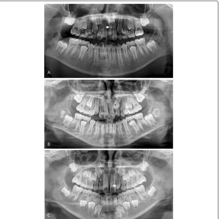
Statistical analysis was based on the dental evaluation age. In the dental panoramic radiographs, the maxillary DDAs were classified into mesiodens and the others, in- cluding impacted maxillary permanent teeth, congenital missing teeth, and peg lateralis, by an expert oral and maxillofacial surgeon (Fig. 1).
Statistical methods
The control, CPP, and PPP groups were determined using a chi-square or Fisher’s exact test for the categor- ical variables or ANOVA for the continuous variables.
Post hoc analysis was performed by Bonferroni correc-
tion. Statistical significance among the groups was evalu-
ated according to the subtypes of maxillary DDAs (total
maxillary DDAs, mesiodens, and other maxillary DDAs
except for mesiodens). One to one propensity score
matching to adjust for age and sex ratio was applied to
the dataset of the control and CPP group. The signifi-
cance of maxillary DDAs in predicting the development
of the PP response was compared using both univariate
and multivariate logistic regression analysis to adjust for
age and sex ratio. The univariate and multivariate odds
ratio with their 95% confidence intervals were calculated
for the subtypes of maxillary DDAs. Two-sided p values
of < 0.05 were considered significant. The analysis was performed using SAS version 9.4 (SAS Institute, Cary, NC) and R 3.5. 1 (Vienna, Austria; http://www.R-project.org/).
Ethics statement
The study was reviewed and approved by the Institu- tional Review Board at Seoul National University Bun- dang Hospital (No. B-1904/535-106). It was granted an exemption of the informed consent due to the retro- spective nature of this study.
Results
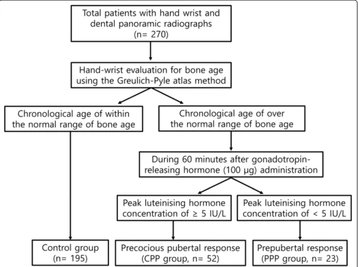
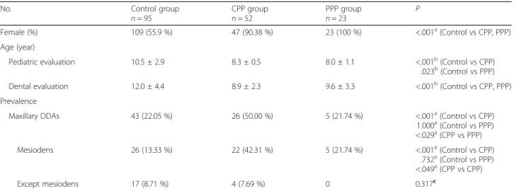
Two hundred and seventy patients (12.3 ± 4.4 years) were enrolled in this study; 195, 52, and 23 were allocated to the control, CPP, and PPP groups, respectively (Fig. 2). The pediatric evalua- tions were performed earlier on average than the dental evaluations in all groups. CPP and PPP groups revealed significant intergroup differences in sex and age compared with control group (Table 1).
Fig. 1 Maxillary dental developmental abnormality. a Mesiodens (arrow). b Impacted maxillary canine (asterisk). c Congenital missing of the lateral
incisor (asterisk) and peg lateralis (arrow)
The prevalence of maxillary DDA was significantly higher in the CPP group (50.00%) than in the control (22.05%) and PPP (21.74%) groups (P < 0.05). Mesiodens was more prevalent in the CPP group (42.31%) than in the control (13.33%) and PPP (21.74%) groups (P <
0.05). But no intergroup difference was observed be- tween the prevalence of the other maxillary DDAs, ex- cept for the mesiodens, in the PPP (0.00%), CPP (7.69%), and control (8.71%) groups (Table 1).
One-to-one propensity score matching was used to ad- just for age and sex ratio, which differed in the control and CPP groups. The prevalence of maxillary DDAs were significantly different in the CPP (50.00%) and con- trol (19.23%) groups (P < 0.001). The prevalence of mesiodens was significantly higher in the CPP group (42.31%) than in the control group (9.53%) (P < 0.001).
On the other hand, the prevalence of the other maxillary DDAs, except for mesiodens, was similar in the control (11.54%) and CPP (7.69%) groups (Table 2).
Univariate analysis and age- and sex-adjusted multi- variate analysis revealed maxillary DDAs (odds ratio,
3.85; 95% confidence interval, 1.92-6.92 and 3.36; 1.60–
7.05, respectively), especially mesiodens (odds ratio, 4.98;
95% confidence interval, 2.46–10.06 and 5.52; 2.29–
13.28, respectively), to be associated with the precocious puberty response (Table 3).
Discussion
In this study, the relationship between maxillary DDAs and PP was firstly analyzed retrospectively. In general, it is important that children with CPP be identified from normal and PPP early because delayed diagnosis and the treatment of CPP leads to a loss of growth potential and psycho-social problems [1, 18]. Many studies have been conducted to identify the screening or predictive factors of CPP. The relationship remains controversial between the CPP and dental factors such as dental maturity [10, 19], dental age [11], malocclusion [12], and mandibular growth pattern [10]. At time of PP onset, many children with mixed-dentition visited dental clinics to have their dental development evaluated. For this dental examin- ation, the patients are radiographed to examine the
Fig. 2 Flow diagram of patient classification
eruption of permanent teeth or identify the DDAs in- cluding supernumerary, impacted, and missing teeth.
DDAs can be diagnosed clearly and efficiently using ra- diographs by the abnormality of shape or number of tooth [17]. The supernumerary tooth—mesiodens—occurs at the time of maxillary permanent tooth germ formation [20]. The enamel portion of the maxillary permanent anter- ior teeth is formed between 3 to 4 years of age, and the age of eruption of these teeth is commonly between 6 to 7 years of age. Therefore, DDA may be a predictive factor in the early diagnosis of CPP because DDA can be identified before the onset of signs of pubertal development.
Embryologically, the pituitary gland is an important struc- ture for the migration of neural crest cells involved in oral formation [21]. The anterior pituitary gland has the same origin as oral neural crest cells. And the posterior pituitary gland has the same mesenchymal origin as the maxillofacial region [22]. The sella turcica forms a bony seat for the pituit- ary gland. Therefore, a sella turcica deformity has been re- ported to be associated with tooth developmental disorders
including mesiodens [23–26]. In addition, an abnormality of the sella turcica has been proven to be related to tooth eruption by an analysis of the eruption timing and eruption disorders of the maxillary teeth according to the nerve distri- bution [22]. On the other hand, the effect of hormones re- mains controversial on tooth maturity or jaw growth. Some studies have reported a relationship between the hormones secreted by the pituitary gland and the development of denti- tion or jaw growth. Kjellberg et al. reported that patients with a GH deficiency exhibited delayed tooth eruption [27], and Cantu et al. reported that GH-deficiency patients showed a delayed bone age but no delay in dental age on the radio- graphs [28]. Others have reported that sexual maturity is not related to dental maturity [29], and a GH treatment has little effect on tooth development [30, 31].
GnRHST is a test method commonly used to diagnose CPP [5]. The test is used to evaluate the activity of the hypothalamic-pituitary axis by measuring the amount of releasing concentration of LH and FSH. GnRHST is Table 1 Demographic characteristics of the subjects and prevalence of maxillary dental developmental abnormalities in the three study groups
No. Control group
n = 95 CPP group
n = 52 PPP group
n = 23 P
Female (%) 109 (55.9 %) 47 (90.38 %) 23 (100 %) <.001
a(Control vs CPP, PPP)
Age (year)
Pediatric evaluation 10.5 ± 2.9 8.3 ± 0.5 8.0 ± 1.1 <.001
b(Control vs CPP)
.023
b(Control vs PPP)
Dental evaluation 12.0 ± 4.4 8.9 ± 2.3 9.6 ± 3.3 <.001
b(Control vs CPP, PPP)
Prevalence
Maxillary DDAs 43 (22.05 %) 26 (50.00 %) 5 (21.74 %) <.001
a(Control vs CPP)
1.000
a(Control vs PPP)
<.029
a(CPP vs PPP)
Mesiodens 26 (13.33 %) 22 (42.31 %) 5 (21.74 %) <.001
a(Control vs CPP)
.732
a(Control vs PPP)
<.049
a(CPP vs CPP)
Except mesiodens 17 (8.71 %) 4 (7.69 %) 0 0.317
cAbbreviations: CPP central precocious puberty, DDAs dental developmental abnormalities, PPP peripheral precocious puberty
a
Post hoc analysis after chi-square test
b
Post hoc analysis after ANOVA
c
Fisher exact test compared with control group
Table 2 Prevalence of maxillary dental developmental abnormalities in the control group and precocious pubertal response group after 1:1 propensity score matching ( n = 52)
No. (%) P
Control group CPP group
Maxillary DDAs 10 (19.23 %) 26 (50.00 %) 0.001
aMesiodens 4 (7.69 %) 22 (45.31 %) < 0.001
aExcept mesiodens 6 (11.54 %) 4 (15.38 %) 1
bAbbreviations: CPP central precocious puberty, DDAs dental
developmental abnormalities
a
Chi-square test
b
Fisher exact test
Table 3 Predictors of the precocious pubertal response, including the univariate and adjusted odd ratio and 95%
confidence limits according to maxillary dental developmental abnormalities
Control group vs CPP group
Odd ratio (95 % CI)
Unadjusted
aAdjusted
bMaxillary DDAs 3.85 (1.92-6.92) 3.36 (1.60-7.05)
Mesiodens 4.98 (2.46-10.06) 5.52 (2.29-13.28)
Except mesiodens 1.39 (0.43-4.44)
Abbreviations: CPP central precocious puberty, CI confidence interval, DDAs dental developmental abnormalities
a
Univariate logistic regression
b
Adjustment of age and sex
considered invasive given the patients’ ages because the test requires two to three consecutive blood tests sepa- rated by intervals of 15–30 min [5]. For this reason, a screening examination is performed by evaluating the bone age using a hand-wrist radiograph [19]. In the present study, the control group included patients whose bone age (as determined using the Greulich-Pyle atlas method) did not exceed the chronological age. Among the patients with an advanced bone age than chrono- logical age, the CPP and PPP groups were classified based on the peak LH above 5 lU/L [4].
The CPP group had a significantly higher prevalence of maxillary DDAs than the control or PPP groups. Stat- istical analysis was performed between the CPP and con- trol groups with an adjustment for sex and age because the proportion of girls was higher in the CPP group at 90.38%, but no sex difference was observed in the con- trol group at 55.90%. The prevalence of maxillary DDAs was higher in the CPP group; regression analysis showed that the odds of maxillary DDAs in this group were 3.36 times higher than in the control group (confidence inter- val; CI, 1.60-7.05). In addition, mesiodens was remark- ably prevalent in the CPP group, and the odds ratio of mesiodens was 4.98 times higher (CI, 2.46-10.06) in the CPP group. After sex and age-adjusted regression ana- lysis, the odds ratio increased to 5.52 times higher (CI, 2.29–13.28) for mesiodens in the CPP group than the control group (Table 3). As a consequence, mesiodens could be considered a strong predictor of the develop- ment of CPP. As the CNS problem becomes a well- known risk factor for PP, CPP boys were frequently found among the CNS problem patients [8]. Similarly, mesiodens could be used to predict CPP in boys who may not be diagnosed. Furthermore, CPP might be de- termined at the gestation period because mesiodens is developed around at the 16th week of gestation [20].
Early diagnosis and treatment of DDA patients are also important for increasing spontaneous tooth eruption and reducing the need for additional orthodontic or sur- gical intervention as well as the development of psycho- social problems [17]. In this study, however, the average dental evaluation age of the patients was slightly later than those of the pediatric evaluation age. The authors indicate that they might miss the opportunity for the early diagnosis of maxillary DDAs because no associa- tions between the DDAs and PP have been established.
Therefore, patients diagnosed with CPP should be re- ferred for a dental examination. Furthermore, it should be noted that maxillary DDA patients were at high risk of developing CPP. Thus, they need to be referred for a pediatric examination.
On the other hand, DDA has a prevalence of 5-19% in mixed-dentition juveniles. In DDA, the prevalence of supernumerary teeth is generally approximately 3% [17].
Compared to previous studies, the results of this study showed a high prevalence of DDA in all three study groups, which can be attributed to the retrospective sin- gle institutional study design. Because not every patient with mesiodens was simultaneously tested for bone age and precocious puberty, it could be possible that the mesiodens patients were not evenly distributed among the groups. In addition, the risk of PP in boys with max- illary DDA could not be evaluated because of the limited number of samples. This study had some limitations, such as small sample size, heterogeneous sex distribu- tion, and retrospective cohort study. Further large-scale or prospective multicenter studies of the association be- tween CPP and maxillary DDAs, particularly mesiodens, will be needed.
Conclusions
Maxillary DDA was associated in the CPP group com- pared with the PPP or control groups. In particular, mesiodens was associated significantly with the PP re- sponse, and could be considered a predictor of CPP de- velopment. A patient diagnosed with CPP needs to be referred for a dental examination, and those identified with mesiodens should be referred for a pediatric exam- ination of CPP.
Abbreviations
CPP: Central precocious puberty; CI: Confidence interval; DDAs: Dental developmental abnormalities; PP: Precocious puberty; PPP: Peripheral precocious puberty
Acknowledgements Not applicable
Authors ’ contributions
Ku JK conceptualized the study and analyzed the data. Kim Y investigated the data and wrote the initial manuscript. Choi SK statistically analyzed the data. Jung HI, Lee NK, and Kim JH co-conceptualized the study. All authors revised and approved the final manuscript.
Funding
No financial or non-financial benefits have been received or will be received from any party related directly or indirectly to the subject of this article.
Availability of data and materials
The datasets used during the current study are available from the corresponding author on reasonable request.
Ethics approval and consent to participate
The study was reviewed and approved by the Institutional Review Board at Seoul National University Bundang Hospital (No. B-1904/535-106) with waiver of the informed consent.
Consent for publication Not applicable
Competing interests Not applicable
Author details
1
Department of Preventive Dentistry and Public Oral Health, Yonsei
University, Seoul, Korea.
2Department of Orthodontics, Section of Dentistry,
Seoul National University Bundang Hospital, Seongnam-si, Korea.
3