HIGHLIGHTS
• Robot-assisted gait training (RAGT) has recently attracted attention.
• There are few studies investigating use of RAGT for the patients with ataxia.
• Patients with ataxia might benefit from Morning Walk®-assisted gait training in walking ability and balance.
Original Article
Received: Oct 27, 2020 Revised: Nov 1, 2020 Accepted: Nov 2, 2020 Correspondence to Dae Yul Kim
Department of Rehabilitation Medicine, Asan Medical Center, University of Ulsan College of Medicine, 88 Olympic-ro 43-gil, Songpa-gu, Seoul 05505, Korea.
E-mail: [email protected]
Chul Jung, Dae Yul Kim, Sara Kwon, Min Ho Chun, JaYoung Kim, Sung Hyun Kim
Morning Walk ® -Assisted Gait Training
Improves Walking Ability and Balance
in Patients with Ataxia: a Randomized
Controlled Trial
ABSTRACT
This study aimed to investigate walking ability and balance improvement of patients with ataxia caused by brain lesions after end-effector type robot (Morning Walk®)-assisted gait training. This study randomly assigned 19 patients to one of two groups: 30 minutes of Morning Walk® training with 1 hour of conventional physiotherapy (Morning Walk® group;
n = 10) or 1.5 hours of conventional physiotherapy (Control group; n = 9). Five treatment sessions per week were given for 3 weeks. The primary outcomes were walking ability and balance, which were assessed by the functional ambulation category (FAC) and Berg Balance Scale (BBS), respectively. The secondary outcomes included 10-meter Walk Test (10mWT), Rivermead Mobility Index (RMI), Motricity Index (MI), and Modified Barthel Index (MBI).
At baseline, there was no statistically significant difference between the two groups except MBI. After the treatment, the Morning Walk® group showed significant improvement in the FAC, BBS, 10mWT, RMI and MBI. The control group showed significant improvement in the BBS, 10mWT, RMI and MBI. Inter-group comparison demonstrated that the ∆FAC, ∆10mWT and ∆RMI of the Morning Walk® group were significantly higher than those of the control group. Our results suggest that the patients with ataxia receiving Morning Walk®-assisted gait training might improve greater in walking ability and balance than those trained with conventional physiotherapy.
Keywords: Ataxia; Robotics; Gait; Postural Balance; Neurologic Rehabilitation
INTRODUCTION
Ataxia is a disorder of muscle activity coordination during voluntary movement, thereby also affecting gait function [1,2]. It could indicate dysfunction of the nervous system that coordinates movement, such as cerebellum or brain stem [1,3]. Several diseases can cause ataxia; hemorrhagic or ischemic stroke is one of the leading causes [1,4].
Gait ataxia is characterized by unsteadiness, variable foot placement, widened step or stance, and abnormal joint torque, and its severity is correlated with the degree of impaired balance [3,5-7]. The balance deficit is not only associated with impaired gait function but also with increased difficulty in performing activities of daily living (ADL) when the deficit is combined
Original Article
Received: Oct 27, 2020 Revised: Nov 1, 2020 Accepted: Nov 2, 2020 Correspondence to Dae Yul Kim
Department of Rehabilitation Medicine, Asan Medical Center, University of Ulsan College of Medicine, 88 Olympic-ro 43-gil, Songpa-gu, Seoul 05505, Korea.
E-mail: [email protected] Copyright © 2020. Korean Society for Neurorehabilitation
This is an Open Access article distributed under the terms of the Creative Commons Attribution Non-Commercial License (https://
creativecommons.org/licenses/by-nc/4.0) which permits unrestricted non-commercial use, distribution, and reproduction in any medium, provided the original work is properly cited.
ORCID iDs Chul Jung
https://orcid.org/0000-0001-5770-9107 Sara Kwon
https://orcid.org/0000-0002-7088-3393 Min Ho Chun
https://orcid.org/0000-0001-8666-7225 JaYoung Kim
https://orcid.org/0000-0002-5010-8090 Sung Hyun Kim
https://orcid.org/0000-0002-1678-3498 Funding
This study is the result of a research project of Future Growth Engine Flagship Project funded by the Minister of Science, ICT and Future Planning and supported by the Korean Institute of Science & Technology Evaluation and Planning (KISTEP).
Chul Jung , Dae Yul Kim, Sara Kwon , Min Ho Chun , JaYoung Kim , Sung Hyun Kim
Department of Rehabilitation Medicine, Asan Medical Center, University of Ulsan College of Medicine, Seoul, Korea
Morning Walk ® -Assisted Gait Training
Improves Walking Ability and Balance
in Patients with Ataxia: a Randomized
Controlled Trial
Conflict of Interest
The authors have no potential conflicts of interest to disclose.
with decreased joint mobility, muscle tone problems, and loss of proprioception [8].
Therefore, the balance training for patients with ataxia is a crucial part of rehabilitation. For these patients, conventional therapy methods, such as the Bobath approach, proprioceptive neuromuscular facilitation, therapist-assisted walking, and Frenkel exercise have been commonly used treatment methods [9-11].
Recently, robot-assisted gait training (RAGT) has attracted attention. Its effectiveness in restoring walking ability of the patients with neurologic diseases including stroke, traumatic brain injury, and spinal cord injury has been reported [12-15]. Compared with conventional training, RAGT has several advantages including increasing reproducible symmetrical gait patterns and reducing the energy expenditure of therapists [16]. Previous studies using end-effector type RAGT for patients with subacute stroke have demonstrated that RAGT combined with conventional physiotherapy could significantly improve walking speed, functional gait, balance and motor power [17-19].
Although there have been several studies proving the effectiveness of RAGT on balance and walking ability, few studies have investigated the use of RAGT for the patients with ataxia.
Among the studies, very limited reports have addressed the effect of end-effector type RAGT on ataxic patients. The aim of this study was to investigate the improvement in the walking ability and balance of the patients with ataxia caused by brain lesions after 3 weeks of end- effector type (Morning Walk® [Hyundai Heavy Industry, Seoul, Korea]) RAGT.
MATERIALS AND METHODS
Participants
We screened the patients admitted to the department of rehabilitation medicine at one tertiary hospital from December 2016 to June 2017. Patients were eligible for the study if the following inclusion criteria were met: 1) age > 18 years, 2) gait disturbance mainly caused by ataxia of the limb or trunk due to brain lesions, 3) FAC score ≥ 2, 4) ability to participate in gait training using the Morning Walk®, and 5) previously an independent walker. Exclusion criteria were as follows: 1) severe communication disorders due to cognitive impairment or aphasia, 2) severe musculoskeletal diseases of lower extremity, 3) psychological instability, 4) body weight > 135 kg or height > 195 cm, and 5) other physical status that could limit physical therapy (severe deformity or contracture of limbs; fractures, open wounds, or pressure ulcers;
severe osteoporosis). The final population size was targeted at 40 patients.
Study design and intervention
This study was a non-blinded, prospective, randomized controlled trial that compared the effects of RAGT with Morning Walk® with those of conventional physiotherapy in the patients with ataxia. The trial was registered at the Clinical Research Information Service (CRIS), Republic of Korea (KCT0003452). The study protocol was approved by the Institutional Review Board (IRB No. 2016-1335) of our hospital. Before the start of the study, all participants were informed of the study purpose, intervention, and possible incidents and they filled out the informed consent form. The patients were assigned to either the Morning Walk® or control group using a random number table. The both group patients received five training treatment sessions per week for 3 weeks (a total of 15 sessions).
The Morning Walk® group received RAGT with Morning Walk® for 30 minutes and conventional physiotherapy for 1 hour per session. The control group received conventional physiotherapy for 1.5 hours per session (additional 30 minutes of conventional physiotherapy instead of 30 minutes of Morning Walk® training). Conventional physiotherapy consisted of 30 minutes of therapist-assisted gait and balance training based on neurodevelopmental treatment and 30 minutes of strengthening exercise using NuStep® (NuStep LLC, Ann Arbor, MI, USA) [20]. In the strengthening exercise using NuStep®, the level of resistance (step 1 to 10) was set according to each patient's initial strength, and then progressed. The strengthening exercise was performed one more time in the control group for additional 30 minutes of conventional physiotherapy.
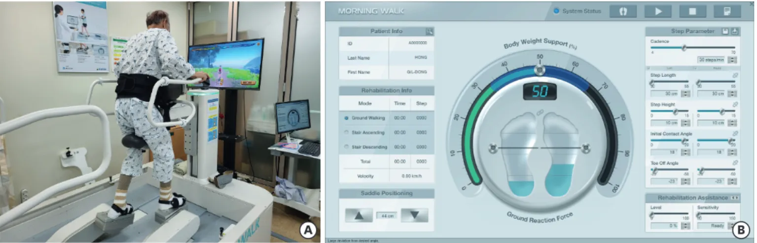
Morning Walk® is a new end-effector type robot developed by Hyundai Heavy Industries and Taeha Mechatronics for lower limb rehabilitation in the patients with gait disturbance [17]. The device was approved in December 2014 by the Food and Drug Administration. It has a saddle supporting the patient's weight and enables the joint movement of the lower limb, including the ankle, knee, and pelvis, according to footplate trajectory. Morning Walk® training started with ground level gait training at a speed of 30–35 steps/min with a step length of 30–35 cm. These training progressed by adjusting the various parameters according to the patient's performance. Additionally, the patients progressed to up and down stair gaits.
Morning Walk® has several differences from one of well-known end effector-type robots, Gait Trainer® (Reha-Stim, Berlin, Germany). Morning Walk® can provide various walking experience for patients, such as ground walking, stair ascending and descending. As well as, a therapist can adjust varied parameters of ground walking; cadence, step height, step length, toe-off angle, and initial contact angle according to the specific status of the patients.
Particularly, a therapist can control the patient's motion of ankle precisely. However, the Gait Trainer® can only change the step length and velocity.
Morning Walk® also provides the amount of “body weight support” of the saddle during real time walking and the graphical information on the monitor of the “ground reaction force”
on each foot. It gives a visual biofeedback to the patients for forming good center f pressure pattern. In addition, its monitor-based virtual reality software shows patient's avatar on a parkway and gait guidance. If the patient steps on the ground with a left or right foot, the light of a stepping-side foot icon turns on (Fig. 1).
A B
Fig. 1. (A) Morning Walk®-assisted gait training with monitor-based virtual reality software. (B) Interface of Morning Walk® for adjusting gait parameter and providing graphical information.
Outcome measures
All participants were examined by a trained evaluator at two-time points: baseline (pre- treatment) and after intervention (post-treatment). The primary outcome measures of walking ability and balance were assessed using the FAC and Berg Balance Scale (BBS). The FAC is a commonly used clinical gait assessment scale with consistent reliability and validity. It distinguishes 6 levels (from 0 to 5) of walking ability based on the required amount of physical support [21,22]. The BBS is used to evaluate the balance and risk of fall. The BBS focuses on static and dynamic balance, which includes 14 tasks with a maximum score of 56 [23,24].
The secondary outcome measures included the 10-meter Walk Test (10mWT), Rivermead Mobility Index (RMI), Motricity Index (MI), and modified Barthel Index (MBI). The timed 10 mWT is a frequently used measure of walking speed (m/s) in the patients with neurologic diseases [25,26]. Patients unable to undergo the 10mWT were considered to have a walking speed of 0 m/s [17,27]. The RMI is a scale consisting of 15 items addressing several aspects of mobility with sum scores ranging from 0 to 15 [28]. The MI is a reliable instrument for measuring the strength and a feasible measure that can determine the overall impairment of patients. The MI of the lower limb comprises hip flexion, knee extension, and ankle dorsiflexion. Six grades of Medical Research Council (MRC) were converted into modified weighted scores; thus, three scores of each movement were summed and added by one. The total score ranged from 0 (complete paresis) to 100 (normal strength) [29]. If there is difference between MI of each side, we used lower value for the MI of each patient. The MBI is one of the common measures of ADL and consists of 10 items: feeding, dressing, personal hygiene, bathing, toilet transfer, bladder control, bowel control, chair/bed transfers, ambulation, and stair climbing. Scores of the MBI ranged from 0 to 100, with higher scores indicating greater independence in ADL. To minimize effects of upper limb function, we additionally analyzed summated scores of items associated with lower limb function in MBI (MBI-Lower extremity, MBI-LE). MBI-LE included transfer, ambulation, and stair climbing categories.
Statistical analysis
Statistical analysis was performed using SPSS version 18 (SPSS Inc., Chicago, IL, USA). Mann- Whitney U test and Fisher's exact test were used to compare the baseline characteristics and initial (pre-treatment) functional states of the two groups. Wilcoxon signed-rank test was performed to identify within-group changes of pre- and post-treatment. Mann-Whitney U test was performed to compare changes of pre- and post-treatment in the Morning Walk® group with those of control group. A p value of less than 0.05 was considered statistically significant in all tests.
RESULTS
Study population
Among the patients admitted to the department of rehabilitation medicine in our hospital from December 2016 to June 2017, 23 patients met the inclusion and exclusion criteria, and they were eligible for the study. Of these, 2 patients refused to be enrolled, and 2 patients were unable to participate in the study because of deconditioning. As a result, 19 patients were assigned to either the Morning Walk® group or control group. During the follow-up period, each 2 patients withdrew from the study; this occurred due to unexpectedly early discharge and transfer to other hospital nearby home. As a result, 8 patients in Morning Walk® group, and 7 patients in control group completed the study. Although total of 15
patients did not meet the targeted size of population, low rate of enrollment limited the study to proceed further. During the intervention, there were no safety issues or adverse events related to Morning Walk® training or conventional physiotherapy (Fig. 2).
Baseline characteristics
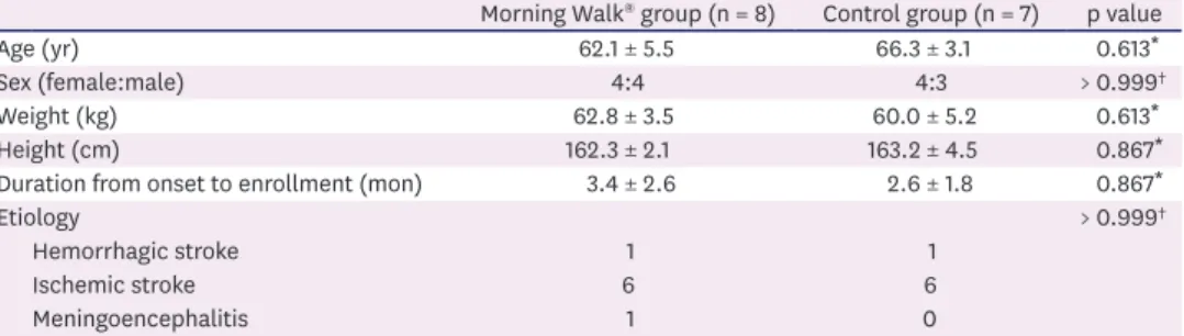
There was no significant difference in the baseline characteristics of the two groups (Table 1).
The Morning Walk® group included the patients with 1 hemorrhagic stroke, 6 ischemic stroke, and 1 meningoencephalitis. Similarly, the control group included the patients with 1 hemorrhagic stroke and 6 ischemic stroke.
The characteristics of each participant were shown in Table 2. According to duration from onset to study enrollment, most patients were in acute stage; each group included one patient with ataxia caused by chronic brain lesions. In addition, all the included patients presented ataxia without definite weakness. To be specific, baseline MI of the patients were assessed as 70 or more, which meant that MRC grades of the lower extremities were generally over than grade of 4. Two patients in Morning Walk® group, and 4 patients in control group exhibited generalized ataxia involving bilateral limbs and trunk. Total of 9 patients developed ataxia caused by cerebellar lesions.
Discontinued intervention (n = 2) - Early discharge
Discontinued intervention (n = 2) - Early discharge
Assessed for eligibility (n = 23)
Morning Walk® group (n = 10) Allocation Control group (n = 9) (0 weeks)
Intervention (3 weeks)
Final assessment
A total of 19 patients were included
Analysed (n = 8) Analysed (n = 7)
4 patients were excluded:
- Medical deconditioning (n = 2) - Declined to participate (n = 2)
Fig. 2. Flow chart of the screening, allocation and follow-ups of the study.
Table 1. Comparison of characteristics between the Morning Walk® and control groups
Morning Walk® group (n = 8) Control group (n = 7) p value
Age (yr) 62.1 ± 5.5 66.3 ± 3.1 0.613*
Sex (female:male) 4:4 4:3 > 0.999†
Weight (kg) 62.8 ± 3.5 60.0 ± 5.2 0.613*
Height (cm) 162.3 ± 2.1 163.2 ± 4.5 0.867*
Duration from onset to enrollment (mon) 3.4 ± 2.6 2.6 ± 1.8 0.867*
Etiology > 0.999†
Hemorrhagic stroke 1 1
Ischemic stroke 6 6
Meningoencephalitis 1 0
Values are expressed as the mean ± SE or numbers.
*Analysis by Mann-Whitney test; †Analysis by Fisher's exact test.
Pre-treatment and post-treatment status
Before the training sessions (pre-treatment), there was no statistically significant difference between the two groups except for the MBI (Table 3). After total of 15 training sessions, both groups showed significant improvement in several outcome measures; the Morning Walk® group showed statistically significant improvement in the FAC, BBS, 10mWT, RMI, MBI, and MBI-LE, whereas the control group showed significant improvement in the BBS, 10mWT, RMI, MBI, and MBI-LE. In control group, three patients and one patient could not perform the 10mWT at the pre- and post-treatment, respectively; therefore, their walking speeds were scored as 0 m/s. Inter-group comparison of changes in the outcome measures demonstrated that the ∆FAC, ∆10mWT, and ∆RMI of the Morning Walk® group were higher than those of the control group with statistical significance (p = 0.029, 0.021 and 0.040, respectively).
Although the improvement in the ∆BBS, ∆MBI, and ∆MBI-LE was greater in the Morning Walk® group (∆BBS, 13.1 ± 3.70; ∆MBI, 20.4 ± 6.92; ∆MBI-LE, 9.8 ± 3.09) than in the control group (∆BBS, 5.9 ± 1.77; ∆MBI, 16.0 ± 5.06; ∆MBI-LE, 8.1 ± 2.36), this difference was not statistically significant (p = 0.152, 0.779, and 0.955, respectively). On the other hand, the Table 2. Baseline characteristics of each participant
No. Sex Age
(yr) Diagnosis Duration*
(mon) Weakness (MI) Ataxia Brain lesions
Rt. Lt. Rt. U/L limbs Trunk Lt. U/L limbs Morning Walk® group
1 M 51 ME 2 100 76 N Y N Diffuse cortex
2 F 44 Hemorrhagic 22 76 76 Y Y Y SAH, IVH
3 F 43 Ischemic 1 92 76 Y N N Rt. cbll
4 F 78 Ischemic 0 76 76 Y Y Y Both cbll
5 M 65 Ischemic 0 100 100 Y N N Lt. midbrain
6 M 58 Ischemic 0 100 76 N N Y Rt. BG-CR
7 F 74 Ischemic 1 76 76 N N Y Both cbll
8 M 84 Ischemic 1 76 76 N Y N Rt. cbll
Control group
1 M 66 Ischemic 1 76 76 Y Y Y Both cbll, Lt. pons, Rt.
midbrain
2 F 62 Ischemic 0 100 76 N Y Y Lt. cbll, pons
3 M 71 Hemorrhagic 13 76 76 Y Y Y Both pons, midbrain
4 M 55 Ischemic 2 76 76 Y Y Y Both cbll
5 F 60 Ischemic 0 76 100 N Y Y Both pons
6 F 78 Ischemic 1 70 70 Y Y Y Both cbll
7 F 74 Ischemic 1 100 100 N N Y Lt. cbll, pons
MI, Motricity Index (Lower extremity); U/L, upper/lower; ME, meningoencephalitis; SAH, subarachnoid hemorrhage; IVH, intraventricular hemorrhage; cbll, cerebellum; BG-CR, basal ganglia-corona radiata.
*Duration was counted as the number of months in the period from onset to enrollment.
Table 3. Outcome measurements of the Morning Walk® and control groups (pre-, post-treatment, and changes [∆Post-pre])
Morning Walk® group (n = 8) Control group (n = 7) p value†
Pre-treatment Post-treatment* ΔPost-pre Pre-treatment Post-treatment* ΔPost-pre
FAC 2.9 ± 0.30 4.4 ± 0.42‡ 1.5 ± 0.33 2.3 ± 0.29 2.7 ± 0.42 0.4 ± 0.20 0.029‡
BBS 30.4 ± 6.19 43.5 ± 6.31‡ 13.1 ± 3.70 20.3 ± 5.00 26.1 ± 6.41‡ 5.9 ± 1.77 0.152
10mWT 0.8 ± 0.83 1.5 ± 0.97‡ 0.7 ± 0.33 0.3 ± 0.27 0.5 ± 0.45‡ 0.2 ± 0.24 0.021‡
RMI 6.8 ± 1.00 10.3 ± 1.10‡ 3.5 ± 0.50 4.4 ± 0.92 6.4 ± 1.19‡ 2.0 ± 0.38 0.040‡
MI 79.0 ± 3.00 87.0 ± 4.25 8.0 ± 4.00 78.6 ± 3.67 88.6 ± 4.57 10.0 ± 4.72 0.867
MBI§ 67.8 ± 8.67 88.1 ± 6.56‡ 20.4 ± 6.92 43.1 ± 8.01 59.1 ± 8.75‡ 16.0 ± 5.06 0.779
MBI-LE 22.8 ± 4.30 32.5 ± 4.03‡ 9.8 ± 3.09 12.0 ± 3.24 20.1 ± 4.33‡ 8.1 ± 2.36 0.955
Values are expressed as the mean ± SE.
FAC, functional ambulatory category; BBS, Berg Balance Scale; 10mWT, 10-meter Walk Test; RMI, Rivermead Mobility Index; MI, Motricity Index (lower extremity);
MBI, modified Barthel Index; MBI-LE, modified Barthel Index-lower extremity.
*Treatment effects in each group were evaluated by Wilcoxon signed-rank test. Statistically significant pre- and post-treatment changes were indicated in the post-treatment column; †∆Post-Pre values between the two groups were analyzed by Mann-Whitney test; ‡p < 0.05; §At baseline (pre-treatment), there were no statistically significant differences in the variables other than the MBI. Inter-group baseline comparison was performed by Mann-Whitney test.
improvement in the ∆MI was slightly greater in the control group (10.0 ± 4.72) than in the Morning Walk® group (8.0 ± 4.00) without statistical significance (p = 0.867).
Considering that the FAC, one of primary outcomes is the categorical variable, the number of participants in each FAC category was also identified for both groups. As for the Morning Walk® group, 3, 3, and 2 patients were firstly classified into FAC of 2, 3, and 4, respectively. After the training sessions (post-treatment), walking ability in 7 of 8 Morning Walk® patients improved, and then 1, 1, and 6 patients were classified into FAC of 2, 3, and 5, respectively. As for the control group at pre-treatment, 6 and 1 patients were classified into FAC of 2 and 4, each. At post- treatment, 2 patients with FAC 2 and 1 patient with FAC 4 progressed to FAC of 3 and 5, each.
DISCUSSION
Several studies have investigated the effects of RAGT for stroke patients with hemiparesis;
however, only a few studies have focused on the patients with ataxia instead of weakness.
To our knowledge, this is the first study to report the effects of Morning Walk®-assisted gait training for the patients with ataxia. In our study, inter-group comparisons demonstrated significantly greater changes in the FAC, RMI, and 10mWT in the Morning Walk® group, suggesting that Morning Walk®-assisted gait training combined with conventional
physiotherapy might have positive effects on the walking ability and balance of ataxic patients.
As a primary outcome, the FAC score represented walking ability of the patients. The walking ability improved in both the Morning Walk® and control groups after the training. The Morning Walk® group showed statistically significant improvement in the FAC score and the change was significantly greater than that of the control group. The FAC score was analyzed with Mann-Whitney and Wilcoxon signed-rank test with reference to the previous report [17].
The FAC score was categorical, but ordinal scale; therefore, it was possible to use the tests.
The recovery of balance control is known to be crucial for regaining walking ability. It is a major obstacle especially for the patients with ataxia [30-33]. The static and dynamic balance assessed by the BBS showed improvement in both the Morning Walk® and control groups after intervention. Although inter-group comparisons did not show any statistically significant difference, greater change in the BBS score was observed in the Morning Walk® group. The recovery of balance control might have contributed to improvement of walking ability in the Morning Walk® group.
The 10mWT and RMI, as secondary outcomes significantly increased in both groups. The Morning Walk® group showed greater change in the 10mWT and RMI than the control group with statistical significance. Three of 7 patients in the control group could not complete the 10mWT, and then their scores were considered as zero. This might have limited the clinical value of the 10mWT; therefore, further studies would be needed to reexamine the parameter.
The lower limb muscle strength is associated with maximum walking speed of patients [34].
The improvement in the MI was observed in both the Morning Walk® and control groups;
however, it was not statistically significant. This result is somewhat different from that in our previous study [17]. The previous study which investigated the effects of Morning Walk®-assisted gait training in the subacute hemiparetic stroke patients showed greater improvement in the MI of the affected side in the Morning Walk® group. This conflicting
result might be driven from the fact that, in our study patients, motor weakness was not the main cause of gait disturbance, and the severity of motor weakness was relatively mild.
The MBI and MBI-LE significantly increased in both groups. Inter-group comparison of changes in the MBI and MBI-LE showed that ∆MBI and ∆MBI-LE were higher in the Morning Walk® group than those in the control group without statistical significance. These results were not much different from those from other parameters; however, considering statistically significant difference of pre-treatment MBI, further studies composed of patients with well- balanced MBI will be needed to validate these results.
A few previous studies reported conflicting results of RAGT in the patients with ataxia.
Hamada et al. [35] reported the use of hybrid assistive limb robot suit improved gait function in 3 patients with ataxia. The rehabilitation program with hybrid assistive limb robot stabilized the center of gravity and shortened the 10mWT. Belas et al. [3] compared the effects of RAGT using the exoskeleton device Lokomat® 5.0 (Hacoma, Zurich, Switzerland) with those of therapist-assisted gait training (TAGT) for chronic stroke patients with ataxia. Each group received 3 rehabilitation sessions per week for 5 months; 2 sessions were conventional physiotherapy, and 1 session was gait training (RAGT or TAGT). Both groups showed statistically significant improvement in balance, functional independence, and general ataxia symptoms; however, inter-group comparisons did not show statistically significant difference.
Based on the principle of body-weight-supported treadmill training, RAGT can be advantageous over conventional physiotherapy as it offers practice in gait with higher intensity and numerous repetitions, thereby achieving functional motor relearning [36-38]. The intensive repetition induces the circuits involved in locomotor movement to be activated, allowing the nervous system to establish and restore links between the motor centers and sensory pathways [39].
This might facilitate the recovery of gait function in the Morning Walk® group.
However, there was no difference of improvement in gait and functional parameters of the patients with ataxia between RAGT using Lokomat® 5.0 and TAGT group [3]. These contrary results might be attributed to various factors; types of robot (end-effector versus exoskeleton), study protocol and onset of disease. End-effector type robot works by applying mechanical forces to the distal segment of limbs. However, exoskeleton-type robotic devices have robot axes aligned with the anatomical axes of the patients. Morning Walk®, as an end-effector type robot has been suggested to reduce the effects of the therapists by allowing relatively easy getting on and off the patient, or by providing partial body weight support through the saddle use [17]. This would lead to an increase in the treatment intensity, compared to RAGT using other exoskeleton robots. In addition, the average duration from the disease onset to enrollment was different between two studies (10.5 and 4.8 years for the Lokomat® 5.0 RAGT and TAGT group, respectively; 3.4 and 2.6 months for the Morning Walk® and control group, respectively). The recovery of motor impairment mostly occurs within 6 months after the stroke event and the rate of recovery slows down over time within the 6-month period [40]. Difference in duration of the disease may lead to different effects of treatment; intense rehabilitation therapy in this early period (i.e., within 6 months) could be more effective also in patients with ataxia.
This study had several limitations. First, the study included only small sample of patients.
The small population limited us to use nonparametric tests with poor statistical power.
At the pre-treatment, the Morning Walk® group were assessed as roughly better than the control group; however, the difference was not statistically significant. This could have been attributed to the small population. Second, both the patients and evaluators were not blinded. The patients knew the group they were assigned to, and the evaluators also knew the assigned group of the patients. Although the evaluators tried to assess the patients objectively, we could not exclude the possibility of bias. Third, this study did not utilize the specific assessment tool for the severity of ataxia. If we utilized the specific tool, such as scale for the assessment and rating of ataxia (SARA), it would have been clear that each patient's gait disturbance was due to ataxia. Instead,-we used the FAC and the BBS for addressing the severity of ataxia. Fourth, the patients in the control group received 30 min of strengthening exercise in place of Morning Walk® assisted gait training; therefore, additional gait training as well as effects of Morning Walk® might have contributed to greater improvement in the Morning Walk® group. Fifth, the duration from stroke onset to study enrollment was slightly heterogeneous. One patient from each group were in chronic phase. This might have acted as a confounding factor. Lastly, as this study assessed outcomes only at baseline and at the end of 3-week treatment, it was unclear whether the therapeutic effects were temporary or sustained. Further study of larger sample size with blinded evaluators and longer follow-up periods would be needed to validate the results of this study.
In conclusion, we compared the effects of end-effector type robot, Morning Walk® assisted gait training plus conventional physiotherapy with those of conventional physiotherapy alone on the gait and balance function in the patients with ataxia. Our results suggest that Morning Walk®-assisted gait training combined with conventional physiotherapy could contribute to further improvement in the walking ability and balance of the patients with ataxia caused by brain lesions.
REFERENCES
1. Brunberg JA; Expert Panel on Neurologic Imaging. Ataxia. AJNR Am J Neuroradiol 2008;29:1420-1422.
PUBMED
2. Winstein CJ, Stein J, Arena R, Bates B, Cherney LR, Cramer SC, Deruyter F, Eng JJ, Fisher B, Harvey RL, Lang CE, MacKay-Lyons M, Ottenbacher KJ, Pugh S, Reeves MJ, Richards LG, Stiers W, Zorowitz RD;
American Heart Association Stroke Council, Council on Cardiovascular and Stroke Nursing, Council on Clinical Cardiology, and Council on Quality of Care and Outcomes Research. Guidelines for adult stroke rehabilitation and recovery: a guideline for healthcare professionals from the American Heart Association/American Stroke Association. Stroke 2016;47:e98-e169.
PUBMED | CROSSREF
3. Belas Dos Santos M, Barros de Oliveira C, Dos Santos A, Garabello Pires C, Dylewski V, Arida RM.
A comparative study of conventional physiotherapy versus robot-assisted gait training associated to physiotherapy in individuals with ataxia after stroke. Behav Neurol 2018;2018:2892065.
PUBMED | CROSSREF
4. Terry JB, Rosenberg RN. Frontal lobe ataxia. Surg Neurol 1995;44:583-588.
PUBMED | CROSSREF
5. Hallett M, Massaquoi SG. Physiologic studies of dysmetria in patients with cerebellar deficits. Can J Neurol Sci 1993;20 Suppl 3:S83-S92.
PUBMED
6. Ilg W, Golla H, Thier P, Giese MA. Specific influences of cerebellar dysfunctions on gait. Brain 2007;130:786-798.
PUBMED
7. Morton SM, Bastian AJ. Relative contributions of balance and voluntary leg-coordination deficits to cerebellar gait ataxia. J Neurophysiol 2003;89:1844-1856.
PUBMED
8. Sawacha Z, Carraro E, Contessa P, Guiotto A, Masiero S, Cobelli C. Relationship between clinical and instrumental balance assessments in chronic post-stroke hemiparesis subjects. J Neuroeng Rehabil 2013;10:95.
PUBMED
9. Garson JG. The Frenkel system of exercises for tabes. BMJ 1911;2:420-421.
PUBMED | CROSSREF
10. Hesse S. Treadmill training with partial body weight support after stroke: a review. NeuroRehabilitation 2008;23:55-65.
PUBMED | CROSSREF
11. Zesiewicz TA, Wilmot G, Kuo SH, Perlman S, Greenstein PE, Ying SH, Ashizawa T, Subramony SH, Schmahmann JD, Figueroa KP, Mizusawa H, Schöls L, Shaw JD, Dubinsky RM, Armstrong MJ, Gronseth GS, Sullivan KL. Comprehensive systematic review summary: treatment of cerebellar motor dysfunction and ataxia: Report of the Guideline Development, Dissemination, and Implementation Subcommittee of the American Academy of Neurology. Neurology 2018;90:464-471.
PUBMED | CROSSREF
12. Calabrò RS, Cacciola A, Bertè F, Manuli A, Leo A, Bramanti A, Naro A, Milardi D, Bramanti P. Robotic gait rehabilitation and substitution devices in neurological disorders: where are we now? Neurol Sci 2016;37:503-514.
PUBMED | CROSSREF
13. Chang WH, Kim YH. Robot-assisted therapy in stroke rehabilitation. J Stroke 2013;15:174-181.
PUBMED | CROSSREF
14. Hwang S, Kim HR, Han ZA, Lee BS, Kim S, Shin H, Moon JG, Yang SP, Lim MH, Cho DY, Kim H, Lee HJ. Improved gait speed after robot-assisted gait training in patients with motor incomplete spinal cord injury: a preliminary study. Ann Rehabil Med 2017;41:34-41.
PUBMED | CROSSREF
15. Sung J, Choi S, Kim H, Lee G, Han C, Ji Y, Shin D, Hwang S, Yun D, Jang H, Kim MJ. Feasibility of rehabilitation training with a newly developed, portable, gait assistive robot for balance function in hemiplegic patients. Ann Rehabil Med 2017;41:178-187.
PUBMED | CROSSREF
16. Swinnen E, Beckwée D, Meeusen R, Baeyens JP, Kerckhofs E. Does robot-assisted gait rehabilitation improve balance in stroke patients? A systematic review. Top Stroke Rehabil 2014;21:87-100.
PUBMED | CROSSREF
17. Kim J, Kim DY, Chun MH, Kim SW, Jeon HR, Hwang CH, Choi JK, Bae S. Effects of robot-(Morning Walk®) assisted gait training for patients after stroke: a randomized controlled trial. Clin Rehabil 2019;33:516-523.
PUBMED | CROSSREF
18. Peurala SH, Airaksinen O, Huuskonen P, Jäkälä P, Juhakoski M, Sandell K, Tarkka IM, Sivenius J. Effects of intensive therapy using gait trainer or floor walking exercises early after stroke. J Rehabil Med 2009;41:166-173.
PUBMED | CROSSREF
19. Tong RK, Ng MF, Li LS. Effectiveness of gait training using an electromechanical gait trainer, with and without functional electric stimulation, in subacute stroke: a randomized controlled trial. Arch Phys Med Rehabil 2006;87:1298-1304.
PUBMED | CROSSREF
20. Hafsteinsdóttir TB, Algra A, Kappelle LJ, Grypdonck MH; Dutch NDT Study Group. Neurodevelopmental treatment after stroke: a comparative study. J Neurol Neurosurg Psychiatry 2005;76:788-792.
PUBMED | CROSSREF
21. Holden MK, Gill KM, Magliozzi MR. Gait assessment for neurologically impaired patients. Standards for outcome assessment. Phys Ther 1986;66:1530-1539.
PUBMED | CROSSREF
22. Mehrholz J, Wagner K, Rutte K, Meissner D, Pohl M. Predictive validity and responsiveness of the functional ambulation category in hemiparetic patients after stroke. Arch Phys Med Rehabil 2007;88:1314- 1319.
PUBMED | CROSSREF
23. Dundar U, Toktas H, Solak O, Ulasli AM, Eroglu S. A comparative study of conventional physiotherapy versus robotic training combined with physiotherapy in patients with stroke. Top Stroke Rehabil 2014;21:453-461.
PUBMED | CROSSREF
24. Miyamoto ST, Lombardi Junior I, Berg KO, Ramos LR, Natour J. Brazilian version of the Berg balance scale. Braz J Med Biol Res 2004;37:1411-1421.
PUBMED | CROSSREF
25. Graham JE, Ostir GV, Fisher SR, Ottenbacher KJ. Assessing walking speed in clinical research: a systematic review. J Eval Clin Pract 2008;14:552-562.
PUBMED | CROSSREF
26. Hirsch MA, Williams K, Norton HJ, Hammond F. Reliability of the timed 10-metre walk test during inpatient rehabilitation in ambulatory adults with traumatic brain injury. Brain Inj 2014;28:1115-1120.
PUBMED | CROSSREF
27. Schwartz I, Sajin A, Fisher I, Neeb M, Shochina M, Katz-Leurer M, Meiner Z. The effectiveness of locomotor therapy using robotic-assisted gait training in subacute stroke patients: a randomized controlled trial. PM R 2009;1:516-523.
PUBMED | CROSSREF
28. Roorda LD, Green JR, Houwink A, Bagley PJ, Smith J, Molenaar IW, Geurts AC. The Rivermead Mobility Index allows valid comparisons between subgroups of patients undergoing rehabilitation after stroke who differ with respect to age, sex, or side of lesion. Arch Phys Med Rehabil 2012;93:1086-1090.
PUBMED | CROSSREF
29. Fayazi M, Dehkordi SN, Dadgoo M, Salehi M. Test-retest reliability of Motricity Index strength assessments for lower extremity in post stroke hemiparesis. Med J Islam Repub Iran 2012;26:27-30.
PUBMED
30. de Sousa Britto HM, de Andrade Mendes L, de Carvalho Moreno C. de Souza e Silva EMG, Lindquist ARR.
Correlation between balance, speed, and walking ability in individuals with chronic hemiparesis. Fisioter Mov 2016;29:87-94.
CROSSREF
31. de Haart M, Geurts AC, Huidekoper SC, Fasotti L, van Limbeek J. Recovery of standing balance in postacute stroke patients: a rehabilitation cohort study. Arch Phys Med Rehabil 2004;85:886-895.
PUBMED | CROSSREF
32. Garland SJ, Willems DA, Ivanova TD, Miller KJ. Recovery of standing balance and functional mobility after stroke. Arch Phys Med Rehabil 2003;84:1753-1759.
PUBMED | CROSSREF
33. Kollen B, van de Port I, Lindeman E, Twisk J, Kwakkel G. Predicting improvement in gait after stroke: a longitudinal prospective study. Stroke 2005;36:2676-2680.
PUBMED | CROSSREF
34. Suzuki K, Nakamura R, Yamada Y, Handa T. Determinants of maximum walking speed in hemiparetic stroke patients. Tohoku J Exp Med 1990;162:337-344.
PUBMED | CROSSREF
35. Hamada O, Samura K, Abe H, Fukuda H, Ogata T, Nonaka M, et al. Three cases with ataxic gait disorder improved by using a hybrid assistive limb robot suit. Jpn J Neurosurg 2015;24:413-418.
36. Hesse S, Konrad M, Uhlenbrock D. Treadmill walking with partial body weight support versus floor walking in hemiparetic subjects. Arch Phys Med Rehabil 1999;80:421-427.
PUBMED | CROSSREF
37. Mao YR, Lo WL, Lin Q, Li L, Xiao X, Raghavan P, Huang DF. The effect of body weight support treadmill training on gait recovery, proximal lower limb motor pattern, and balance in patients with subacute stroke. BioMed Res Int 2015;2015:175719.
PUBMED | CROSSREF
38. McCain KJ, Pollo FE, Baum BS, Coleman SC, Baker S, Smith PS. Locomotor treadmill training with partial body-weight support before overground gait in adults with acute stroke: a pilot study. Arch Phys Med Rehabil 2008;89:684-691.
PUBMED | CROSSREF
39. Wallard L, Dietrich G, Kerlirzin Y, Bredin J. Effects of robotic gait rehabilitation on biomechanical parameters in the chronic hemiplegic patients. Neurophysiol Clin 2015;45:215-219.
PUBMED | CROSSREF
40. Lee KB, Lim SH, Kim KH, Kim KJ, Kim YR, Chang WN, Yeom JW, Kim YD, Hwang BY. Six-month functional recovery of stroke patients: a multi-time-point study. Int J Rehabil Res 2015;38:173-180.
PUBMED | CROSSREF
![Table 3. Outcome measurements of the Morning Walk ® and control groups (pre-, post-treatment, and changes [∆Post-pre])](https://thumb-ap.123doks.com/thumbv2/123dokinfo/5408832.220791/7.892.62.829.153.460/table-outcome-measurements-morning-control-groups-treatment-changes.webp)
