Vol. 16, No. 12 pp. 8836-8843, 2015
미성숙 감귤 과피 초임계 추출물의 성분 분석과 자궁암세포 성장억제효능
문정용1, 송연우2, 현호봉2, 김소미1,2*
1제주대학교 아열대원예산업연구소, 2제주대학교 생명공학부
Chemical Composition and Antiproliferative Activity of Supercritical Extract of Immature Citrus Peel in
human cervical carcinoma HeLa cells
Jeong Yong Moon
1, YeonWoo Song
2, Ho Bong Hyun
2, Somi Kim Cho
1,2*1Subtropical Horticulture Research Institute, Jeju National University
2Faculty of Biotechnology, College of Applied Life Sciences, Jeju National University
요 약 본 연구의 목적은 제주도 재래감귤인 팔삭과 이예감 초임계 추출물에 대한 인간 자궁암 세포인 HeLa 세포에서의 성장억제 효능을 탐색하고, 각 추출물에서의 유효 활성 성분을 분석하는데 있다. HeLa 세포에서의 성장억제 및 세포사멸 효능을 탐색하기 위해 MTT assay와 Hoechst 33342 염색을 수행하였으며, 성분분석은 가스크로마토그래피 질량분석기 (GC/MS)를 이용하였다. 두 종류의 미성숙 감귤 과육, 과피 초임계 추출물에 대한 HeLa 세포의 성장 억제 효능을 비교해 본 결과, 과피 추출물은 팔삭과 이예감 모두 비슷한 세포사멸 효능을 나타냈다. 과피 추출물을 처리한 세포에서는 농도 의존 적으로 성장률이 억제되었으며, 처리 농도 400 μg/mL에서 팔삭과 이예감 추출물이 각각 87.16%와 92.95%의 세포사멸 효능 을 나타냈으나, 과육 추출물의 세포 성장 억제 효능은 처리 농도 400 μg/mL까지 관찰되지 않았다. Hoechst 33342 염색을 통해 apoptotic body 형성을 현미경으로 관찰한 결과, 100, 200 μg/mL 과육 초임계 추출물을 처리한 세포에서는 apoptpotic body를 관찰할 수 없었으나, 동일한 농도의 과피 추출물을 처리한 세포의 경우에서는 apoptotic body가 현저하게 증가하는 것을 확인 할 수 있었다. GC/MS 분석을 통해 미성숙 팔삭 과육과 과피 초임계 추출물에서 각각 27개와 31개의 화합물을 검출하였으며, 미성숙 이예감 과육과 과피 초임계 추출물에서는 각각 27개와 29개의 화합물을 검출하였다. 팔삭 과피 초임계 추출물에는 1,1,4,4-Tetramethyl- 2-tetralone(20.86%), alloimperatorin(8.15%), limonene (11.23%), auraptene(7.29%) 등이 주로 함유되어 있었으며, 이예감 과피 초임계 추출물에는 limonene(22.19%), linalool(11.23%), γ-sitosterol(9.12%) 등이 주로 함유되 어 있었다.
Abstract This study was performed to investigate the antiproliferative activities of supercritical extracts from phalsak(Citrus hassaku Hort ex Tanaka) and yeagam(Citrus iyo Hort. ex Tanaka) against human cervical carcinoma HeLa cells and the chemical compositions of the extracts. The anticancer properties of supercritical extracts were demonstrated using the MTT assay and Hoechst 33342 staining and the compositional analyses were conducted by using gas chromatography-mass spectrometry(GC-MS). The peel extracts of both species exhibited similar antiproliferative effect. The antiproliferative activity of the flesh extracts was not detected up to 400 μg/mL, whereas peel extracts of phalsak and yeagam reduced cell viability with 87.16% and 92.95% at 400 μg/mL, respectively. There was a dramatic increase of the apoptotic body formation in the cell treated with peel extracts while no apoptotic body formation detected in the cell treated with flesh extracts at 100, 200 μg/mL. By GC-MS analysis, 27 and 31 kinds of compounds identified in flesh and peel of phalsak, while 27 and 29 kinds of compounds were identified in flesh and peel of yeagam, respectively. 1,1,4,4-Tetramethyl- 2-tetralone(20.86%), alloimperatorin(8.15%), limonene(11.23%), and auraptene(7.29%) were major in peel of phalsak, whereas limonene(22.19%), linalool(11.23%), and γ -sitosterol(9.12%) were major in peel of yeagam. som
Keywords : Antiproliferative activity, Apoptosis, HeLa cell, Phalsak, Supercritical Extract, Yeagam
본 논문은 지식경제부, 한국산업기술평가관리원, 중소기업청 중소기업기술혁신개발사업의 연구결과입니다.
*Corresponding Author : Somi Kim Cho(Jeju National Univ.) Tel: +82-64-756-3351 email: [email protected] Received November 10, 2015
Accepted December 4, 2015
Revised December 3, 2015 Published December 31, 2015
1. 서론
제주도에서 자생하는 재래감귤은 한방학적으로 효능 이 탁월하여 일부 아시아 나라들에서 전통적으로 의약제 로 사용되어져 왔다[1]. 또한 의약제로서의 사용뿐만 아 니라, 샐러드 드레싱, 소스, 잼과 식초를 만드는 등 요리 재료로서도 사용되고 있다. 이러한 감귤에 대한 연구는 의학적, 산업적 중요성에 따라 성분 분석 및 생리활성에 대한 다양한 연구들이 수행되었다. 역학적 연구 결과들 을 통해 감귤의 과일 또는 주스 섭취가 암을 포함한 많 은 질병의 위험도를 낮추는 것으로 나타났으며[2-4], 이 러한 감귤의 효능은 다양한 성분 중에서도 특히 비타민, 식이섬유, 카로티노이드, 플라보노이드, 지질과 에센셜 오일로부터 기인한 것으로 생각되어 진다[5-7]. 이와 더 불어 다른 식물에는 상대적으로 드물지만 감귤에 특이적 인 몇 가지 주요 성분들에 대한 항암 활성들이 보고되고 있으며[8-12], 다양한 감귤 품종에 대해서도 생리활성 연구들이 보고되고 있다[13, 14]. 본 연구에 사용되어진 Citrus hassaku Hort ex Tanaka와 Citrus iyo Hort. ex Tanaka는 동남아시아와 일부 동양 문화권에 분포되어 있으며, 한국에서는 각각 팔삭과 이예감으로 알려져 있 다[15]. 또한, 이러한 감귤류에서 성숙과에 대한 연구는 많은 반면, 미성숙과에 대한 연구가 미비한 실정이다. 하 지만, Lim 등[16]과 Kang 등[17]에 의하면 오히려 미성 숙과에 대한 효능이 성숙과보다 뛰어나다고 보고함에 따 라 본 연구에서도 제주도 재래감귤인 팔삭과 이예감의 미성숙과를 이용하여 연구를 수행하였다.
감귤 주스 가공 공장에서는 매년 방대한 양의 부산물 (e.g. 과피, 펄프와 씨; 감귤박)이 발생하게 되는데, 이러 한 부산물에는 다양한 생리활성 물질들이 포함되어 있 다. 따라서 과피와 펄프 등을 고부가가치 상품으로써 활 용하기 위하여 이들의 항암 활성과 성분 조성에 대한 관 심이 높아지고 있다[18, 19]. 이러한 감귤박의 효과적인 활용 방안을 위한 연구와[20, 21], 열수와 에탄올 추출방 법에 따른 감귤박의 항산화 활성 및 영양 성분들의 함량 을 비교 분석한 연구 결과가 보고된 바 있다[22]. 한편 이산화탄소를 이용한 초임계 추출은 상온 근처의 온화한 조건에서 추출을 수행할 수 있으며, 독성, 가연성, 추출 대상물질과의 반응성 및 부식성이 없다[23]. 따라서, 이 러한 초임계 추출의 장점 때문에 국내에서도 다양한 식 물로부터의 활성 탐색과 생리활성 성분을 추출하려는 많
은 연구들이 수행되고 있다[24-28]. 초임계 추출법은 감 귤로부터 에센셜 오일과 polymethoxylated flavonoid를 추출하는 데에도 수율적인 면에서 가장 유용한 방법으로 보고된 바 있으나[29-31], 미성숙 감귤 초임계추출물에 대한 성분 분석과 항암효능에 대한 연구는 아직 제한적 이다. 자궁경부암은 전 세계적으로 여성에게서 세 번째 로 진단되고 있으며[32], 우리나라 여성에게서 발생하는 악성 종양 중 발생률이 5위를 차지한다[33]. 자궁경부암 의 치료법은 보편적으로 수술치료법, 방사선요법, 항암 화학요법 등이 사용되고 있으나, 다양한 부작용이 유발 되어 최근에는 인체에 안전한 천연물로부터 항암 물질을 탐색하는 연구가 많이 진행되고 있다[34-37]. 따라서 본 연구는 미성숙 팔삭과 이예감의 과육과 과피 초임계추출 물에 의한 인간 자궁암 세포(HeLa)에서의 세포증식 억 제 효능을 탐색하였고, GC/MS를 이용하여 이들 초임계 추출물들의 유효 활성 성분들을 분석하였다.
2. 실험 재료 및 방법
2.1 재래감귤
본 실험에 사용된 제주 재래감귤인 미성숙 팔삭과 이 예감은 (e)제주영농조합으로부터 10월에 시료를 확보한 후 농약 및 기타 불순물을 제거하기 위해 물로 2회 세척 후 과육과 과피를 분리하여 동결건조 하였다. 동결건조 된 과육과 과피를 각각 마쇄시킨 뒤, 2시간 동안 1L extractor로 300 bar, 50℃에서 초임계 추출법을 사용하 여 추출하였다(팔삭 과육 초임계 추출물: PF, 팔삭 과피 초임계 추출물: PP, 이예감 과육 초임계 추출물: YF, 이 예감 과피 초임계 추출물: YP). 이후 추출물들은 상온에 서 암 조건에 보관하여 사용 시 DMSO에 녹여 사용하였 다.
2.2 시약
DMEM medium, trypsin/EDTA, fetal bovine serum(FBS), antibiotics, Hoechst 33342 dye는 Invitrogen(USA)에서 구입하여 사용하였으며, 3-(4,5-dimethylthiazol-2-yl)-2,5-diphenyltetrazolium bromide(MTT)는 Amresco(USA)에서, poopidium iodide(PI)는 Sigma Chemical Co(USA)사에서 구입하였 다.
2.3 세포 배양
실험에 사용한 인간 자궁암 세포인 HeLa는 10%
heat-inactivated FBS, 1% antibiotics가 첨가된 DMEM 배지를 사용하여 5% CO2 인큐베이터에서 배양하였다.
2.4 세포 생존율 측정
HeLa 세포에서 MTT assay를 통해 세포의 생존율을 측정하였다. 다양한 농도의 추출물을 48시간동안 세포 와 배양한 뒤, 5 mg/mL의 MTT solution을 4시간 동안 처리하였다. 배지를 제거하고 DMSO에 formazan crystal 을 녹인 뒤, 570 nm에서 흡광도를 측정하였다[38].
2.5 Hoechst33342 염색
60 mm cell culture dish에 HeLa 세포를 3×10⁴ cells/mL 농도로 넣어 하루 배양한 뒤, 추출물을 처리하 고 24시간 더 배양하였다. 24시간이 지난 후, 10 μM의 Hoechst 33342 dye 넣어 37℃에서 10분간 염색시켜 형 광 현미경을 이용하여 세포를 관찰하였다[39].
2.6 초임계 추출물의 대사체 분석
팔삭, 이예감 초임계 추출물들의 대사체 분석을 위해 서는 Shimadzu GC–MS(Model QP-2010, ultra plus, Shimadzu Co., Kyoto,Japan) 기기를 사용하였으며, RTX-5MS(30 m long, 0.25 mm internal diameter and 0.25 mm, film thickness) 컬럼을 사용하였다. Carrier gas는 helium(1.0 mL/min)을 사용하였고, column의 온 도는 60℃에서 2분간 대기한 후 분당 5℃로 250℃까지 승온 한 뒤 분당 8℃로 310℃까지 승온하여 5분간 유지 하였다. Injector 온도는 250℃로 하였고, 이온화는 EI 모드로 하였으며, voltage는 70 eV로 설정하였다.
GC/MS로 얻어진 각 화합물의 질량 스펙트럼은 발표된 논문의 것과 WILEY 7 라이브러리를 참고하여 수행하 였으며, 추가적인 확인을 위해 online NIST library와 m a s s s p e c t r a l d a t a b o o k s 과 비교 분석했다.
2.7 통계분석
결과분석은 4회 반복수행 평균값과 표준편차로 나타 내었으며, 유의성 검증은 version 14의 Statistical Package for the Social Sciences를 이용하여 나타내었다 (유의수준 *p < 0.01).
(A)
(B)
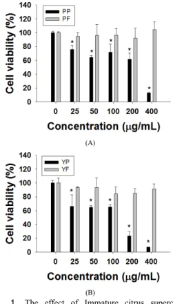
Fig. 1. The effect of Immature citrus supercritical extracts on HeLa cell viability as determined by the MTT assay. (A) Phalsak; PP, phalsak peel; PF, phalsak flesh, (B) Yeagam; YP, yeagam peel; YF, yeagam flesh. Data correspond to the mean ± standard deviation (SD) from four independent experiments.
3. 실험 결과 및 고찰
3.1 초임계 추출물의 자궁암 세포 증식 억제 효능
미성숙 재래감귤 초임계 추출물들의 세포증식 억제 효능을 확인하기 위하여 HeLa 세포에서의 성장률을 MTT assay를 통해 확인하였다. 48 시간동안 추출물들 을 처리한 결과, 과육 추출물에서는 최대 농도에서도 세 포의 성장률이 control과 비교하여 큰 차이를 보이지 않 았으나, 팔삭과 이예감의 과피 추출물에서는 농도가 증 가함에 따라 세포의 성장률이 감소됨을 확인 할 수 있었 다 Fig. 1. 또한 IC50 값은 팔삭 과피 추출물(252.7 μ g/mL), 이예감 과피 추출물(113.0 μg/mL)로 HeLa 세포 에서의 세포 성장 억제 효능이 이예감 과피 추출물이 더
(A) (B)
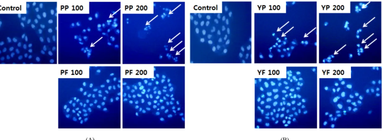
Fig. 2. The changes of the nuclear morphology of HeLa cells upon treatment with immature citrus supercritical extracts(μg/mL). (A) Phalsak; PP, phalsak peel; PF, phalsak flesh, (B) Yeagam; YP, yeagam peel; YF, yeagam flesh.
뛰어남을 알 수 있었고, 이를 통해 재래감귤 미성숙 팔삭 과 이예감 과피 초임계 추출물들의 HeLa 세포에 대한 암세포의 성장 억제 효능을 확인 할 수 있었다.
3.2 초임계 추출물에 의한 자궁암세포 apoptosis 유도
Apoptosis(세포사멸)는 programed cell death(세포예 정사)라 불리며, 발달과정, 항상성 유지 및 노화에서 중 요하게 작용하는 기작 중의 하나이다. 세포 내에서 apoptosis 신호를 받게 되면 세포막 수용체를 통한 extrinsic 경로와 미토콘드리아를 통한 intrinsic 경로를 통하여 caspase들을 활성화시킴에 따라 결국 세포가 죽 게 된다. Apoptosis가 진행될 때 나타나는 현상으로는 DNA 분절과 핵 응축이 나타난다[40]. 미성숙 재래감귤 초임계 추출물들을 100, 200 μg/mL로 처리한 뒤, Hoechst 33342로 염색하여 세포사멸의 특징 중의 하나 인 핵 응축 현상을 형광 현미경을 통해 확인한 결과, 과 육 추출물은 200 μg/mL 농도에서도 control과 비교하여 큰 차이를 보이지 않은 반면에, 과피 추출물들을 처리하 였을 경우 농도가 높아짐에 따라 apoptotic body의 형성 증가를 확인 할 수 있었다 Fig. 2.
3.3 GC/MS를 이용한 초임계 추출물 대사체 분석
미성숙 팔삭 과육, 과피 초임계 추출물에서 각각 27개 와 31개의 화합물이 검출되었다. 과피 추출물의 주요 화 합물로는 1,1,4,4-Tetramethyl- 2-tetralone(20.86%)이 가 장 큰 함량을 차지하고 있었으며, alloimperatorin(8.15%),
limonene (7.98%), auraptene(7.29%) 등이 확인되었다 [Table 1]. 한편, 미성숙 이예감 과육, 과피 초임계 추출 물에는 각각 27개와 29개의 화합물이 검출되었다. 과피 추출물 중 가장 함유량이 많은 화합물로는 limonene(22.19%) 으로 확인되었으며, linalool(11.23%)과 γ-sitosterol(9.12%) 등도 큰 비중을 차지하고 있음을 알 수 있었다 Table 1.
이전 연구들에서 limonene과 linalool 및 γ-sitosterol 은 항암 효능을 가진다는 보고가 있었으며[41-44], auraptene 은 항암 활성[45-48] 뿐만 아니라 라디칼 소거능[49] 및 항염증[50] 활성이 있다고 보고된 바 있다. 따라서 팔삭 과 이예감 과피 초임계 추출물에 포함되어 있는 주요 화 합물들간의 시너지 효과로 HeLa 세포에서의 세포 성장 억제 효능을 가진다고 사료된다.
4. 결론
본 연구에서는 제주 재래감귤인 미성숙 팔삭과 이예 감 초임계 추출물에 대한 항암 효능과 이들 추출물에서 의 유효 활성 성분들을 GC/MS로 분석하기 위하여 과육 과 과피를 나누어 추출물을 제조하였다. MTT assay를 통하여 인간 자궁암 세포의 성장률을 측정한 결과 팔삭 과 이예감 모두에서 과피 초임계 추출물이 효과적으로 암세포의 성장을 억제하였으며, Hoechst 33342 염색을 통하여 apoptotic body의 형성 증가를 확인함으로써 세 포사멸(apoptosis)이 유도됨을 확인 할 수 있었다. 자궁 암세포 성장억제 효능을 나타내는 과피 초임계 추출물에 서의 주요 화합물들을 GC/MS를 이용하여 분석한 결과,
No. RI a) Name of the compound b) Area % c)
phalsak yeagam
peel flesh peel flesh
1 1030 Sylvestrene 4.24 -d) - -
2 1039 Limonene 7.98 8.03 22.19 3.6
3 1061 γ-Terpinene 4.86 3.28 - -
4 1098 Linalool 1.79 1.48 11.23 1.42
5 1191 α-Terpineol 1.61 - 1.60 -
6 1201 Decanal 0.72 - 0.48 -
7 1278 Perillal - - 0.29 -
8 1341 d-Elemene - - 5.02 0.31
9 1362 Limonene oxide - - - 0.53
10 1363 Neryl acetate 1.05 - 0.38 -
11 1378 Geranyl acetate 1.09 - 1.13 -
12 1391 β-Elemene 0.88 - 1.55 -
13 1403 Decyl acetate 0.4 - - -
14 1426 β-Caryophyllen - - 1.61 -
15 1456 Nerolidol - - 1.17 -
16 1461 α-Humulene - - 0.72 -
17 1485 Germacrene-D 4.17 - 4.46 0.44
18 1498 α-Selinene 1.67 - - -
19 1502 Bicyclogermacrene - - 0.35 -
20 1504 Farnesene 1.77 - - -
21 1511 2,4-Di-tert-butylphenol - 0.35 - 0.77
22 1526 δ-Cadinene 0.46 - 0.92 -
23 1552 Elemol 0.31 - 1.06 -
24 1563 Nerolidol - - 0.24 -
25 1606 4-(2,2,3,3-tetramethylbutyl)-phenol - - - 0.27
26 1699 β-Sinensal - - 1.31 -
27 1757 α-Sinensal - - 2.66 -
28 1786 Ethyl myristate 0.2 - 0.29 -
29 1919 Methyl palmitate - 0.27 - 0.22
30 1960 Hexadecanoic acid - - - 4.21
31 1984 Ethyl palmitate 0.26 - 0.36 -
32 2098 Heneicosane - - - 0.62
33 2135 Ethyl linoleate - 2.80 - 2.52
34 2139 Octadecanoic acid - 1.17 - 2.58
35 2148 Osthol 0.66 - - -
36 2189 Docosane - 0.65 - 1.52
37 2231 7-Methoxy-8-(2-formyl-2-methylpropyl)coumarin 1.68 - - -
38 2241 Xanthotoxol 0.84 - - -
39 2251 1,1,4,4-Tetramethyl-2-tetralone 20.86 - - -
40 2253 3-Methyldocosane - 3.42 - 4.89
41 2291 Tricosane - 5.37 - 12.10
42 2300 7-Methoxy-8-(2-oxo-3-methylbutyl)coumarin 1.37 - - -
43 2354 Methyltricosane - 0.79 - -
44 2364 9-Butyldocosane - 3.05 - -
45 2390 Tetracosane - 2.08 - 3.89
46 2454 2-Methyltetracosane - 4.82 - 6.59
47 2474 Pentacosanol - - - 0.44
48 2491 Pentacosane - 5.94 - 9.11
49 2565 3-Methylpentacosane - 2.83 - -
50 2590 Hexacosane - 0.98 - 1.46
51 2633 Alloimperatorin 8.15 - - -
52 2653 Auraptene 7.29 0.54 - -
53 2653 δ-Methylhexacosane - 1.73 - -
54 2690 Heptacosane - 2.14 - 2.51
55 2798 Octacosane - 1.03 - -
56 2829 Squalene - 1.26 1.19 0.67
57 3000 Triacontane - 0.28 - -
58 3000< γ-Tocopherol 0.73 - 1.33 -
59 3000< α-tocopherol 2.41 0.86 4.24 -
60 3000< 4',5,6,7,8-Pentamethoxyflavone 1.78 - 4.68 0.5
61 3000< δ 5-Ergostenol 1.87 9.6 2.18 5.54
62 3000< Stigmasterol 1.34 2.71 2.86 2.05
63 3000< γ-Sitosterol 4.38 16.65 9.12 11.72
64 3000< 3',4',5,6,7,8-Hexamethoxyflavone 0.24 - 1.59 0.3
Total identified compounds 87.06 84.11 86.21 80.78
a) retention indices relative to C7-C30 n-alkanes calculated on Rtx-5MS capillary column.
b) supercritical CO2 extract of fruit compounds tentatively identified based on retention index and elution order as well as the fragmentation pattern described in the literature.
c) relative peak area percentage (peak area relative to the total peak area %) d) not detected
Table 1. Compounds from the immature citrus supercritical extracts identified by gas chromatography-mass spectrometry (GC-MS).
팔삭 추출물에서는 1,1,4,4-Tetramethyl-2-tetralone (20.86%), alloimperatorin(8.15%), limonene(7.98%), auraptene(7.29%) 등이 나타났으며, 이예감 추출물에서의 주요 화합물로는 limonene(22.19%), linalool(11.23%)과 γ-sitosterol(9.12%) 등이 함유되어 있음을 알 수 있었다. 이전 연구에서 팔삭 과 이예감 성숙 초임계 추출물에 대한 성분 분석 결과 팔삭에서의 주요 화합물로는 (Z)-9-Octadecenoic acid(17.79%), auraptene (16.00%), hexadecanoic acid(6.74%), tricosane (5.44%) 순으로 나타났으며, 이예감 초임계 추출물에서 는 limonene(12.14%), (Z)-9-Octadecenoic acid(8.78%), γ-sitosterol(5.38%), hexadecanoic acid(4.92%) 순으로 나타났다[51]. 이를 바탕으로 본 연구에서는 미성숙과에 대해 과육과 과피를 분리하여 항암 효능 탐색 및 GC/MS를 이용하여 성분분석 하였다. 그 결과, 과피 초 임계 추출물들에서 더 많은 활성 화합물들이 검출되었으 며, 이는 HeLa 세포에 대한 항암 효능을 나타내는데 기 여할 것으로 사료된다.
References
[1] M. Kubo, H. Matsuda. N. Tomohiro, S. Harima,
“Historical and Pharmalogical Study of Citrus hassaku.
Yakushigaku Zasshi,” The Journal of Japanese History of Pharmacy, vol. 40, no. 1, pp. 47-51, 2005.
[2] O. Benavente-garcia, J. Castillo, “Update on Uses and Properties of Citrus Flavonoids: New Findings in Anticancer, Cardiovascular, and Anti-inflammatory Activity,” Journal of Agricultural and Food Chemistry, vol. 56, pp. 6185-6205, 2008.
[3] G. K. Jayaprakasha, K. K. Mandadi, S. M. Poulose, Y.
Jadegoud, G. A. Nagana Gowda, B. S. Patil, “Novel Triterpenoid from Citrus aurantium L. Possesses Chemopreventive Properties Against Human Colon Cancer Cells,” Bioorganic and Medicinal Chemistry, vol.
16, no. 11, pp. 5939-5951, 2008.
[4] W. Y. Huang, Y. Z. Cai, Y. Zhang, “Natural Phenolic Compounds from Medicinal Herbs and Dietary Plants:
Potential Use for Cancer Prevention,” Nutrition and Cancer, vol. 62, no. 1, pp. 1-20, 2009.
[5] S. Ejaz, A. Ejaz, K. Matsuda, W. L. Chae, “Limonoids as Cancer Chemopreventive Agents,” Journal of the Science of Food and Agriculture, vol. 86, pp. 339-345, 2006.
[6] R. Patil Jaiprakash, G. K. Jayaprakasha, K. N.
Chidambara Murthy, S. E. Tichy, M. B. Chetti, B. S.
Patil, “Apoptosis-mediated Proliferation Inhibition of Human Colon Cancer Cells by Volatile Principles of Citrus aurantifolia,” Food Chemistry, vol. 114, pp.
1351-1358, 2009.
[7] A. Hardin, G. P. Crandall, T. Stankus, “Essential Oils and Antioxidants Derived from Citrus by-products in
Food Protection and Medicine: an Introduction and Review of Recent Literature,” Journal of Agricultural and Food Information, vol. 11, no. 2, pp. 99-122, 2010.
[8] K. R. Park, D. W. Nam, H. M. Yun, S. G. Lee, H. J.
Jang, G. Sethi, S. K. Cho, K. S. Ahn, “β-Caryophyllene oxide Inhibits Growth and Induces Apoptosis Through the Suppression of PI3K/AKT/mTOR/S6K1 Pathways and ROS-mediated MAPKs Activation,” Cancer Letters, vol. 312, no. 2, pp. 178-188, 2011.
[9] P. L. Crowell, C. E. Elson, H. H. Bailey, A. Elegbede, J. D. Haag, M. N. Gould, “Human Metabolism of the Experimental Cancer Therapeutic Agent d-limonene,”
Cancer Chemotherapy and Pharmacology, vol. 35, no. 1, pp. 31-37, 1994.
[10] A. Murakami, W. Kuki, Y. Takahashi, H. Yonei, Y.
Nakamura, Y. Ohto, H. Ohigashi, K. Koshimizu,
“Auraptene, a Citrus Coumarin, Inhibits 12-O-tetradecanoylphorbol-13-acetateinduced Tumor Promotion in ICR Mouse Skin, Possibly Through suppression of superoxide generation in leukocytes,”
Japanese Journal of Cancer Research, vol. 88, pp.
4443-4452, 1997.
[11] N. Yoshimizu, Y. Otani, Y. Saikawa, T. Kubota, M.
Yoshida, T. Furukawa, K. Kumai, K. Kameyama, M.
Fujii, M. Yano, T. Sato, A. Ito, M. Kitajima,
“Anti-tumour Effects of Nobiletin, a Citrus Flavonoid, on Gastric Cancer Include: Antiproliferative Effects, Induction of Apoptosis and Cell Cycle Deregulation,”
Alimentary Pharmacology and Therapeutics, vol. 20, pp.
95-101, 2004.
[12] C. Chaumontet, C. Droumaguet, V. Bex, C. Heberden, I.
Gaillard-sanchez, P. Martel, “Flavonoids (apigenin, tangeretin) Counteract Tumor Promoter-induced Inhibition of Intercellular Communication of Rat Liver Epithelial Cells,” Cancer Letters, vol. 114, no. 1-2, pp. 207-210, 1997.
[13] H. Kim, J. Y. Moon, A. Mosaddik, S. K. Cho,
“Induction of Apoptosis in Human Cervical Carcinoma HeLa Cells by Polymethoxylated flavone-rich Citrus grandis Osbeck (Dangyuja) Leaf Extract,” Food and Chemical Toxicology, vol. 48, pp. 2435-2442, 2010.
[14] J. Y. Moon, H. Kim, M. Cho, W. Y. Chang, C. T. Kim, S. K. Cho, “Induction of Apoptosis in SNU-16 Human Gastric Cancer Cells by The Chloroform Fraction of Extracts of Dangyuja (Citrus grandis) Leaves,” Journal of the Korean Society for Applied Biological Chemistry, vol. 52, pp. 168-175, 2009.
[15] Y. Takahash, N. Inaba, S. Kuwahara, W. Kuki, K.
Yamane, A. Murakami, “Rapid and Convenient Method for Preparing Aurapten-Enriched Product from Hassaku Peel Oil: Implications for Cancer-Preventive Food Additives,” Journal of Agricultural and Food Chemistry, vol. 50, pp. 3193-3196, 2002.
[16] H. K. Lim, J. Y. Moon, H. Kim, M. Cho, S. K. Cho,
“Induction of Apoptosis in U937 Human Leukaemia Cells by the Hexane Fraction of an Extract of Immature Citrus grandis Osbeck Fruits", Food Chemistry, vol. 114, pp. 1245-1250, 2009.
[17] G. J. Kang, S. C. Han, J. W. Ock, H. K. Kang, E. S.
Yoo, “Anti-Inflammatory Effect of Quercetagetin, an Active Component of Immature Citrus unshiu, in HaCaT Human Keratinocytes", Biomolecules and Therapeutics, vol. 21, no. 2, pp. 138-145, 2013.
[18] G. A. Moore, “Oranges and Lemons: Clues to The Taxonomy of Citrus From Molecular Markers,”
TRENDS in Genetics, vol. 17, pp. 536-540, 2001.
[19] M. Saidani, W. Dhifi, B. Marzouk, “Lipid Evaluation of Some Tunisian Citrus Seeds,” Journal of Food Lipids, vol. 11, pp. 242-250, 2004.
[20] J. W. Kim, Y. J. Jeon, J. H. Lee, S. C. Lee, “Effect of Far-infrared Irradiation and Heat Treatment on the Antioxidant Activity of Extracts from Citrus pomaces,”
Journal of the Korean Society for Applied Biological Chemistry, vol. 49, pp. 60-64, 2006.
[21] S. J. Yang, I. C. Jung, Y. H. Moon, “Physicochemical Properties and Sensory Characteristics of Korean Native Beef Loin Fed With Citrus Byproduct,” Journal of Life Sciences, vol. 17, pp. 540-545, 2007.
[22] Y. W. Song, K. S. Moon, S. K. Cho, “Antioxidant Activity and Nutrient Content of Ethanol and Hot-Water Extracts of Citrus unshiu Pomace,” Journal of the Korean Society of Food Science and Nutrition, vol. 42, no. 9, pp. 1345-1350, 2013.
[23] M. V. Palmer, S. S. T. Ting, “Application of Supercritical Fluid Technology in Food Processing,”
Food chemistry, vol. 52, pp. 345-352, 1995.
[24] S. H. Jung, K. S. Chang, K. H. Ko, “Physiological Effects of Curcumin Extracted by Supercritical Fluid from Turmeric (Curcuma longa L.),” Korean Journal of Food Science and Technology, vol. 36, no. 2, pp.
317-320, 2004.
[25] E. J. Lee, S. A. Yang, H. D. Choi, H. G. Im, K. Whang, I. S. Lee, “Comparison of Gingerols in Various Fractions and the Antioxidant Effects of Supercritical Fluid Extracts from Ginger,” Korean Journal of Food Science and Technology, vol. 43, no. 4, pp. 469-474, 2011.
[26] T. O. Jang, Y. H. Yoo, Y. C. Hwang, H. K. Kim, H. C.
Woo, “Total Polyphenol Content and Antioxidative Activities of Mistletoe (Viscum album) Extracts by Supercritical Carbon Dioxide,” Journal of the Korean Society of Food Science and Nutrition, vol. 39, no. 1, pp. 20-24, 2010.
[27] I. D. Kim, R. H. Kwon, Y. Y. Heo, H. J. Jung, H. Y.
Kang, B. J. Ha, “Supercritical Extraction of Oriental Herb : Anti-aging and Anti-wrinkle Effects,” Korean Journal of Biotechnology and Bioengineering, vol. 23, no. 6, pp. 529-534, 2008.
[28] B. Kim, S. M. Lee, T. Y. Hwang, H. S. Kim,
“Anti-oxidative and skin barrier effects of natural plants with a supercritical extract,” Korean Journal of Food Preservation, vol. 20, no. 5, pp. 597-601, 2013.
[29] E. D. Porta, D. Reverehon, D. C. Barth, “Mandarin and Lime Peel Oil Processing by Supercritical CO2
Desorption; Deterpenation and High Molecular Weight Compound Elimination,” Journal of Essential Oil Research, vol. 9, 515-522, 1997.
[30] S. Li, T. Lambros, Z. Wang, R. Goodnow, C. T. Ho,
“Efficient and Scalable Method in Isolation of Polymethoxyflavones From Orange Peel Extract by Supercritical Fluid Chromatography,” Journal of Chromatography B, Vol. 846, pp. 291-297, 2007.
[31] A. C. Atti-santos, M. Rossato, L. A. Serafini, E. Cassel, P. Moyna, “Extraction of Essential Oils From Lime
(Citrus latifolia Tanaka) by Hydrodistillation and Supercritical Carbon Dioxide,” Brazilian Archives of Biology and Technology, Vol. 48, pp. 155-160, 2005.
[32] A. Jemal, F. Bray, M. M. Center, J. Ferlay, E. Ward, D.
Forman, “Global cancer statistics,” CA: A Cancer Journal for Clinicians, vol. 61, no. 2, pp. 69-90, 2011.
[33] K. W. Jung, Y. J. Won, H. J. Kong, C. M. Oh, H. Cho, D. H. Lee, K. H. Lee, “Cancer Statistics in Korea:
Incidence, Mortality, Survival, and Prevalence in 2012.,”
Cancer Research and Treatment, vol. 47, no. 2, pp.
127-141, 2015.
[34] H. H. Kim, G. Min, “Inhibitory Effects of S-allylcysteine on Cell Proliferation of Human Cervical Cancer Cell Line, HeLa,” Journal of Life Science, vol.
25, no. 4, pp. 397-405, 2015.
[35] J. H. Park, J. S. Kim, E. J. Yun, K. S. Song, K. S. Seo, H. Kim, Y. J. Jung, W. H. Yun, K. Lim, H. D. Hwang, J. I. Park, “Cytocidal Effects of TALP-32 on Human Cervical Cancer Cell HeLa,” Toxicological Research, vol. 22, no. 4, pp. 315-322, 2006.
[36] B. O. Cho, H. W. Ryu, Y. K. So, C. H. Jin, M. W.
Byun, W. G. Kim, I. Y. Jeong, “Ishige sinicola Extracts Induce Apoptosis via Activation of a Caspase Cascade in Human HeLa Cells,” Journal of Korean Society of Food Science and Nutrition, vol. 41, no. 7, pp. 901-906, 2012.
[37] M. S. Lee, “Cisplatin and Extract of Tissue Cultured Mountain Ginseng-Induced Apoptosis in Human Cervial Cancer Cells,” Korean Journal of Microscopy, vol. 40, no. 3, pp. 133-138, 2010.
[38] T. Mosmann, “Rapid colorimetric assay for cellular growth and survivals: application to proliferation and cytotoxicity assays,” Journal of Immunological Methods, Vol. 65, pp. 55-63, 1983.
[39] Brady HJM. Apoptosis Methods and Protocols. Humana Press, Totowa, 2004.
[40] R. Vidya Priyadarsini, R. Senthil Murugan, S. Maitreyi, K. Ramalingam, D. Karunagaran, S. Nagini, “The Flavonoid Quercetin Induces Cell Cycle Arrest and Mitochondria-mediated Apoptosis in Human Cervical Cancer (HeLa) Cells Through p53 Induction and NF-kappaB Inhibition,” European Journal of Pharmacology, Vol. 649, no. 1-3, pp. 84-91, (2010).
[41] M. R. Khan, S. M. Mlungwana, “γ-Sitosterol, a cytotoxic sterol from Markhamia zanzibarica and Kigelia africana,” Fitoterapia, vol. 70, pp. 96-97, 1999.
[42] J. D. Haag, M. J. Lindstrom, M. N. Gould,
“Limonene-induced Regression of Mammary Carcinomas,”
Cancer Research, vol. 52, no. 14, pp. 4021-4026, 1992.
[43] M. Y. Chang, Y. L. Shen, “Linalool Exhibits Cytotoxic Effects by Activating Antitumor Immunity,” Molecules, vol. 19, no. 5, pp. 6694-706, 2014.
[44] Y. Ting, Y. S. Chiou, M. H. Pan, C. T. Ho, Q. Huang,
“In vitro and in vivo Anti-cancer Activity of Tangeretin Against Colorectal Cancer was Enhanced by Emulsion-based Delivery System,” Journal of Functional Foods, vol. 15, pp. 264-273, 2015.
[45] A. Murakami, W. Kuki, Y. Takahashi, H. Yonei, Y.
Nakamura, Y. Ohto, H. Ohigashi, K. Koshimizu,
“Auraptene, a Citrus Coumarin, Inhibits
12-O-tetradecanoylphorbol-13-acetate-induced Tumor Promotion in ICR Mouse Skin, Possibly Through Suppression of Superoxide Generation in Leukocytes,”
Japanese Journal of Cancer Research, vol. 88, no. 5, pp.
443-452, 1997.
[46] T. Tanaka, K. Kawabata, M. Kakumoto, K. Matsunaga, H. Mori, A. Murakami, W. Kuki, Y. Takahashi, H.
Yonei, K. Satoh, A. Hara, M. Maeda, T. Ota, S.
Odashima, K. Koshimizu, H. Ohigashi,
“Chemoprevention of 4-nitroquinoline 1-oxide-induced Oral Carcinogenesis by Citrus Auraptene in Rats,”
Carcinogenesis, vol. 19, no. 3, pp. 425-431, 1998.
[47] K. Kawabata, T. Tanaka, T. Yamamoto, A. Hara, A.
Murakami, K. Koshimizu, H. Ohigashi, G. D. Stoner, H.
Mori, “Suppression of N-nitrosomethylbenzylamine-induced Rat Esophageal Tumorigenesis by Dietary Feeding of Auraptene,” Journal of Experimental and Clinical Cancer Research, vol. 19, no. 1, pp. 45-52, 2000.
[48] A. Hara, K. Sakata, Y. Yamada, T. Kuno, N. Kitaori, T.
Oyama, Y. Hirose, A. Murakami, T. Tanaka, H. Mori,
“Suppression of Beta-catenin Mutation by Dietary Exposure of Auraptene, a Citrus Antioxidant, in N, N-diethylnitrosamine-induced Hepatocellular Carcinomas in Rats” Oncology Reports, vol. 14, no. 2, pp. 345-351, 2005.
[49] A. Murakami, H. Ohigashi, “Cancer-preventive Anti-oxidants that Attenuate Free Radical Generation by Inflammatory Cells,” The Journal of Biological Chemistry, vol. 387, no. 4, pp. 387-392, 2006.
[50] T. Tanaka, K. Kawabata, M. Kakumoto, A. Hara, A.
Murakami, W. Kuki, Y. Takahashi, H. Yonei, M. Maeda, T.
Ota, S. Odashima, T. Yamane, K. Koshimizu, H. Ohigashi,
“Citrus Auraptene Exerts Dose-dependent Chemopreventive Activity in Rat Large Bowel Tumorigenesis: the Inhibition Correlates With Suppression of Cell Proliferation and Lipid Peroxidation and With Induction of Phase II Drug-metabolizing Enzymes,” Cancer Research, vol. 58, no. 12, pp. 2550-2556, 1998.
[51] R. Gyawali, J. Y. Moon, D. H. Jeon, H. J. Kim, Y. W.
Song, H. B. Hyun, T. H. Kang, K. S. Moon, S. Jeong, J. C. Kim, K. S. Ahn, S. L. Cho, “Chemical Composition and Antiproliferative Activity of Supercritical CO2 Extracts from Citrus Fruits,” Food Science and Technology Research, vol. 18, no. 6, pp.
813-823, 2012.
김 소 미(Somi Kim) [정회원]
•1982년 3월 ~ 1989년 2월 : 서울 대학교 식품공학과 학사 및 석사
•1991년 9월 ~ 1997년 5월 : 오클 라호마 주립대 생화학/분자생물학 박사
•1997 ~ 1998 : 미시간 주립대 Parke-Davis Pharmaceutical Research Division Post-Doc.
•2004년 9월 ~ 현재 : 제주대학교 생명자원과학대학 교수
<관심분야>
생명과학, 융합바이오
문 정 용(Jeong Yong Moon) [정회원]
•2007년 3월 ~ 2009년 2월 : 제주 대학교 생명공학과 이학석사
•2009년 3월 ~ 2013년 2월 : 제주 대학교 생명공학과 이학박사
•2013년 3월 ~ 현재 : 제주대학교 아열대원예산업연구소 전임연구원
<관심분야>
생명과학, 융합바이오
송 연 우(YeonWoo Song) [준회원]
•2012년 3월 ~ 현재 : 제주대학교 응용생명공학과 석·박사 통합 과정
<관심분야>
생명과학, 융합바이오
현 호 봉(Hyun Ho Bong) [준회원]
•2014년 3월 ~ 현재 : 제주대학교 응용생명공학 전공 석사 과정
<관심분야>
생명과학, 융합바이오