How to Avoid Graft-Tunnel Length Mismatch in Modified Transtibial Technique for Anterior Cruciate Ligament Reconstruction Using Bone-
Patellar Tendon-Bone Graft
Dukhwan Ko, MD, Hyeung-June Kim, MD, Seong-Hak Oh, MD, Byung-June Kim, MD, Sung-Jae Kim, MD*
Department of Orthopedic Surgery, Konkuk University School of Medicine, Chungju,
*Department of Orthopedic Surgery, Yonsei Sarang Hospital, Seoul, Korea
Background: We conducted this study to determine the optimal length of patellar and tibial bone blocks for the modified trans- tibial (TT) technique in anterior cruciate ligament (ACL) reconstruction using the bone-patellar tendon-bone (BPTB) graft.
Methods: The current single-center, retrospective study was conducted in a total of 64 patients with an ACL tear who underwent surgery at our medical institution between March 2015 and February 2016. After harvesting the BPTB graft, we measured its length and that of the patellar tendon, patellar bone block, and tibial bone block using the arthroscopic ruler and double-checked measurements using a length gauge. Outcome measures included the length of tibial and femoral tunnels, inter-tunnel distance, length of the BPTB graft, patellar tendon, patellar bone block, and tibial bone block and graft-tunnel length mismatch. The total length of tunnels was defined as the sum of the length of the tibial tunnel, inter-tunnel distance and length of the femoral tunnel.
Furthermore, the optimal length of the bone block was calculated as (the total length of tunnels – the length of the patellar tendon) / 2. We analyzed correlations of outcome measures with the height and body mass index of the patients.
Results: There were 44 males (68.7%) and 20 females (31.3%) with a mean age of 31.8 years (range, 17 to 65 years). ACL recon- struction was performed on the left knee in 34 patients (53%) and on the right knee in 30 patients (47%). The optimal length of bone block was 21.7 mm (range, 19.5 to 23.5 mm). When the length of femoral tunnel was assumed as 25 mm and 30 mm, the optimal length of bone block was calculated as 19.6 mm (range, 17 to 21.5 mm) and 22.1 mm (range, 19.5 to 24 mm), respectively.
On linear regression analysis, patients’ height had a significant correlation with the length of tibial tunnel (p = 0.003), inter-tunnel distance (p = 0.014), and length of patellar tendon (p < 0.001).
Conclusions: Our results indicate that it would be mandatory to determine the optimal length of tibial tunnel in the modified TT technique for ACL reconstruction using the BPTB graft. Further large-scale, multi-center studies are warranted to establish our re- sults.
Keywords: Anterior cruciate ligament reconstruction, Modified transtibial technique, Bone-patellar tendon-bone grafts, Graft- tunnel length mismatch, Bone block length
Copyright © 2018 by The Korean Orthopaedic Association
This is an Open Access article distributed under the terms of the Creative Commons Attribution Non-Commercial License (http://creativecommons.org/licenses/by-nc/4.0) which permits unrestricted non-commercial use, distribution, and reproduction in any medium, provided the original work is properly cited.
Clinics in Orthopedic Surgery • pISSN 2005-291X eISSN 2005-4408 Received December 7, 2017; Accepted July 30, 2018
Correspondence to: Hyeung-June Kim, MD
Department of Orthopedic Surgery, Konkuk University Chungju Hospital, Konkuk University School of Medicine, 82 Gugwon-daero, Chungju 27376, Korea Tel: +82-43-840-8250, Fax: +82-43-844-7300, E-mail: [email protected]
Incorrect creation of bone tunnels is one of the most com- mon causes of failure of anterior cruciate ligament (ACL) reconstruction.1) Their accurate location is an essential factor for placing the intra-articular graft of an appropriate length as well as preventing impingement, limited motion, flexion contracture, laxity, and early failure.2,3)
Conventional transtibial (TT) techniques for ACL reconstruction have limitations in creating femoral tun- nels on the anatomical footprint of the ACL. Therefore, they have been modified, and thus termed as modified TT techniques. The modified techniques have been reported to be useful in creating oblique femoral and tibial tunnels while preserving the native femoral footprint of the ACL.4-7) It re- mains problematic, however, that more oblique and short- er tibial tunnels should be created for drilling femoral and tibial footprints through their centers.8) Due to the shorter tibial tunnel, the bone-patellar tendon-bone (BPTB) graft of patellar and tibial bone blocks (25–30 mm) becomes longer than the tunnel. Graft-tunnel length mismatch with BPTB grafts occurs accordingly. This has been described as a potential complication of the modified TT techniques for ACL reconstruction using the BPTB graft, which may lead to graft extrusion, failure of interference screw fixa- tion as well as decreased stiffness, micromovement, and laxity of the graft.9-11) To avoid this, a shorter BPTB graft is required for the modified TT techniques in ACL re- construction. Still, there is a paucity of data regarding the optimal length of patellar and tibial bone blocks. Given the above background, we conducted this study to determine the optimal length of patellar and tibial bone blocks for the modified TT techniques in ACL reconstruction using the BPTB graft.
METHODS
Patients and Study Setting
The current single-center, retrospective study was con- ducted in a total of 64 patients with an ACL tear who un- derwent surgery at Konkuk University Chungju Hospital between March 2015 and February 2016. Inclusion criteria for the current study are as follows: (1) patients with avail- able follow-up data; (2) patients who underwent primary ACL reconstruction with the autologous BPTB graft; and (3) patients who underwent surgery using the modified TT techniques. Exclusion criteria for the current study are as follows: (1) patients who had a past history of knee sur- gery, revision ACL reconstruction, or multi-ligamentous reconstruction surgery; (2) patients lost to follow up; and (3) patients who were deemed to be ineligible for study participation according to our judgment.
We therefore enrolled a total of 64 patients in the current study. The study was approved by the Institutional Review Board of Konkuk University Chungju Hospital (IRB No. KUCH-2013-048). Informed consent was waived due to its retrospective nature.
Operative Procedure
Standard arthroscopic ACL reconstruction was performed using the autologous BPTB graft via the modified TT technique by the senior author (DK). All the tibial tunnels were created using a tibial drill guide (Arthrex, Naples, FL, USA), which was set at 50° and intra-articularly posi- tioned at the center of the footprint of ACL. To create the anatomical femoral tunnel, the tibial drill guide was placed more horizontally and then directed to the common intra- articular entry point at the center of the tibial footprint of ACL. Subsequently, the extra-articular starting point was placed on the cortex of the medial tibia midway between the tibial tuberosity and the posteromedial margin of tibia, which was just superior to the pes anserinus and anterior to the medial collateral ligament. Then, with the knee joint placed in varus and external rotation, attempts were made to create the femoral tunnel in the anatomical femoral footprint of the ACL.
Measurement of the Length of the Tunnel
After harvesting the BPTB graft, we measured its length and that of the patellar tendon, patellar bone block and tibial bone block using the arthroscopic ruler and double- checked measurements using a length gauge. Both the femoral tunnel and the tibial tunnel were evaluated for the total length, the intra-articular, inter-tunnel distance (Fig. 1). After the passage and fixation of autologous BPTB graft, the length of protruded graft, such as graft-tunnel length mismatch, was measured using the ruler. All mea- surements were recorded.
Patient Evaluation and Criteria
We postoperatively analyzed the position of intra- articular openings of femoral and tibial tunnels on three- dimensional (3D) multi-detector computed tomography scans using the GE Lightspeed 16-slice scanner (Toshiba, Otawara, Japan), as previously described.12) We evaluated baseline and clinical characteristics of the patients, such as age, sex, height, and bone mass index (BMI) through a retrospective analysis of medical records.
In the current study, outcome measures include the length of tibial and femoral tunnels, inter-tunnel distance, length of the BPTB graft, the patellar tendon, the patellar bone block, and the tibial bone block, and graft-tunnel
length mismatch. Moreover, the total length of tunnels was defined as a sum of the length of the tibial tunnel, inter- tunnel distance, and length of the femoral tunnel. Further- more, the optimal length of the bone block was calculated as (the total length of tunnels – the length of the patellar tendon) / 2. Finally, we analyzed correlations of outcome measures with the height and BMI of the patients.
Data Analysis
All data was expressed as mean ± standard deviation. Each outcome measure was analyzed as compared with the corresponding one described in the literature. To identify correlations of measurements with height and BMI of the patients, both a correlation analysis and a linear regres- sion analysis were performed. Statistical analysis was performed using the IBM SPSS ver. 20.0 (IBM Corp., Ar- monk, NY, USA). Statistical significance was set at p < 0.05.
RESULTS
Baseline and Clinical Characteristics of the Patients The clinical series consists of 44 male (68.7%) and 20 fe- male (31.3%) patients with a mean age of 31.8 years (range,
17 to 65 years). The operated side was left in 34 patients (53%) and right in 30 patients (47%).
Outcome Measures
The mean length of femoral tunnel was 29.2 mm (range, 27 to 30 mm). The mean length of tibial tunnel was 33.7 mm (range, 28 to 40 mm). The mean inter-tunnel distance was 23.9 mm (range, 22 to 29 mm). The mean total length of tunnels was 86.8 mm (range, 80 to 96 mm) (Table 1).
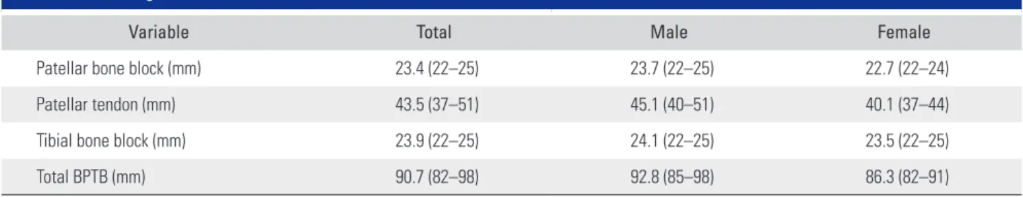
The mean length of patellar tendon was 43.5 mm (range, 37 to 51 mm). The mean length of patellar bone block was 23.4 mm (range, 22 to 25 mm). The mean length of tibial bone block was 23.9 mm (range, 22 to 25 mm). The mean length of BPTB graft was 90.7 mm (range, 82 to 98 mm) (Table 2).
Graft-Tunnel Length Mismatch
There were 52 cases (81.3%) of graft-tunnel length mis- match with a mean length of 3.9 mm (range, 0 to 8 mm) (Table 3).
Optimal Length of Bone Block
The optimal length of bone block was 21.7 mm (range,
A B C
FT
IT
TT
Fig. 1. (A) Measurement of femoral tunnel (FT) length, inter-tunnel distance (IT), tibial tunnel (TT) length, and the total length (FT + IT + TT). (B) Tunnel length measurement was done using an arthroscopic ruler. (C) The tunnel length gauge was used to double-check the tunnel length.
Table 1. Mean Length of Tunnels in Modified Transtibial Anterior Cruciate Ligament Reconstruction
Variable Total Male Female
Femoral tunnel (mm) 29.2 (27–30) 29.5 (27–30) 28.7 (27–30)
Inter-tunnel distance (mm) 23.9 (22–29) 24.2 (22–29) 23.3 (22–26)
Tibial tunnel (mm) 33.7 (28–40) 35.1 (30–40) 30.9 (28–34)
Total tunnel (mm) 86.8 (80–96) 88.7 (82–96) 82.8 (80–86)
Values are presented as mean (range).
19.5 to 23.5 mm). When the length of femoral tunnel was assumed as 25 mm and 30 mm, the optimal length of bone block was calculated as 19.6 mm (range, 17 to 21.5 mm) and 22.1 mm (range, 19.5 to 24 mm), respectively (Table 4 and Fig. 2).
Correlations of Patients’ Height and BMI with the Length of Tibial Tunnel, Inter-tunnel Distance, the Length of Patellar Tendon, and the Extent of Graft- Tunnel Length Mismatch
On linear regression analysis, patients’ height had a sig- nificant correlation with the length of tibial tunnel (p = 0.003), inter-tunnel distance (p = 0.014), and length of patellar tendon (p < 0.001). But it had no significant cor- relation with the extent of graft-tunnel length mismatch (p
= 0.753). Patients’ BMI had no significant correlation with the length of tibial tunnel (p = 0.271), inter-tunnel distance (p = 0.219), length of patellar tendon (p = 0.936), and the extent of graft-tunnel length mismatch (p = 0.856).
DISCUSSION
The results of the study can be summarized as follows: (1) The mean length of tibial tunnel was 33.7 mm (range, 28 to 40 mm), which is significantly shorter that of the tradi- tional TT technique (42.1 mm9) and 51.62 mm13)). (2) The total mean length of tunnels was 86.8 mm, and the mean length of BPTB graft was 90.7 mm. The mean length of Table 2. Mean Length of BPTBs
Variable Total Male Female
Patellar bone block (mm) 23.4 (22–25) 23.7 (22–25) 22.7 (22–24)
Patellar tendon (mm) 43.5 (37–51) 45.1 (40–51) 40.1 (37–44)
Tibial bone block (mm) 23.9 (22–25) 24.1 (22–25) 23.5 (22–25)
Total BPTB (mm) 90.7 (82–98) 92.8 (85–98) 86.3 (82–91)
Values are presented as mean (range).
BPTB: bone-patellar tendon-bone.
Table 3. Graft-Tunnel Length Mismatch
Variable Total Male Female
Graft-tunnel length mismatch (mm) 3.9 (0–8) 4.1 (0–8) 3.5 (0–7)
Values are presented as mean (range).
Table 4. Ideal Length of Each Bone Block
Variable Total Male Female
This study (mm) 21.7 21.8 21.4
Femoral tunnel length, 30 mm (mm) 22.1 22.1 22.0
Femoral tunnel length, 25 mm (mm) 19.6 19.6 19.5
Ideal length of each bone block = [total tunnel length (femoral tunnel + inter-tunnel distance + tibial tunnel) – patellar tendon length] / 2.
Total length
Femoral tunnel Inter-tunnel Tibial tunnel
Patellar bone Patellar tendon Tibial bone
Fig. 2. Ideal length of patellar bone block and tibial bone block of bone- patellar tendon-bone graft in anterior cruciate ligament reconstruction:
[total tunnel length (femoral tunnel + inter-tunnel distance + tibial tunnel) – patellar tendon length] / 2.
patellar and tibial bone blocks was 21.7 mm. (3) The graft- tunnel length mismatch was a mean of 3.9 mm.
Based on the above results, it can be inferred that graft-tunnel length mismatch can be prevented by adjust- ing the length of patellar and tibial bone blocks for the modified TT technique using the BPTB graft. We there- fore propose that when the length of the femoral tunnel is 30 and 25 mm, the length of patellar and tibial bone blocks should correspond to 22.1 and 19.6 mm, respectively.
To date, various measurement techniques and math- ematical formulas have been used to avoid the graft-tunnel length mismatch. These include “graft – 50” formula,
“patellar tendon + 2” formula, and “patellar tendon + 7°”
rule.14-16) But each of these methods has its own merits and demerits: “graft – 50” formula can make an unacceptable tunnel length; “patellar tendon + 2” formula is technically difficult and may require measurement of proposed tibial tunnel length and adjustment before the selection of the optimal angle for the placement of the guide; and “patel- lar tendon + 7°” rule may result in an unacceptable steep tibial tunnel with a tibial guide angle of > 55°. Moreover, the “patellar tendon + 7°” rule cannot be used to appropri- ately place the guide pin on the femoral footprint because its angle is relatively steeper. It would, therefore, be man- datory not only to create anatomical tibial and femoral tunnels but also to harvest the BPTP graft of an appropri- ate length. The BPTP graft is usually harvested before the creation of tunnels in ACL reconstruction. This makes it possible to adjust the length of BPTP graft by cutting the patellar and tibial bone blocks with optimal length.
To date, several attempts have been made to allow bitunnel interference screws and thereby to avoid graft- tunnel length mismatch. These include recession of the femoral bone plug,17) free bone block placement,18) flip- ping of the tibial bone plug,19) use of soft tissue interfer- ence screws,20) and rotation of graft.21) The disadvantages include poor biomechanical stability and technical errors, which can be resolved with use of the BPTB graft with an appropriate length. In our series, the length of patellar tendon was 43.1 mm, shorter than that in previous reports (48.4 mm,9) 45.48 mm,13) and 52.6 mm22)). Presumably,
this might arise from differences in the morphotype and ethnicity.
Recent efforts have been made to preoperatively avoid graft-tunnel length mismatch, according to which not only patient-related factors,23) such as height and weight, but also preoperative imaging studies of the pa- tellar tendon24) should be considered in identifying the appropriate length of the overall graft construct. Indeed, both patients’ height and the length of patellar tendon measured on magnetic resonance imaging scans are closely associated with the overall length of the graft. In the current study, we found that the patients’ height had a significant correlation with the length of tibial tunnel (p <
0.01), inter-tunnel distance (p < 0.05) and length of patel- lar tendon (p < 0.001).
There are some limitations of the current study.
First, we performed the modified TT technique with the tibial guide placed in an oblique position (50°) based on intra-articular references of the PCL, medial tibial spine, and anterior horn of lateral meniscus as well as offset femoral aimer depending on the direction of tibial tunnel.
Despite successful outcomes, it has been suggested that the aperture of the tibial tunnel be displaced posterior to the intra-articular references in the native ACL footprint. This may shorten inter-tunnel distances.25) Second, we analyzed the position of the tunnel based on CT scans rather than assessment of the native ACL footprint in a poor resource setting. The method that evaluates the native ACL foot- print requires magnetic resonance images of the contralat- eral knee and their 3D reconstruction, but this is consid- ered to be practically impossible in current clinics.
In conclusion, our results indicate that it would be mandatory to determine the optimal length of tibial tun- nel in the modified TT technique for ACL reconstruction using the BPTB graft. Further large-scale, multi-center studies are warranted to establish our results.
CONFLICT OF INTEREST
No potential conflict of interest relevant to this article was reported.
REFERENCES
1. Getelman MH, Friedman MJ. Revision anterior cruciate ligament reconstruction surgery. J Am Acad Orthop Surg.
1999;7(3):189-98.
2. Fineberg MS, Zarins B, Sherman OH. Practical consider- ations in anterior cruciate ligament replacement surgery.
Arthroscopy. 2000;16(7):715-24.
3. Sommer C, Friederich NF, Muller W. Improperly placed anterior cruciate ligament grafts: correlation between ra- diological parameters and clinical results. Knee Surg Sports Traumatol Arthrosc. 2000;8(4):207-13.
4. Harner CD, Honkamp NJ, Ranawat AS. Anteromedial portal technique for creating the anterior cruciate ligament femoral tunnel. Arthroscopy. 2008;24(1):113-5.
5. Fu FH, van Eck CF, Tashman S, Irrgang JJ, Moreland MS.
Anatomic anterior cruciate ligament reconstruction: a changing paradigm. Knee Surg Sports Traumatol Arthrosc.
2015;23(3):640-8.
6. Chhabra A, Diduch DR, Blessey PB, Miller MD. Recreating an acceptable angle of the tibial tunnel in the coronal plane in anterior cruciate ligament reconstruction using external landmarks. Arthroscopy. 2004;20(3):328-30.
7. Howell SM, Hull ML. Checkpoints for judging tunnel and anterior cruciate ligament graft placement. J Knee Surg.
2009;22(2):161-70.
8. Heming JF, Rand J, Steiner ME. Anatomical limitations of transtibial drilling in anterior cruciate ligament reconstruc- tion. Am J Sports Med. 2007;35(10):1708-15.
9. Shaffer B, Gow W, Tibone JE. Graft-tunnel mismatch in endoscopic anterior cruciate ligament reconstruction: a new technique of intraarticular measurement and modified graft harvesting. Arthroscopy. 1993;9(6):633-46.
10. Spindler KP, Bergfeld JA, Andrish JT. Intraoperative com- plications of ACL surgery: avoidance and management.
Orthopedics. 1993;16(4):425-30.
11. L'Insalata JC, Klatt B, Fu FH, Harner CD. Tunnel expansion following anterior cruciate ligament reconstruction: a com- parison of hamstring and patellar tendon autografts. Knee Surg Sports Traumatol Arthrosc. 1997;5(4):234-8.
12. Lorenz S, Elser F, Mitterer M, Obst T, Imhoff AB. Radiologic evaluation of the insertion sites of the 2 functional bundles of the anterior cruciate ligament using 3-dimensional com- puted tomography. Am J Sports Med. 2009;37(12):2368-76.
13. Denti M, Bigoni M, Randelli P, et al. Graft-tunnel mismatch in endoscopic anterior cruciate ligament reconstruction: in- traoperative and cadaver measurement of the intra-articular graft length and the length of the patellar tendon. Knee Surg Sports Traumatol Arthrosc. 1998;6(3):165-8.
14. Miller MD, Olszewski AD. Cruciate ligament graft intra- articular distances. Arthroscopy. 1997;13(3):291-5.
15. Kenna B, Simon TM, Jackson DW, Kurzweil PR. Endoscopic
ACL reconstruction: a technical note on tunnel length for interference fixation. Arthroscopy. 1993;9(2):228-30.
16. Miller MD, Hinkin DT. The "N + 7 rule" for tibial tunnel placement in endoscopic anterior cruciate ligament recon- struction. Arthroscopy. 1996;12(1):124-6.
17. Taylor DE, Dervin GF, Keene GC. Femoral bone plug reces- sion in endoscopic anterior cruciate ligament reconstruc- tion. Arthroscopy. 1996;12(4):513-5.
18. Fowler BL, DiStefano VJ. Tibial tunnel bone grafting: a new technique for dealing with graft-tunnel mismatch in endo- scopic anterior cruciate ligament reconstruction. Arthros- copy. 1998;14(2):224-8.
19. Morgan CD, Kalman VR, Grawl DM. Definitive landmarks for reproducible tibial tunnel placement in anterior cruciate ligament reconstruction. Arthroscopy. 1995;11(3):275-88.
20. Grawe B, Smerina A, Allen A. Avoiding graft-tunnel length mismatch in anterior cruciate ligament reconstruction: the single-bone plug technique. Arthrosc Tech. 2014;3(3):e417- 20.
21. Auge WK 2nd, Yifan K. A technique for resolution of graft- tunnel length mismatch in central third bone-patellar tendon-bone anterior cruciate ligament reconstruction. Ar- throscopy. 1999;15(8):877-81.
22. Hogerle S, Letsch R, Sievers KW. ACL reconstruction by pa- tellar tendon: a comparison of length by magnetic resonance imaging. Arch Orthop Trauma Surg. 1998;117(1-2):58-61.
23. Brown JA, Brophy RH, Franco J, et al. Avoiding allograft length mismatch during anterior cruciate ligament recon- struction: patient height as an indicator of appropriate graft length. Am J Sports Med. 2007;35(6):986-9.
24. McAllister DR, Bergfeld JA, Parker RD, Grooff PN, Valdevit AD. A comparison of preoperative imaging techniques for predicting patellar tendon graft length before cruciate liga- ment reconstruction. Am J Sports Med. 2001;29(4):461-5.
25. Kopf S, Forsythe B, Wong AK, et al. Nonanatomic tun- nel position in traditional transtibial single-bundle ante- rior cruciate ligament reconstruction evaluated by three- dimensional computed tomography. J Bone Joint Surg Am.
2010;92(6):1427-31.