Stiffness and Elasticity of the Masticatory and Facial Expression Muscles in Patients with the Masticatory Muscle
Pain
Yeon-Shin Kim, D.D.S.,M.S.D., Ki-Suk Kim, D.D.S.,M.S.D.,Ph.D., Mee-Eun Kim, D.D.S.,M.S.D.,Ph.D.
Dept. of Oral Medicine, Dankook University School of Dentistry
This study aimed to assess stiffness and elasticity of the masticatory muscle in the patients with the masticatory muscle pain using a tactile sensor and to investigate whether the masticatory muscle pain affects the facial expression muscles.
From those who visited Department of Oral Medicine in Dankook University Dental Hospital, 27 patients presenting with unilateral muscle pain and tenderness in the masseter muscle (Ms) were selected (mean age: 36.4±13.8 years).
Exclusion criterion was those who also had temporomandibular joint (TMJ) disorders or any neurological pain. Muscle stiffness and elasticity for the muscles of mastication and facial expression was investigated with the tactile sensor (Venustron, Axiom Co., JAPAN) and the muscles measured were the Ms, anterior temporal muscle (Ta), frontalis (Fr), inferior orbicularis oculi (Ooci), zygomaticus major (Zm), superior and inferior orbicularis oris (Oors, Oori) and mentalis (Mn). t-tests was used to compare side difference in muscle stiffness and elasticity. Side differences were also compared between diagnostic groups (local muscle soreness (LMS) vs myofascial pain syndrome (MPS) and between acute (< 6M) and chronic (≥ 6M) groups.
This study showed that Ms and Zm at affected side exhibited significantly increased stiffness and decreased elasticity as compared to the unaffected side.(p<0.05) There was no significant difference between local muscle soreness and myofascial pain syndrome groups and between acute and chronic groups. The results of this study suggests that masticatory muscle pain in Ms can affect muscle stiffness and elasticity not only for Ms but also for Zm, the facial expression muscle.
Key words: Masticatory muscle pain, Facial expression muscle, Stiffness, Elasticity, Tactile sensor
Corresponding author : Mee-Eun Kim Dept. of Oral Medicine,
Dankook University School of Dentistry Sinbu-dong San 7-1 Cheonan 330-716 Tel: 82-41-550-1915
Fax: 82-41-553-7169
E-mail: [email protected] Received: 2009-06-17
Revised: 2009-07-13 Accepted: 2009-07-29
I. INTRODUCTION
Noninflammatory muscle pain is the most common
type of somatic pain, and it emanates from the
skeletal muscles, tendons, and fascia. Muscle pain is
far more complex than simple overuse and fatigue.
1)Etiology of masticatory muscle pain includes various
types of events which can arise from either local or
systemic factors. Local factors represent any events
that acutely alter sensory or proprioceptive input in
the masticatory structures, for example, the fracture
of a tooth or the placement of a restoration in supraocclusion, and trauma like excessive or unaccustomed use of masticatory structures. One of the most commonly recognized systemic factors is emotional stress.
2-4)Carlson et al.
5)indicated that persons with masticatory muscle pain reported greater fatigue, disturbed sleep, depression, anxiety, menstrual symptoms, and less self-deception than normal controls.
Pain decreases the biting strength of masticatory muscles by 33% to 50% and induces a feeling of muscular weakness.
6)Palpable muscle tenderness and the fact that movement and functioning modify the pain and stiffness are clinical indications of the presence of muscle pain.
7)Masticatory muscle activity is coordinated with perioral muscle during chewing.
8)The facial musculature performs a variety of complex and important orofacial functions such as speech, mastication, swallowing and the mediation of emotional and affective states.
9)The facial muscles were active during mastication, displaying both tonic and phasic activity. The timing and amplitude of facial muscle EMG activity cycle, the side on which chewing took place, the size of the bolus, and whether or not lip to lip contact was made.
10)Therefore, it is thought that masticatory muscle pain may affect the facial expression muscles.
Evaluation of muscle condition in relation of muscle function or activity was reported in many studies. Muscle condition can be evaluated with various methods including digital palpation, algometry, electromyography (EMG), determination of bite force and chewing ability. Several studies have looked at the EMG frequency spectrum in the jaw-closing muscles during a fatiguing task.
11-13)However, it is difficult to measure muscle fatigue of the masticatory muscles and facial expression muscles to have different functions through set movement. Therefore, evaluation of the facial expression muscles using EMG would be a complicated and time-consuming task in clinical setting.
While EMG can mesure functional change through
movement, tactile sensor can measure muscle stiffness and elasticity in a static condition. A newly-introduced tactile sensor is thought to be an effective instrument indicating muscle stiffness and elasticity. Katayama et al.
14)evaluated muscle fatigue in the masseter muscle using tactile sensor and EMG and concluded the muscle stiffness and elasticity measured with the tactile sensor more accurately reflect muscle fatigue than EMG. Some authors investigated successfully masticatory muscles and facial expression muscles with a tactile sensor.
There existed few studies concerning muscle evaluation with the parameters of muscle stiffness and elasticity in the patients with masticatory muscle pain. Therefore, the purposes of this study were to assess stiffness and elasticity of the masticatory muscles in the patients with the masticatory muscle pain using a tactile sensor and to investigate whether masticatory muscle pain affects the facial expression muscles.
Ⅱ. MATERIALS AND METHODS 1. Subjects
Of those who visited department of Oral Medicine in Dankook University Dental Hospital, 27 subjects with unilateral muscle pain and tenderness in the masseter muscle (Ms) were selected for this study.
7 of them also presented with pain and tenderness in the anterior temporal muscle (Ta). The mean age of the subjects was 36.4±13.8 years and a ratio of male to famale was 1:2
The subjects were diagnosed as local muscle soreness (LMS) or myofascial pain syndrome (MPS) of Ms based on RDC/TMD.
15)Exclusion criteria were those who had pain or tenderness of Ms or Ta bilaterally; temporomandibular joint (TMJ) problems;
bony change on x-ray examination; combined with any neurologic pain. None of the subjects had systemic disease which could influence muscle condition.
All the subjects were divided into LMS group and
MPS groups according to their diagnosis and acute(<
6M) and chronic(≥ 6M) groups based on duration of their symptoms.
2. Tactile sensor system
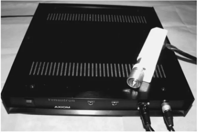
A Tactile sensor system used for this study was Venustron
Ⓡ(Axiom Co. Ltd., Japan).
16)(Fig 1) With computer control, this motor-driven sensor unit automatically presses down on surfaces such as those of muscle and skin to provide a continuous stream of simultaneous stiffness, pressure, and depression data in real time.
3. Measurement of muscle stiffness and elasticity
The masticatory muscles to be measured in this study included the Ms and Ta, and the facial expression muscles comprised frontalis(Fr), inferior orbicularis oculi(Ooci), zygomaticus major(Zm), superior orbicularis oris(Oors), inferior orbicularis oris(Oori) and mentalis(Mn) muscles.
Each subject was instructed to sit on a dental unit chair with head support in upright position. Ta and Ms were identified by manual palpation while each subject was asked to relax and clench alternatively and their thickest area was selected to be measured.
Measuring sites for different facial expression muscles were chosen on the basis of anatomical textbooks and other studies.
17-20)Measuring points
Fig. 1. The tactile sensor system(Venustron
Ⓡ, Axiom Co Ltd, Japan).
were marked on the skin over involved muscles with a pen. Measurements of muscle stiffness and elasticity with tactile sensor were performed on the muscles of mastication and facial expression in relaxed condition.
While the subjects were in light contact in their teeth, the probe of the tactile sensor was placed perpendicularly over the marked point over the skin, followed by computer-controlled movement including gently pressing straight down on the muscle for a second and retracting. The distance moved by the sensor probe for each muscle was determined separately in regards with the thickness of muscle and relation with adjacent structures; 8mm for Ms and Zm, 5mm for Oors and Oori, and 3mm for the rest of them. The examinations were performed bilaterally for each muscle for comparison between affected and unaffected sides.
4. Statistical Analysis
Paired t-test was used to compare muscle stiffness and elasticity between affected and unaffected sides of each muscle. The difference between LMS and MPS and between acute and chronic groups was compared by unpaired t-tests. The significance of the differences was assessed at the 0.05 level of probability.
Fig. 2. Diagram of masticatory and facial muscles
examined for the study.
Ⅲ. RESULTS
A hysteresis curve composed of two parts is obtained when the tactile sensor pushes down (bottom part) and then retracts (top) against the muscles to be measured. The sole op the tangent of the hysteresis curve (∆f/∆x) is defined as stiffness of the muscle being measured and the distance between the two parts as its elasticity. The higher value of stiffness indicates decrease of stiffness of target material and the lower value dose increase of that.
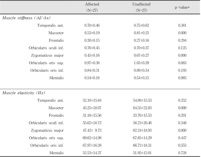
16)Table 1 shows the muscle stiffness and elasticity in the patients with unilateral masticatory muscle
Affected (N=27)
Unaffected
(N=27) p-value*
Muscle stiffness (∆f/∆x)
Temporalis ant. 0.70±0.46 0.75±0.62 0.381
Masseter 0.53±0.19 0.81±0.25 0.000
Frontalis 0.26±0.15 0.27±0.16 0.294
Orbicularis oculi inf. 0.76±0.45 0.70±0.37 0.125
Zygomaticus major 0.45±0.16 0.67±0.27 0.000
Orbicularis oris sup. 0.97±0.38 1.03±0.39 0.083
Orbicularis oris inf. 0.84±0.31 0.89±0.34 0.193
Mentalis 0.54±0.18 0.54±0.15 0.985
Muscle elasticity (Hz)
Temporalis ant. 52.10±15.64 54.80±15.53 0.252
Masseter 45.25±18.07 64.55±22.03 0.000
Frontalis 31.16±15.56 33.70±15.53 0.201
Orbicularis oculi inf. 55.62±18.72 58.23±20.46 0.348
Zygomaticus major 47.42± 9.73 62.24±18.93 0.000
Orbicularis oris sup. 69.62±14.98 67.85±14.29 0.447
Orbicularis oris inf. 67.97±16.28 66.72±18.31 0.553
Mentalis 52.53±14.37 51.95±15.01 0.728
* paired t-test
Table 1. Side difference in muscle stiffness and elasticity for patients with masticatory muscle pain.
pain in Ms. Significant difference between the
affected and unaffected sides was found in Ms and
Zm. The stiffness of Ms was 0.53±0.19 at the affected
side and 0.81±0.25 at the unaffected side (p<0.005)
and its elasticity was 52.84±20.35 at the affected side
and 64.55±22.03 at the unaffected side.(p<0.005,
Table 1) Zm also exhibited side difference in muscle
stiffness and elasticity in the patients with
masticatory muscle pain.(p<0.000, Table 1) The
stiffness of Zm was 0.45±0.16 at the affected side and
0.67±0.27 at the unaffected side (p<0.005) and its
elasticity was 47.42±9.73 at the affected side and
62.24±18.93 at the unaffected side.(p<0.005, Table 1)
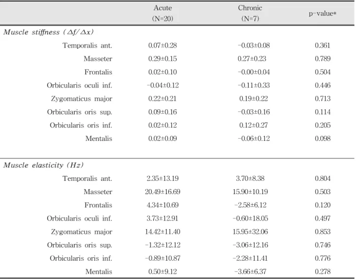
When dividing into acute and chronic group based
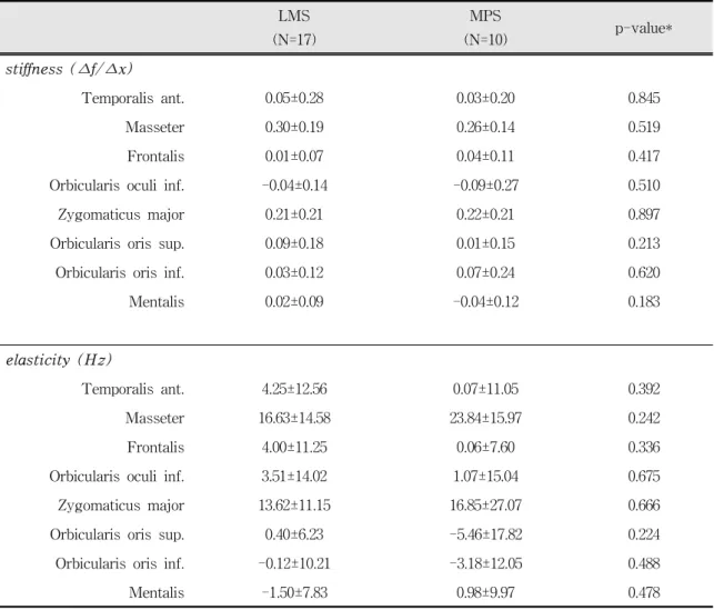
on pain duration of 6 month, there was no significant side difference in the muscle stiffness and elasticity between the groups.(Table 2) Comparison between LMS and MPS was shown in Table 3. Significant side difference in the stiffness and elasticity of the muscles of mastication and facial expression was not found between LMS and MPS groups.
Ⅳ. DISCUSSIONS
Manual palpation of a painful or sensitive masticatory muscle is a common examination method frequently used by clinicians for the evaluation of the patients with masticatory muscle
Acute (N=20)
Chronic
(N=7) p-value*
Muscle stiffness (∆f/∆x)
Temporalis ant. 0.07±0.28 -0.03±0.08 0.361
Masseter 0.29±0.15 0.27±0.23 0.789
Frontalis 0.02±0.10 -0.00±0.04 0.504
Orbicularis oculi inf. -0.04±0.12 -0.11±0.33 0.446
Zygomaticus major 0.22±0.21 0.19±0.22 0.713
Orbicularis oris sup. 0.09±0.16 -0.03±0.16 0.114
Orbicularis oris inf. 0.02±0.12 0.12±0.27 0.205
Mentalis 0.02±0.09 -0.06±0.12 0.098
Muscle elasticity (Hz)
Temporalis ant. 2.35±13.19 3.70±8.38 0.804
Masseter 20.49±16.69 15.90±10.19 0.503
Frontalis 4.34±10.69 -2.58±6.12 0.120
Orbicularis oculi inf. 3.73±12.91 -0.60±18.05 0.497
Zygomaticus major 14.42±11.40 15.95±32.06 0.853
Orbicularis oris sup. -1.32±12.12 -3.06±12.16 0.746
Orbicularis oris inf. -0.89±10.87 -2.28±11.41 0.776
Mentalis 0.50±9.12 -3.66±6.37 0.278
* unpaired t-test
Table 2. Comparison of side difference in muscle stiffness and elasticity between acute and chronic groups.
pain. Other methods include measurements and evaluation of algometry, EMG, bite force, and tactile sensor. Tactile sensor among these has recently been used as an objective clinical examination method. A study by Oh et al
21)on the reliability of the tactile sensor system indicated that tactile sensor had high intra-and inter-reliability in measuring muscle stiffness and elasticity of anterior temporalis, masseter and upper trapezius muscles and that the sensor was a highly reproducible, effective and easy-to-use instrument for quantitative evaluation of the muscles in head and neck region.
This study aimed to assess masticatory muscle in
the patients with the masticatory muscle pain using
by two parameters of the muscle stiffness and elasticity with a tactile sensor. Because of inaccessibility of a tactile sensor to the medial and lateral pterygoid muscles, the subjects in this study confined to those who had pain and tenderness in the Ms and some of them also had Ta pain. In addition, muscle stiffness and elasticity measured with the tactile sensor can be affected by individual characteristics including skin thickness, content of subcutaneous fat, etc, which cam be variable in from person to person.
20)Therefore, to avoid error due to individual variation through comparison between the affected and unaffected sides in each subject, we selected the patients with unilateral symptoms.
LMS (N=17)
MPS
(N=10) p-value*
Muscle stiffness (∆f/∆x)
Temporalis ant. 0.05±0.28 0.03±0.20 0.845
Masseter 0.30±0.19 0.26±0.14 0.519
Frontalis 0.01±0.07 0.04±0.11 0.417
Orbicularis oculi inf. -0.04±0.14 -0.09±0.27 0.510
Zygomaticus major 0.21±0.21 0.22±0.21 0.897
Orbicularis oris sup. 0.09±0.18 0.01±0.15 0.213
Orbicularis oris inf. 0.03±0.12 0.07±0.24 0.620
Mentalis 0.02±0.09 -0.04±0.12 0.183
Muscle elasticity (Hz)
Temporalis ant. 4.25±12.56 0.07±11.05 0.392
Masseter 16.63±14.58 23.84±15.97 0.242
Frontalis 4.00±11.25 0.06±7.60 0.336
Orbicularis oculi inf. 3.51±14.02 1.07±15.04 0.675
Zygomaticus major 13.62±11.15 16.85±27.07 0.666
Orbicularis oris sup. 0.40±6.23 -5.46±17.82 0.224
Orbicularis oris inf. -0.12±10.21 -3.18±12.05 0.488
Mentalis -1.50±7.83 0.98±9.97 0.478
* unpaired t-test
Table 3. Comparison of side difference in muscle stiffness and elasticity between local muscle soreness (LMS) and myofascial pain syndrome (MPS).
This study showed that Ms and Zm exhibited
significantly increased stiffness and decreased
elasticity at the affected side compared with the
unaffected side.(p<0.05, Table 1) Some studies
showed the relation of masticatory muscles and
facial expression muscles. Wohlert and Goffman
9)reported that perioral muscle activity was positively
correlated during the oral task including lip
protrusion, chewing, and speech. In addition, Inada et
al
22)reported that the stiffness of the muscles of
mastication and facial expression increased with
increase in the strength of their contraction. Kim et
al
23)investigated effects of oral parafunction on the
stiffness and elasticity in the muscles of the
mastication and facial expression and concluded that not only the masticatory muscles but also facial expression muscles could be considerably influenced by parafunctional activities such as unilateral clenching, jaw thrust and lip bracing. According to the results of their study, experimentally-induced unilateral clenching highly increased muscle stiffness of Ms, which resulted in significant difference as compared with relaxation and other parafunction including jaw thrusting and lip bracing. Zm also changed in a similar manner. These findings somewhat explain the finding that Zm as well as Ms showed significant increase of muscle stiffness and decrease of elasticity at the affected side of patients with unilateral masseter muscle pain. From these studies, it is assumed that Zm is positively correlated with Ms, suggesting that clinicians’ attention needs to be paid to both the masticatory and facial expression muscles, although no significant side difference was found in other facial expression muscles in this study.
Kim et al also showed that unilateral clenching with a force of 50 kg affected not only clenching side but also non-clenching side, exhibiting no side difference with high correlation coefficients.
However, significant side difference in Ms and Zm was found in the subjects with unilateral masticatory muscle pain in this study. Although some of the subjects reported oral habit such as clenching and bruxism in this study, a relation of muscle stiffness and elasticity with the oral habit was not assessed.
A further study needs to be performed in order to evaluate a relation of the muscles of mastication and facial expression with severity of oral habit.
Except for Ms and Zm, no significant difference was found in the other muscles. While all the subjects in this study had pain and tenderness in Ms, the subject with pain and tenderness in Ta were 7 patients. Significant side difference in Ta was not found for them. This finding may be partly due to small-sized sample in this study. A further study with a large population needs to be performed in the near future.
Ⅴ. CONCLUSIONS
Patients with unilateral masticatory muscle pain showed increased stiffness and decreased elasticity of Ms and Zm at affected side compared with the unaffected .(p<0.05) There was no significant difference between LMS and MPS and between acute and chronic groups. Based on the result of this study, it is assumed that masticatory muscle pain in Ms can affect muscle stiffness and elasticity not only for Ms but also for Zm, the facial expression muscle.
REFERENCES
1. Okeson JP. Bell’s orofacial pains 6
thed., Chicago, 2005, Quintessence, pp.287-311.
2. Selye H. Stress without distress. Philadelphia, 1974, Lippincott, pp.32.
3. Schiffman EL, Fricton JR, Haley D. The relationship of occlusion, parafunctional habits and recent life events to mandibular dysfunction in a non-patient population.
J Oral Rehabil 1992;19:201-223.
4. McCreary CP, Clark GT, Merril RL, Flack V, Oakley ME. Psychological distress and diagnostic subgroups of temporomandibular disorder patients. Pain 1991;44:29-34.
5. Carlson CR, Okeson JP, Falace DA et al. Comparison of psychologic and physiologic functioning between patients with masticatory muscle pain and matched controls. J Orofac Pain 1993;7:15-22.
6. Molin C. Vertical isometric muscle forces of the mandible. A comparative study of subjects with and without manifest mandibular pain dysfunction syndrome. Acta Odontol Scand 1972;30:485-499.
7. Wolff HG. Headache and Other Head Pain, 2
nded, New York, 1963, Oxford University Press.
8. Nozomi T, Toshimichi I, Kazunori Y. Electromyo- graphic activity of lower lip muscles when chewing with the lips in contact and apart. Angle Orthod 2004;74:31-36.
9. Wohlert AB. Goffman L. Human perioral muscle activation patterns. J Speech Hear Res 1994;37:1032- 1040.
10. Schieppati M, Di Francesco G, Nardone A. patterns of activity of perioral facial muscles during mastication in man. Exp Brain Res 1989;77:103-112.
11. Naeije M, Zorn H. Changes in the power spectrum of
the surface electromyogram of the human masseter
muscle due to local muscle fatigue. Archs oral Biol 1983;26:409-412.
12. Palla S, Ash MM. Power spectral analysis of the surface electromyogram of human jaw muscles during fatigue. Archs oral Biol 1981;26:547-553.
13. Lindstrom L. Hellsing G. Masseter muscle fatigue in man objectively quantified by analysis of myoelectric signals. Archs oral Biol 1983;28:197-301.
14. Katayama H, Inada J. Evaluation of muscle fatigue in the masseter muscle using a tactile sensor. Journal of the Osaka Odontology Society 2000;63:23-32.
15. Plesh O, Sinisi SE, Crawford PB et al. Diagnoses based on the Research Diagnostic Criteria for Temporo- mandibular Disorders in a biracial population of young women. J Orofac Pain 2005;19:65-75.
16. www.axiom-j.co.jp
17. Rohen JW, Yokochi C, Lutjen-Drecoll E et al. Color atlas of anatomy. 4th ed. Philadelphia, 1998, Lippincott Williams and Wilkins.
국문초록
저작근통 환자에서 저작근 및 안면표정근의 경도와 탄성도 평가