Visuomotor Coordination Deficits of Ipsilateral Upper Limb in Stroke Patients with Shoulder Pain
Purpose: The purpose of this study was to investigate whether ipsilateral shoulder pain affects the sensorimotor function of the same side shoulder in patients with stroke.
Methods: Thirty stroke patients, who were divided into the ipsilateral shoulder pain group (n=15) and the ipsilateral shoulder non-pain group (n=15). Subjects were evaluated on performance of a tracking task, joint reposition test and 9-Hole pegboard test for sensorimotor functions, and Fugl-Meyer test and Motricity Index for functional ability of the contralateral side.
Results: In comparison of the two groups, significant differences in performance on functional ability, including the Fugl-Meyer test(both upper and lower limb) and Motricity Index(only lower limb) were observed (p<0.05). With regard to sensorimotor functions, the ipsilateral shoulder pain group were observed significantly poor scores on the Accuracy Index, joint reposition score and 9-Hole pegboard test, when compared with the ipsilateral shoulder non-pain group (p<0.05).
Conclusion: We found that ipsilateral shoulder pain could impede accurate performance of a movement and result in deteriorated proprioception of the ipsilateral shoulder. Therefore, careful evaluation and appropriate therapeutic intervention are essential for stroke patients who suffer from ipsilateral shoulder pain.
Key Words:Motor deficits, Shoulder pain, Sensorimotor function, Stroke Sung-Min Son1, Kyoung Kim2, Na-Kyung Lee3
1Department of Physical Therapy, College of Rehabilitation Science, Daegu University, 2Department of Physical Therapy, College of Reha- bilitation Science, Daegu University, 3Department of Physical Therapy, College of Rehabilitation Science, Daegu University
The Journal of Korean Society of Physical Therapy Original articles
I. Introduction
More than 60% of patients with chronic stroke have dysfunction in their upper extremities, with only 5% showing complete functional recovery.1,2 Upper-limb dysfunction in stroke is characterized by paresis, loss of manual dexterity, and movement abnormalities that may have a considerable impact on performance of activities of daily living.3,4 And, altered movement patterns in the contralateral as well as the ipsilateral upper limb to the damaged hemisphere have
Copylight ⓒ 2014 The Korea Society of Physical Therapy
This is an Open Access article distribute under the terms of the Creative Commons Attribution Non-commercial License (Http:// creativecommons.org/license/by-nc/3.0.) which permits unrestricted non-commercial use, distribution,and reproduction in any medium, provided the original work is properly cited.
Received May 16, 2014 Revised Jun 10, 2014 Accepted Jun 13, 2014
Corresponding author Na-Kyung Lee, dlskrud@hanmail.net
been well demonstrated.5-7 Although contralateral motor deficits are generally emphasized after unilateral stroke, evidence of more subtle ipsilateral deficits after damage to the left or right hemisphere has also been reported.8-10
Several recent studies have reported that ipsilateral deficits affected activities of daily living, which influenced the independent performance of functional ability in stroke patients.8,11,12 In addition, it is generally assumed that the ipsilateral side will be used more in order to compensate for the decreased use of the contralateral side after stroke.13 All of these factors affect positioning of the shoulder, shoulder stability, and vulnerability to soft-tissue injury, and can impede rehabilitation, as well as initiate a cycle in which the ipsilateral shoulder condition can deteriorate, which may eventually lead to development of shoulder pain.
Previous studies have reported that ipsilateral control of
upper limb trajectory of unilateral stroke patients was impaired and exhibited deficits in fine position accuracy.14 However, these studies are based predominantly on ipsilateral motor deficit in stroke patients; no studies have investigated its effects on sensorimotor function in the ipsilateral upper limbe in stroke patients with ipsilateral shoulder pain. Several studies have investigated the effect of pain on sensorimotor function in musculoskeletal injury patients.15-17 However, the effects of ipsilateral shoulder pain on sensorimotor function in stroke patients have not been well suggested. The purpose of this study was to assess the differences in upper extremity sensorimotor function between individuals with and those without ipsilateral shoulder pain in stroke patients.
II. Methods
1. Subject
We recruited 15 stroke patients with shoulder pain of the ipsilateral side to the damaged hemisphere and the same number of patients without shoulder pain. Finally, 30 chronic stroke patients participated in this study. In order to control the influences of sensorimotor function according to handedness, only right-handed patients, as verified by the Modified Edinburgh Handedness Inventory, were included.
Criteria for inclusion were as follows: (1) unilateral brain injury due to hemorrhage or infarction through computed tomography (CT) or magnetic resonance imaging (MRI), (2) no symptoms of unilateral neglect or hemianopsia, (3) no arthropathy or orthopaedic surgery on the non-affected upper-limb, (4) no sensory or motor deficit on the non- affected upper-limb, and (5) no impairment of cognitive function (>24 point by mini-mental status examination).
If the pain on ipsilateral shoulder was not associated with stroke, the subject was excluded. All of them understood the purpose of this study and provided their written informed consent prior to their participation. The study was conducted according to the Declaration of Helsinki, and ethical approval was granted by the Institutional review board of a university hospital.
2. Procedure
For assessment of ipsilateral shoulder pain, pain was assessed a structured interview and by asking the patients whether they were experiencing muscle or joint pain during rest or during passive or active motion. Functional ability of the contra-lesional side was assessed using the Fugl-Meyer test and all participants underwent a manual strength examination. Fugl-Meyer Test has been widely recognized as a clinical measure of body function impairment after stroke. The maximum motor performance score is 66 points for upper extremity and 34 points for lower extremity.18 A manual strength examination on the contralateral side to the damaged brain is calibrated using Motricity Index scores. The Motricity Index for upper and lower extremity is based on ability to muscular activation, motion of limb segment during a range of motion, and resistance of examiner force. Motricity Index of upper extremity included three motions: shoulder abduction, elbow flexion, and pinch grasp. Lower extremity also included three motions: hip flexion, knee extension, and ankle dorsiflexion.19 Each score for upper and lower extremity involved addition of scores to the sum of three motions, and total scores ranged from 0 (complete paresis) to 100 (normal strength).
The study involved testing sensorimotor function of the ipsilateral shoulder in stroke patients. Examination of sensorimotor function was performed using a custom-made rotator machine with a built-in potentiometer for use by the ipsilateral upper-limb, which was permitted to rotate at the vertical axis. Participants were seated in front of a table, and held a custom-made machine at nearly 90° elbow flexion. The tracking task was assessed by internal rotation and external rotation of the ipsilateral shoulder joint. Participants were instructed to track the target white sine wave as accurately as possible, which was displayed for 15 seconds on the monitor.
The target sine wave was to appear in various amplitudes within 1.5-3 Hz and a range of velocity as a white line, and the response sine wave that was performed by participants was indicated as red a line. The response sine wave was drawn up toward the superior apex as the shoulder was rotated externally, and toward the inferior apex as the shoulder was rotated internally. After sufficient explanation, all participants practiced one demonstration, and were given three trials, in
sequence. The scores were averaged. The accuracy of the tracking task was calibrated as an Accuracy Index (AI).
The shoulder joint position sense was evaluated with a position-reposition test. Participants held a custom-made rotator machine with a built-in potentiometer on the table using their non-affected upper limb, which was allowed to rotate at the vertical axis. The digital signal of potentiometer was transferred to a personal laptop computer, and the signal was changed to angular degrees. Patients were instructed to actively reproduce the same joint position that was passively positioned by the evaluator, in the shoulder external and internal rotation on the vertical axis. The joint reposition error between passively positioned angle and actively repositioned angle was measured. The mean value through three trials was adopted.
Nine-hole pegboard test was used to measure fine motor dexterity of the non-affected upper limb. Patients were instructed to insert and remove nine dowels (9 mm in
diameter and 32 mm long) from holes in the pegboard with their upper limb of the non-affected side. Time measurement began when the first peg was placed in a hole and ended when the last peg was placed.
3. Statistical analysis
Independent t-test was used for determination of which variables differed between the two groups, in terms of age, time since onset, Fugl-Meyer test, Motricity Index, AI, joint reposition score, and 9-hole pegboard test. Statistical software SPSS 12.0 was used for statistical analysis, and p < 0.0 5 was used as the criterion for statistical significance.
III. Result
A summary of the baseline demographic characteristics of subjects in the ipsilateral shoulder pain group and non- pain group is shown in Table 1. There were no significant
Ipsilateral shoulder pain group (n=15) Ipsilateral shoulder non-pain group (n=15)
Age (years) 60.06 ± 8.68 57.53 ± 8.39
Time since stroke (month) 22.00 ± 13.51 21.20 ± 13.64
Ratio of gender (Male/Female) 6/9 5/10
Stroke type (Hemorrhage/infarct) 8/7 9/6
Damaged hemisphere (Rt/Lt) 10/5 10/5
Damaged hemisphere (Rt/Lt) 4.5 ± 1.55
Ipsilateral shoulder pain group (n=15)
Ipsilateral shoulder non-pain group
(n=15) p
Fugl Meyer test of affected side
Upper Extremity 20.20 ± 17.28 37.40 ± 21.34 0.022
Lower Extremity 16.20 ± 7.15 22.33 ± 8.26 0.038
Motricity Index of affected side
Upper Extremity 31.26 ± 10.91 39.00 ± 14.38 0.109
Lower Extremity 44.73 ± 6.36 50.67 ± 7.49 0.027
Accuracy Index of non-affected side 25.58 ± 9.08 31.13 ± 4.16 0.002
Joint Reposition Error of non-affected side 5.58 ± 1.75 4.26 ± 1.40 0.030
9-Hole Pegboard Test of non-affected side 28.97 ± 6.72 23.73 ± 6.85 0.044
Table 1. The general characteristics of subjects
Table 2. Dependent variables of pain group and non-pain group Mean ± S.D.
VAS: visual analogue scale
Mean ± S.D.
differences in demographic characteristics between the pain group and the non-pain group with regard to age, time since onset of stroke, ratio of gender, and stroke type. And ratio of damaged brain hemisphere was also the same in the two groups.
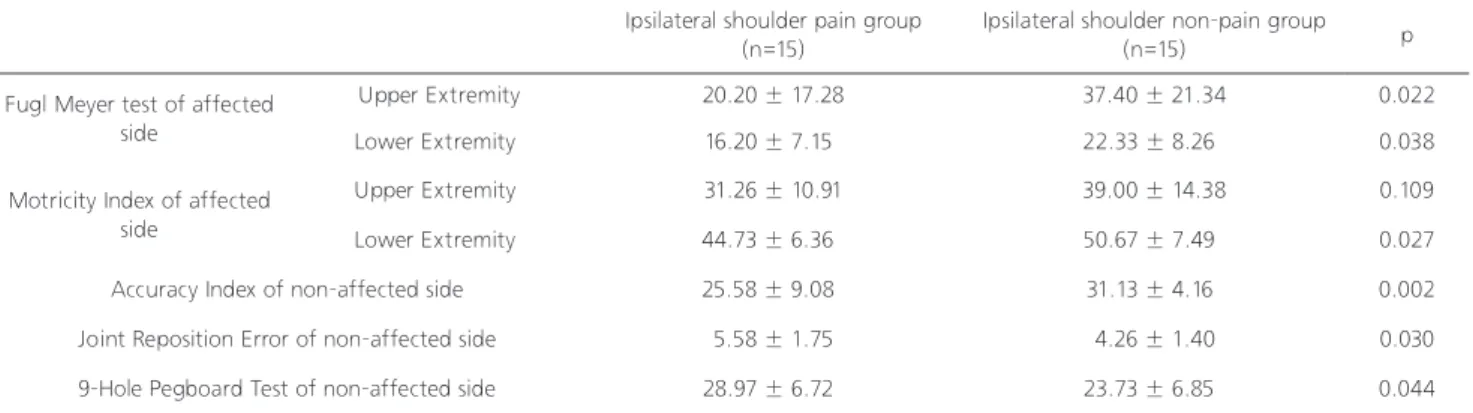
Table 2 shows the mean score on the Fugl-Meyer test and Motricity Index associated with functional ability, which indicated severity of the contralateral side. Pain group showed less functional ability than non-pain group. Significant differences in score for upper and lower extremity of Fugl Meyer test and lower extremity of Motricity Index were observed between the two groups(p<0.05).
A lower Accuracy Index was observed in the pain group, compared with the non-pain group. Joint reposition score and 9-Hole pegboard test in pain group were showed a larger variable value. Comparison of the two groups showed significant difference in terms of Accuracy index, joint reposition error, and 9-Hole pegboard test (p<0.05).
IV. Discussion
In this study, we attempted to determine whether sensorimotor function of the ipsilateral shoulder had deteriorated in stroke patients with same side shoulder pain.
Our main finding was that subjects in the ipsilateral shoulder pain group had more difficulty in performance of visuomotor coordination, joint reposition error and 9-hole pegboard test, compared with those in the ipsilateral shoulder non-pain group. In addition, shoulder pain group showed less functional ability than non shoulder pain group. Based on these results, ipsilateral shoulder pain to the damaged hemisphere could impede sensorimotor function in the same shoulder joint.
Our study indicated that muscle strength and functional ability, including Motricity index score and Fugl-Meyer test were significant lower value in pain group, when compared with non-pain group. It would be interpreted that the occurrence of ipsilateral shoulder pain was influenced by muscle strength and functional ability of contralateral side to damaged brain, in particular, lower limb is more the significant relation than upper limb. Previous studies related to the post-polio and spinal cord injury patients have reported
that the upper-limb pain could be induced by problems, which increased mechanical load or strain on soft tissues and joint due to overusing to compensate for motor weakness.
In this current study, we observed a significantly poor sensorimotor function, such as accuracy index, joint reposition score and 9-hole pegboard test, in the ipsilateral shoulder pain group, compared with the ipsilateral shoulder non-pain group. This finding was in agreement with those of previous studies,20-22 even though the area of pain and type of tracking task differed from those of our study. These previous studies reported that diminished motor function was associated with neck pain. In addition, with respect to proprioception, several studies also have suggested a strong association between pain and proprioception.21,23,24 Niessen et al.23 and Safran et al.,24 who is a studies of normal subject, reported that subjects with shoulder pain had poorer proprioception, compared with shoulder non-pain group.
The mechanism involved in sensorimotor dysfunction, such as decreased fine motor control according to pain, is not known. It is possible that deficit of motor performance is effects of information processing or motor planning modifications driven by higher centers due to features of pain related factors, such as fear, stress, or attention-demanding requirements. Sandlund et al.21 reported poorer sensorimotor function in subjects with neck pain, compared with non- pain subjects, however, no association was observed between psychological attention and sensorimotor function. Thus, the mechanisms for decreased accuracy index might be responsible for unconscious motor control process, rather than the psychological portion.
Sequentially, proprioceptive information plays an important role in successful motor control process.25 If deficit of proprioceptive information is presented, performance of accurately movement will be more difficult. Proprioceptive deficits should lead to an uncoordinated pattern of muscle activation and recruitment. Warner et al.26 reported that alternation in scapulothoracic motion in subjects with instability and impingement of shoulder was related to uncoordinated muscle firing pattern due to a proprioceptive deficit. Several studies have also demonstrated an association of altered motor control with proprioceptive deficits.21,27,28
Therefore, our finding suggests that ipsilateral shoulder pain is diminished performance of accurately movement in relation to impaired proprioception of the shoulder.
The clinical implication of our finding is that musculoskeletal pain of the ipsilateral shoulder indicates significant deterioration in sensorimotor function in stroke patients.
When it think that most stroke patients is performed activities of a daily living by using ipsilateral upper limb, ipsilateral upper limb is of considerable importance for performing daily functional activities. Ipsilateral upper limb pain will lead to the restrictions of functional abilities. Moreover, sensorimotor deficits due to pain of ipsilateral upper limb can be further aggravated the restrictions of functional abilities. Therefore, it is important to provide careful evaluation and appropriate therapeutic intervention for stroke patients with ipsilateral shoulder pain. When interpreting the data, several aspects of this study should be taken into account and could be raised as limitations. First, the number of patients included in this study is limited. In addition, stroke patients form a heterogeneous group; therefore, caution must be taken when drawing generalization from these data. Second, our study was restricted to a proximal single joint and a specific laboratory task, therefore, we were not aware that sensorimotor dysfunction, as demonstrated by our finding, could have an impact on ability to perform tasks of daily living. Conduct of future studies to include the factors mentioned above will be required.
References
1. Nakayama H, Jorgensen HS, Raaschou HO et al. Compensation in recovery of upper extremity function after stroke:
The copenhagen stroke study. Arch Phys Med Rehabil.
1994;75(8):852-7.
2. Parker VM, Wade DT, Langton Hewer R. Loss of arm function after stroke: Measurement, frequency, and recovery. Int Rehabil Med. 1986;8(2):69-73.
3. Kwon YH, Kim CS. Comparison of Motor Function and Skill between Stroke Patients with Cerebellar and Non- cerebellar Lesion in Sub-acute Stage. J Korean Soc Phys Ther.
2012;24(6):423-7.
4. Sveen U, Bautz-Holter E, Sodring KM et al. Comparison of motor function and skill between stroke patients with cerebellar
and non-cerebellar lesion in sub-acute stage. Disabil Rehabil.
1999;21(8):372-7.
5. Meskers CG, Koppe PA, Konijnenbelt MH et al. Kinematic alterations in the ipsilateral shoulder of patients with hemiplegia due to stroke. Am J Phys Med Rehabil. 2005;84(2):97-105.
6. Price CI, Rodgers H, Franklin P et al. Glenohumeral subluxation, scapula resting position, and scapula rotation after stroke: A noninvasive evaluation. Arch Phys Med Rehabil.
2001;82(7):955-60.
7. Yamaguchi K, Sher JS, Andersen WK et al. Glenohumeral motion in patients with rotator cuff tears: A comparison of asymptomatic and symptomatic shoulders. J Shoulder Elbow Surg. 2000;9(1):6-11.
8. Chestnut C, Haaland KY. Functional significance of ipsilesional motor deficits after unilateral stroke. Arch Phys Med Rehabil.
2008;89(1):62-8.
9. Haaland KY, Delaney HD. Motor deficits after left or right hemisphere damage due to stroke or tumor. Neuropsychologia.
1981;19(1):17-27.
10. Haaland KY, Harrington DL. Hemispheric asymmetry of movement. Curr Opin Neurobiol. 1996;6(6):796-800.
11. Kim SH, Pohl PS, Luchies CW et al. Ipsilateral deficits of targeted movements after stroke. Arch Phys Med Rehabil.
2003;84(5):719-24.
12. Wetter S, Poole JL, Haaland KY. Functional implications of ipsilesional motor deficits after unilateral stroke. Arch Phys Med Rehabil. 2005;86(4):776-81.
13. Michielsen ME, Selles RW, Stam HJ et al. Quantifying nonuse in chronic stroke patients: A study into paretic, nonparetic, and bimanual upper-limb use in daily life. Arch Phys Med Rehabil.
2012;93(11):1975-81.
14. Schaefer SY, Haaland KY, Sainburg RL. Hemispheric specialization and functional impact of ipsilesional deficits in movement coordination and accuracy. Neuropsychologia.
2009;47(13):2953-66.
15. Leitner C, Mair P, Paul B et al. Reliability of posturographic measurements in the assessment of impaired sensorimotor function in chronic low back pain. J Electromyogr Kinesiol.
2009;19(3):380-90.
16. Treleaven J. Sensorimotor disturbances in neck disorders affecting postural stability, head and eye movement control-part 2: Case studies. Man Ther. 2008;13(3):266-75.
17. Uthaikhup S, Jull G, Sungkarat S et al. The influence of neck pain on sensorimotor function in the elderly. Arch Gerontol Geriatr. 2012;55(3):667-72.
18. Fugl-Meyer AR, Jaasko L, Leyman I et al. The post-stroke hemiplegic patient. 1. A method for evaluation of physical performance. Scand J Rehabil Med. 1975;7(1):13-31.
19. Demeurisse G, Demol O, Robaye E. Motor evaluation in vascular
hemiplegia. Eur Neurol. 1980;19(6):382-9.
20. Hass CJ, Bishop MD, Doidge D et al. Chronic ankle instability alters central organization of movement. Am J Sports Med.
2010;38(4):829-34.
21. Sjolander P, Michaelson P, Jaric S et al. Sensorimotor disturbances in chronic neck pain--range of motion, peak velocity, smoothness of movement, and repositioning acuity.
Man Ther. 2008;13(2):122-31.
22. Woodhouse A, Vasseljen O. Altered motor control patterns in whiplash and chronic neck pain. BMC Musculoskelet Disord.
2008;9(1):90.
23. Niessen MH, Veeger DH, Meskers CG et al. Relationship among shoulder proprioception, kinematics, and pain after stroke. Arch Phys Med Rehabil. 2009;90(9):1557-64.
24. Safran MR, Borsa PA, Lephart SM et al. Shoulder proprioception
in baseball pitchers. J Shoulder Elbow Surg. 2001;10(5):438-44.
25. Sainburg RL, Ghilardi MF, Poizner H et al. Control of limb dynamics in normal subjects and patients without proprioception. J Neurophysiol. 1995;73(2):820-35.
26. Warner JJ, Micheli LJ, Arslanian LE et al. Scapulothoracic motion in normal shoulders and shoulders with glenohumeral instability and impingement syndrome. A study using moire topographic analysis. Clin Orthop Relat Res. 1992(285):191-9.
27. Anderson VB, Wee E. Impaired joint proprioception at higher shoulder elevations in chronic rotator cuff pathology. Arch Phys Med Rehabil. 2011;92(7):1146-51.
28. Cordo P, Carlton L, Bevan L et al. Proprioceptive coordination of movement sequences: Role of velocity and position information.
J Neurophysiol. 1994;71(5):1848-61.