• 검색 결과가 없습니다.

As diabetes prevalence is increasing annually, diabetic foot ulcer (DFU) occurs in approximately 15% of patients with

N/A
N/A
Protected

Academic year: 2021

Share "As diabetes prevalence is increasing annually, diabetic foot ulcer (DFU) occurs in approximately 15% of patients with "

Copied!
8
0
0

로드 중.... (전체 텍스트 보기)

전체 글

(1)

diabetes and infection is usually accompanied with DFU, therefore antibiotic treatment is often required. 1) Since the need for admission care is inevitable in most patients as- sociated with infection, the economic burden of medical expenses is growing annually. 2) In patients with diabetes, since immunity weakens with decreased function of neutro- phil, soft tissue infections can worsen quickly and may be associated with osteomyelitis and sepsis without adequate antibiotic therapy. 3) Recent studies have reported that anti- biotic-resistant bacteria in patients with DFU are becoming

INTRODUCTION

As diabetes prevalence is increasing annually, diabetic foot ulcer (DFU) occurs in approximately 15% of patients with

This is an Open Access article distributed under the terms of the Creative Commons Attribution Non-Commercial License (http://creativecommons.org/licenses/

CC

by-nc/4.0) which permits unrestricted non-commercial use, distribution, and reproduction in any medium, provided the original work is properly cited.

Copyright 2021 Korean Foot and Ankle Society. All rights reserved. ⓒ

Purpose: The present study aimed to develop guidelines regarding initial choice of antibiotics for diabetic foot ulcers (DFU) by investi- gating bacterial isolates.

Materials and Methods: This study included 223 DFU patients that visited a single tertiary hospital and underwent bacterial culture between January 2016 and February 2020. The study was conducted in two parts: 1) to compare bacterial isolates and wound healing ac- cording to comorbidities such as chronic kidney disease (CKD) and peripheral artery disease (PAD), and 2) to compare bacterial isolates according to wound depth using the Wagner classification.

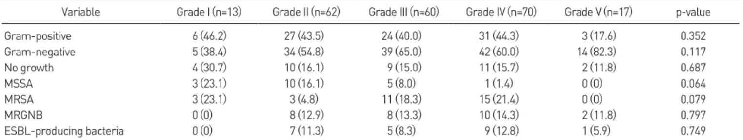
Results: Of the 223 patients, 43 had CKD (group A), 56 had PAD (group B), 30 had CKD and PAD (group C), and 94 had none of these comorbidities (group D). The isolation rate for multidrug-resistant gram-negative bacteria (MRGNB) and gram-negative to gram- positive bacteria ratio were highest in group C (p=0.018, p=0.038), and the proportion that achieved wound healing was lowest in group C (p<0.001). In the second part of the study, subjects were classified into 5 grades by wound depth using the Wagner classification; 13 grade I, 62 grade II, 60 grade III, 70 grade IV, and 17 grade V. No significant difference was observed between these grades in terms of isolation rates or gram-negative to gram-positive bacteria ratios.

Conclusion: This study suggests antibiotics that cover gram-negative bacteria including MRGNB produces better results in the presence of CKD and PAD and that initial antibiotic choice should be based on the presence of CKD and PAD rather than wound depth.

Key Words: Diabetic foot ulcer, Bacterial culture, Wagner grade, Resistant bacteria, Antibiotics

단일 3차 의료기관에 내원한 당뇨병성 족부병변 환자의 창상 배양검사를 통한 세균 검출 현황

정성윤, 이명진, 이승엽, 이상윤

동아대학교 의과대학 정형외과학교실

The Current Status of Bacterial Identification by Wound Culture for Diabetic Foot Lesions in a Single Tertiary Hospital in South Korea

Sung Yoon Jung, Myoung Jin Lee, Seung Yup Lee, Sang Yoon Lee Department of Orthopedic Surgery, College of Medicine, Dong-A University, Busan, Korea

Received February 17, 2021 Revised March 28, 2021 Accepted April 5, 2021 Corresponding Author: Myoung Jin Lee

Department of Orthopedic Surgery, College of Medicine, Dong-A University, 26 Daesingongwon-ro, Seo-gu, Busan 49201, Korea

Tel: 82-51-240-2898, Fax: 82-51-254-6757, E-mail: [email protected] ORCID: https://orcid.org/0000-0002-4388-7196

Financial support: None.

Conflict of interest: None.

(2)

www.jkfas.org increasingly prevalent. 4,5) The use of high-end antibiotics is

difficult in primary care and small-scale clinics because it is restricted by health insurance regulations.

Diabetes is considered as a strong risk factor for chronic kidney disease (CKD), and DFU associated with CKD has also been reported to affect the need for lower-extremity ampu- tation in diabetic patients. 6-8) Furthermore, CKD is known as an independent risk factor for the development of peripheral artery disease (PAD). 9) PAD is currently estimated to be pres- ent in more than half the patients with DFU. And presence of PAD is associated with increased risk of non-healing ulcers, infection and major limb amputation as well as an elevated risk of cardiovascular morbidity and overall mortality. For this reason, PAD has gained much attention as a prognosis of DFU patients. 10,11) Therefore, the comorbidity of CKD and PAD is a critical factor to be considered in the treatment of DFU patients and collaborative care with a multidisci- plinary approach is crucial. However, in primary care clinics, prompt management and appropriate antibiotic selection at the beginning of symptom onset have not frequently been performed. Our institution has treated DFU through inter- disciplinary and integrated care in the diabetic foot center (orthopedics, endocrinology, cardiology, infectious diseases, and radiology). This study aimed to facilitate the selection of empirical antibiotics for DFU by comparing bacterial isolates and wound healing according to presence of comorbidities

such as CKD and PAD, and by comparing bacterial isolates according to wound depth by Wagner classification. 12)

MATERIALS AND METHODS

1. Patients

1) Data collection and ethics approval

This study retrospectively reviewed 223 DFU patients who underwent bacterial culture tests at the initial visit of the dia- betic foot center and had accurate diagnosis code on elec- tric medical records in our hospital between January 2016 and February 2020 and proceeded after receiving written informed consent from all of patients. This study was per- formed after gaining IRB approval from investigators’ institu- tion (DAUHIRB-21-009).

2) Bacterial culture

Bacterial culture tests for DFU were performed on the first day of visit to our diabetic foot center before antibiotic administration. Bacterial cultures were harvested from the deep tissue of DFU after being sterilized with alcohol and betadine. Bacterial identification, and antibiotic susceptibil- ity tests were carried out. The results of bacterial identifica- tion were classified as usual pathogens on the basis of the International Working Group on the Diabetic Foot (IWGDF)

Table 1.

Table 1. International Working Group on the Diabetic Foot (IWGDF) 2019 Update Guideline

Infection severity Additional factor Usual pathogen Potential empirical regimen

Mild No complicating features Gram-positive cocci S-S pen

First generation cephalosporin

β-lactam allergy or intolerance Gram-positive cocci Clindamycin; fluoroquinolone; T/S; macrolide; doxycycline Recent antibiotic exposure Gram-positive cocci

+gram-negative rod

Amoxicillin/Clavulanate; ampicillin/Sulbactam; fluoroquinolone;

T/S

High risk for MRSA MRSA Linezolid; T/S; doxycycline; macrolide

Moderate or severe No complicating features Gram-positive cocci

±gram-negative rod

Amoxicillin/clavulanate; ampicillin/sulbactam, second/third generation cephalosporin

Recent antibiotics Gram-positive cocci

±gram-negative rod

Ticarcillin/clavulanate; piperacillin/tazobactam; third generation cephalosporin; group 1 carbapenem

Macerated ulcer or warm climate

Gram-negative rod including Pseudomonas

Ticarcillin/clavulanate; piperacillin/tazobactam; S-S

pen+ceftazidime; S-S pen+ciprofloxacin; group 2 carbapenem Ischemic limb/necrosis/gas

forming

Gram-positive cocci

±gram-negative rod±anaerobes

Second/third generation cephalosporin+clindamycin or metronidazole

MRSA risk factors MRSA Consider adding, or substituting with, glycopeptides; linezolid;

daptomycin; T/S (±rifampin); doxycycline Risk factors for resistant

gram-negative rod

Extended-spectrum β-lactamase- producing organism

Carbapenems; fluoroquinolone; aminoglycoside and colistin

MRSA: methicillin-resistant Staphylococcus aureus, S-S pen: semisynthetic penicillinase-resistant penicillin, T/S: trimethoprim-sulfamethoxazole.

(3)

2019 update guideline on the diagnosis and treatment of foot infection in persons with diabetes (Table 1). According to IWGDF, many studies demonstrated that the most com- mon pathogen in infection of diabetic foot is Staphylococ- cus aureus. And many studies about patients in Asia showed that aerobic gram-negative bacilli are often isolated either alone or in combination with gram-positivie cocci and often resistant to multiple commonly used antibiotics. Based on this studies, we classified bacterial isolates into methicillin- sensitive Staphylococcus aureus (MSSA), methicillin-resistant Staphylococcus aureus (MRSA), multidrug-resistant gram- negative bacteria (MRGNB), and extended spectrum beta- lactamase (ESBL)-producing bacteria. MRGNB included ESBL-producing bacteria, multidrug resistant Pseudomonas aeruginosa and multidrug resistant Acinetobacter baumannii.

Although when result of bacterial culture shows no growth of bacteria, biofilm formed by multibacterial colonization could be confounders, 13) in clinical practice, because we have to select antibiotics according to that result, we could not help but regard result of no growth in bacterial culture as an ab- sence of bacterial in this study.

3) Treatment

At first visit, wound cultures were obtained before ad- ministration of empirical intravenous antibiotics. Then, in- travenous antibiotics were selected according to the results of bacterial culture. All patients diagnosed with PAD on computed tomography angiography underwent percutane- ous transluminal angioplasty (PTA). Of those, patients on dialysis with CKD underwent dialysis immediately after PTA.

In patients with CKD but not on dialysis, appropriate hydra- tion and N-acetylcysteine were administered before and after PTA. Wound debridement was done for patients with residual wound infection or necrotic tissue after PTA and taking time to fully demarcate.

2. Comparisons of bacterial isolates and the ratio of wound healing to amputation of patients according to presence of comorbidities

The patients were classified into four groups according to comorbidities: 1) patients associated with CKD (group A); 2) patients associated with PAD (group B); 3) patients associ-

with none of the conditions (group D). PAD was diagnosed when ankle-brachial index was less than 0.9 in the affected side or lesions were clearly seen in computed tomography angiography of lower limbs. CKD was diagnosed when glo- merular filtration rate was less than 60 mL/min/1.73 m 2 . Bacterial the isolation rates and the ratio of wound healing were compared among groups. The ratio of wound healing was judged through comparison with the ratio of patients who have simply performed any kinds of amputations. And the ratio of isolation rates of gram-negative to gram-positive bacteria was compared among groups because most physi- cians empirically prescribe the antibacterial coverage of only gram positive bacteria and the anaerobes, we thought that comparing the isolation rates of gram-negative to gram-pos- itive bacteria could be an initial consideration in antibiotics selection.

3. Comparisons of bacterial isolates according to wound depth by Wagner classification

Wound depth was graded according to the Wagner classifi- cation system (Table 2). Bacterial isolation rates and the ratio of isolation rates of gram-negative to gram-positive bacteria were compared among grades.

4. Statistical analysis

All statistical analyses were performed using IBM SPSS ver.

22.0 (IBM Corp., Armonk, NY, USA). The isolation rates for gram-positive bacteria, gram-negative bacteria, non-isola- tion rate, MSSA, MRSA, MRGNB, and ESBL-producing bac- teria were obtained in each group. Chi-square test was used to compare the isolation ratio of gram-negative to gram- positive bacteria among groups according to the presence

Table 2.

Table 2. Wagner Classification of Diabetic Foot Ulcers

Grade Denomination Description

0 Foot at risk Thick calluses, bone deformities, clawed toes, and prominent metatarsal heads I Superficial ulcers Total destruction of the thickness of the

skin

II Deep ulcers Penetrates through skin, fat and ligaments, but not affect bone III Abscessed deep ulcers Limited necrosis in toes or the foot IV Limited gangrene Localized gangrene

V Extensive gangrene Necrosis of the complete foot,

(4)

www.jkfas.org of comorbidities and the ratio of isolation rates of gram-

negative to gram-positive bacteria among grades according to Wagner classification. Additionally, Fisher’s exact test was used to test differences in the isolation rates of bacteria and the ratio of wound healing between groups according to the presence of comorbidities and the isolation rates of bacteria among grades according to Wagner classification. Differ- ences were considered statistically significant at p<0.05.

RESULTS

1. Demographics

Of all 223 DFU patients, gram-positive bacteria were cul- tured from 85 (38.1%) patients and gram-negative bacteria from 122 (54.7%). The mean age was 66 years (34∼99 years), and 168 (75.3%) male and 55 (24.7%) female. A total of 223 patients were divided into four groups consisting of 43 (19.3%) in the group A, 56 (25.1%) in the group B, 30 (13.4%) in the group C, and 94 (42.1%) in the group D. The average Wagner grade was 3.07±1.06. Grade according to wound depth was grade I in 13 (5.8%) patients, grade II in 62 (27.8%), grade III in 60 (26.9%), grade IV in 70 (31.4%), and grade V in 17 (7.6%) (Table 3).

2. Results of bacterial identification

Of all 223 patients, gram-positive bacteria were detected in 85 (38.1%) patients, gram-negative bacteria in 122 (54.7%), and no isolation in 36 (16.1%). There were 41 (18.4%) patients

having more than two species of bacteria coexisting, and 20 (8.9%) having both gram-positive and gram-negative bacte- ria. Among gram-positive bacteria, MRSA was detected from 32 (14.3%) patients and MSSA from 19 (8.5%). MRGNB were detected in 28 (12.6%) patients. Of these multidrug-resistant gram-negative bacteria, multidrug-resistant P. aeruginosa was isolated from 3 (1.3%) patients, multidrug-resistant A.

baumannii from 4 (1.8%), and ESBL-producing bacteria from 22 (9.9%) (Table 4).

3. Correlation of bacterial isolates and the ratio of wound healing with the presence of comorbidities There were no statistical significance in differences of the isolation rate of gram-positive and gram-negative bacteria and the non-isolation rate of bacteria among groups accord- ing to the presence of comorbidities (Fig. 1). In the isolation rates of MSSA, MRSA, MRGNB, and ESBL-producing bacteria, there was a statistical significant difference only in MRGNB among groups (p=0.018; Table 5, Fig. 2). And there was sta- tistical significance in the isolation ratio of gram-negative to

Table 4.

Table 4. Bacterial Species Cultured from Diabetic Foot Ulcer

Variable Value

Gram-positive bacteria 85 (38.1)

Methicilin-sensitive Staphylococcus aureus 19 (8.5) Methicilin-resistant Staphylococcus aureus 32 (14.3)

Streptococcus agalactiae 11 (4.9)

Streptococcus dysgalactiae 7 (3.1)

Methicilin-sensitive coagulase-negative Streptococcus 9 (4.0) Methicilin-resistant coagulase-negative Streptococcus 16 (7.2)

Enterococcus faecalis 8 (3.6)

Enterobacter cloacae 8 (3.6)

Others 4 (1.8)

Gram-negative bacteria 122 (54.7)

Extended spectrum beta-lactamase producing bacteria 22 (9.9)

Escherichia coli 22 (9.9)

Pseudomonas aeruginosa 22 (9.9)

Multidrug-resistant Pseudomonas aeruginosa 3 (1.3)

Acinetobacter baumannii 3 (1.3)

Multidrug-resistant Acinetobacter baumannii 4 (1.8)

Proteus mirabilis 13 (5.8)

Proteus vulgaris 5 (2.2)

Stenotrophomonas maltophilia 4 (1.8)

Serratia marcescens 5 (2.2)

Klebsiella pneumoniae 8 (3.6)

Morganella morganii 4 (1.8)

Citrobacter koseri 3 (1.3)

Others 17 (7.6)

No growth 36 (16.1)

Values are presented as number (%).

Table 3.

Table 3. Demographic Characteristics of Diabetic Foot Ulcer Patients

Variable Value

Age (yr) 66 (34~99)

Sex

Male 168 (75.3)

Female 55 (24.7)

Comorbidities

CKD 43 (19.3)

PAD 56 (25.1)

CKD+PAD 30 (13.4)

No CKD & PAD 94 (42.1)

Wagner grade

I 13 (5.8)

II 62 (27.8)

III 60 (26.9)

IV 70 (31.4)

V 17 (7.6)

Values are presented as mean (range) or number (%).

CKD: chronic kidney disease, PAD: peripheral artery disease.

(5)

gram-positive bacteria among groups (p=0.038; Table 6).

The ratio of wound healing was lowest in group C (7 in wound healing, 23 in amputation) and highest in group D (55 in wound healing, 39 in amputation). There was a statistical significance in difference between the ratio of wound heal- ing of groups (p<0.001; Table 7).

4. Correlation of bacterial isolates with wound depth There were no statistical significance in differences of the isolation rate of gram-positive and gram-negative bacteria and the non-isolation rate of bacteria among grades ac- cording to the Wagner classification (Fig. 3). And there were no statistical significance in differences of the isolation rate of MSSA, MRSA, MRGNB, ESBL-producing bacteria and the

Table 5.

Table 5. Results of Diabetic Foot Ulcer Cultures by Each Disease Group and Chi-Square Analysis

Variable Total (n=223) Group A (n=43) Group B (n=56) Group C (n=30) Group D (n=94) p-value

Wagner grade 3.06±1.06 3.28±0.94 3.18±1.05 3.31±1.11 2.89±1.11

Gram-positive 85 (38.1) 18 (32.1) 14 (32.6) 10 (33.3) 43 (45.7) 0.259

Gram-negative 122 (54.7) 32 (57.1) 21 (48.8) 21 (70.0) 48 (51.1) 0.254

No growth 36 (16.1) 11 (19.6) 8 (18.6) 4 (13.3) 13 (13.8) 0.731

MSSA 19 (8.5) 3 (5.4) 4 (9.3) 2 (6.7) 10 (10.6) 0.773

MRSA 32 (14.3) 4 (7.1) 7 (16.3) 6 (20.0) 15 (16.0) 0.284

MRGNB 28 (12.6) 8 (14.3) 4 (9.3) 9 (30.0) 7 (7.4) 0.018*

ESBL-producing bacteria 22 (9.9) 5 (8.9) 4 (9.3) 7 (23.3) 6 (6.4) 0.084

Values are presented as mean±standard deviation or number (%).

MSSA: methicillin-sensitive Staphylococcus aureus, MRSA: methicillin-resistant Staphylococcus aureus, MRGNB: multidrug-resistant gram-negative bacteria, ESBL: extended spectrum beta-lactamase.

*Statistically significant (p<0.05).

Table 6.

Table 6. Comparative Analysis of Isolation Ratio of Gram-Negative to Gram-Positive of Group A, B, C with Group D

Group A (n=43) Group B (n=56) Group C (n=30) Group D (n=94) p-value

Gram-positive/gram-negative (n) 18/32 14/21 10/21 43/48 0.038

Table 7.

Table 7. Comparative Analysis of Ratio of Wound Healing among Groups by Comorbidities

Total (n=223) Group A (n=43) Group B (n=56) Group C (n=30) Group D (n=94) p-value Group A Group B Group C Group D

80 70 60 50 40 30 20 10

%

0

Gram positive Gram negative No growth

Figure 1.

Figure 1. This figure demonstrates isolation rate of each comorbidity group.

Isolation rate of gram-positive, gram-negative, and non-isolation rate of each comorbidity group (group A~D).

Group A Group B Group C Group D 35

30 25 20 15 10 5

%

0

MSSA MRSA MRGNB ESBL

Figure 2.

Figure 2. This figure demonstrates isolation rate of resistant bacteria of each comorbidity group. Isolation rate of MSSA, MRSA, MRGNB, and ESBL-producing bacteria of each comorbidity group (group A~D). MSSA:

methicillin-sensitive Staphylococcus aureus, MRSA: methicillin-resistant

Staphylococcus aureus, MRGNB: multidrug-resistant gram-negative bacte-

ria, ESBL: extended spectrum beta-lactamase.

(6)

www.jkfas.org isolation ratio of gram-negative to gram-positive bacteria

among grades (Tables 8, 9, Fig. 4).

DISCUSSION

The treatment failure of DFU infection at the initial period may lead to lower extremity amputation and sepsis because immunity, in particular, weakens with decreased function of neutrophils. Proper antibiotic treatment appropriate for bacterial isolates identified is the most critical intervention along with surgical debridement and irrigation. 14) Based on the results of bacterial culture and antibiotic susceptibility tests, initial treatment with appropriate antibiotic selection is important. However in clinical practice, it can take several

days to get the results of a bacterial culture in primary care clinics. For such a reason as mentioned, the choice of initial antibiotics is usually empirically determined, but inadequate initial antibiotics administration can lead to confoundation for treatment. Therefore we thought it is necessary to de- velop guidelines for initial antibiotics selections.

The subjects of the present study were 223 patients includ- ing 168 (75.3%) male and 55 (24.7%) female with a male-to- female ratio of 3:1. There is almost no difference between male and female in prevalence of diabetes, but male patients were more likely to develop DFU and Benotmane et al. 15) sug- gested that higher prevalence in male were attributable to Table 8.

Table 8. Results of Diabetic Foot Ulcer Cultures by Wagner Classification

Variable Grade I (n=13) Grade II (n=62) Grade III (n=60) Grade IV (n=70) Grade V (n=17) p-value

Gram-positive 6 (46.2) 27 (43.5) 24 (40.0) 31 (44.3) 3 (17.6) 0.352

Gram-negative 5 (38.4) 34 (54.8) 39 (65.0) 42 (60.0) 14 (82.3) 0.117

No growth 4 (30.7) 10 (16.1) 9 (15.0) 11 (15.7) 2 (11.8) 0.687

MSSA 3 (23.1) 10 (16.1) 5 (8.0) 1 (1.4) 0 (0) 0.064

MRSA 3 (23.1) 3 (4.8) 11 (18.3) 15 (21.4) 0 (0) 0.079

MRGNB 0 (0) 8 (12.9) 8 (13.3) 10 (14.3) 2 (11.8) 0.797

ESBL-producing bacteria 0 (0) 7 (11.3) 5 (8.3) 9 (12.8) 1 (5.9) 0.749

Values are presented as number (%).

MSSA: methicillin-sensitive Staphylococcus aureus, MRSA: methicillin-resistant Staphylococcus aureus, MRGNB: multidrug-resistant gram-negative bacteria, ESBL: extended spectrum beta-lactamase.

Table 9.

Table 9. Comparative Analysis of Isolation Rate of Gram-Positive and Gram-Negative Bacteria among Groups by Wagner Classification

Grade I (n=13) Grade II (n=62) Grade III (n=60) Grade IV (n=70) Grade V (n=17) p-value

Gram-positive/gram-negative (n) 6/5 27/34 24/39 31/42 3/14 0.266

Grade I 25

20

15

10

5

%

0

MSSA MRSA MRGNB ESBL

Grade II Grade III Grade IV Grade V Figure 4.

Figure 4. This figure demonstrates isolation rate of resistant bacteria among grades by Wagner classification. Isolation rate of MSSA, MRSA, MRGNB, and ESBL-producing bacteria of each grade by Wagner classifica- tion (grade I~V). MSSA: methicillin-sensitive Staphylococcus aureus, MRSA:

methicillin-resistant Staphylococcus aureus, MRGNB: multidrug-resistant gram-negative bacteria, ESBL: extended spectrum beta-lactamase.

Grade I 90

80 70 60 50 40 30 20 10

%

0

Gram positive Gram negative No growth

Grade II Grade III Grade IV Grade V Figure 3.

Figure 3. This figure demonstrates isolation rate among grades by Wagner

classification. Isolation rate of gram-positive, gram-negative, and non-

isolation rate of each grade by Wagner classification (grade I~V).

(7)

lifestyle habits, increased plantar pressure with higher levels of physical activity, and smoking rate.

In other previous domestic studies, isolation rates for gram-positive bacteria were high, ranging between 63.7%∼

76.4%, while isolation rates for gram-negative bacteria ranged between 33.3%∼63.6% in bacterial culture tests for DFU. 16,17) Of all gram-positive bacteria isolated, S. aureus was the most commonly detected pathogen at rates of 39.8%∼

46.3%. Escherichia coli was the most commonly found gram- negative bacteria at isolation rates of 13.7%∼38.8%. Isolation rates for P. aeruginosa ranged between 7.8% and 14.9%. 18) In the current study, the isolation rates of gram-negative bacteria were higher than those of gram-positive bacteria in all groups. The difference in bacterial culture results is thought that patients with more severe ulcer were reviewed in this study because our institution is a tertiary hospital and has a diabetic foot center, so there are many high-risk patients including patients on dialysis for CKD and patients with PAD. In a previous study of Arag ón-Sánchez et al., 19) the isolation rate of higher blood creatine and urea and pres- ence of peripheral artery occlusion could be a predisposing factor to gram-negative diabetic foot osteomyelitis. In this study, there was statistical significance in the isolation ratio of gram-negative to gram-positive bacteria among groups.

The difference in the isolation ratio of gram-negative to gram-positive bacteria by group of different comorbidities is anticipated to be resulted from change in immune mecha- nism and slow tissue recovery mechanism induced by altered tissue osmolarity and decreased peripheral circulation.

This study was limited in certain aspects. First, the control of blood glucose level was not taken into consideration. And type 1 and type 2 diabetes were not differentiated. Second, this study could not consider variability from other underly- ing diseases such as peripheral neuropathy or weakened im- mune systems and thoroughly examine a history of antibiotic use before visiting our hospital. Third, patients were ana- lyzed by wound depth according to the Wagner classification and no comparison was done by ulcer size. Fourth, this study is likely to include DFU patients with possibilities of having chronic ulcers or resistant isolates because the subjects were limited to diabetic patients who visited the tertiary hospital.

Therefore, selection bias could arise.

tive and the isolation rate of multiple resistant bacteria were highest in patients with CKD and PAD. In contrast, there were no significant differences according to wound depth.

CONCLUSION

This study suggests the use of antibiotics that can cover gram-negative bacteria including MRGNB is considered to lead to better results in the presence of CKD and PAD and initial choice of antibiotics should be considered according to the presence of CKD and PAD rather than wound depth.

ORCID

Sung Yoon Jung, https://orcid.org/0000-0003-0090-6729 Seung Yup Lee, https://orcid.org/0000-0001-6410-4771 Sang Yoon Lee, https://orcid.org/0000-0003-1424-0040

REFERENCES

. Lavery LA, Armstrong DG, Wunderlich RP, Tredwell J, Boulton AJ.

Diabetic foot syndrome: evaluating the prevalence and incidence of foot pathology in Mexican Americans and non-Hispanic whites from a diabetes disease management cohort. Diabetes Care. 003; 6: 435- 8. doi: 0. 337/diacare. 6.5. 435.

. Apelqvist J, Ragnarson-Tennvall G, Persson U, Larsson J. Diabetic foot ulcers in a multidisciplinary setting. An economic analysis of primary healing and healing with amputation. J Intern Med. 994; 35:463-7 . doi: 0. /j. 365- 796. 994.tb0 04.x.

3. Fisher TK, Scimeca CL, Bharara M, Mills JL Sr, Armstrong DG. A step- wise approach for surgical management of diabetic foot infections. J Vasc Surg. 0 0;5 (3 Suppl):7 S-5S. doi: 0. 0 6/j.jvs. 0 0.06.0 . 4. Ertugrul BM, Oncul O, Tulek N, Willke A, Sacar S, Tunccan OG, et

al. A prospective, multi-center study: factors related to the manage- ment of diabetic foot infections. Eur J Clin Microbiol Infect Dis.

0 ;3 : 345-5 . doi: 0. 007/s 0096-0 - 574- .

5. Eleftheriadou I, Tentolouris N, Argiana V, Jude E, Boulton AJ. Meth- icillin-resistant Staphylococcus aureus in diabetic foot infections.

Drugs. 0 0;70: 785-97. doi: 0. 65/ 538070-000000000-00000.

6. Ihm CG. Diabetic nephropathy. Korean J Med. 000;59:367-76.

7. Kaminski M, Frescos N, Tucker S. Prevalence of risk factors for foot ulceration in patients with end-stage renal disease on haemodialysis.

Intern Med J. 0 ;4 :e 0-8. doi: 0. /j. 445-5994. 0 .0 605.x.

8. Margolis DJ, Hofstad O, Feldman HI. Association between renal failure and foot ulcer or lower-extremity amputation in patients with diabe- tes. Diabetes Care. 008;3 : 33 -6. doi: 0. 337/dc07- 44.

9. Leskinen Y, Salenius JP, Lehtimäki T, Huhtala H, Saha H. The preva-

(8)

www.jkfas.org Kidney Dis. 00 ;40:47 -9. doi: 0. 053/ajkd. 00 .34885.

0. Prompers L, Huijberts M, Apelqvist J, Jude E, Piaggesi A, Bakker K, et al. High prevalence of ischaemia, infection and serious comorbidity in patients with diabetic foot disease in Europe. Baseline results from the Eurodiale study. Diabetologia. 007;50: 8- 5. doi: 0. 007/s00 5- 006-049 - .

. Kang MJ, Choi SH, Im S, Kong HS, Park MS, Heo CY, et al. The im- pact of peripheral arterial disease on the treatment and amputation of diabetic foot ulcer. J Korean Soc Vasc Surg. 008; 4: 3-8.

. Calhoun JH, Cantrell J, Cobos J, Lacy J, Valdez RR, Hokanson J, et al. Treatment of diabetic foot infections: Wagner classifi- cation, therapy, and outcome. Foot Ankle. 988;9: 0 -6. doi:

0. 77/ 07 007880090030 .

3. Kirby AE, Garner K, Levin BR. The relative contributions of physical structure and cell density to the antibiotic susceptibility of bacteria in biofilms. Antimicrob Agents Chemother. 0 ;56: 967-75. doi:

0. 8/AAC.06480- .

4. Bernstein BH, Tam H. Combination of subatmospheric pressure

dressing and gravity feed antibiotic instillation in the treatment of postsurgical diabetic foot wounds: a case series. Wounds. 005; 7:37- 48.

5. Benotmane A, Mohammedi F, Ayad F, Kadi K, Azzouz A. Diabetic foot lesions: etiologic and prognostic factors. Diabetes Metab.

000; 6: 3-7.

6. Song JY. Antimicrobial therapy in diabetic foot infections. J Korean Diabetes. 0 ; :83-7. doi: 0.4093/jkd. 0 . . .83.

7. Seo YB, Noh JY, Huh JY, Lee J, Song JY, Han SK, et al. Diabetic foot infection: microbiologic analysis based on deep tissue biopsy. Infect Chemother. 007;39: 37-4 .

8. Park SJ, Jung HJ, Shin HK, Kim E, Lim JJ, Yoon JW. Microbiology and antibiotic selection for diabetic foot infections. J Korean Foot Ankle Soc. 009; 3: 50-5.

9. Aragón-Sánchez J, Lipsky BA, Lázaro-Martínez JL. Gram-negative

diabetic foot osteomyelitis: risk factors and clinical presentation. Int J

Low Extrem Wounds. 0 3; :63-8. doi: 0. 77/ 5347346 34774 3.

수치

Table 4. Bacterial Species Cultured from Diabetic Foot Ulcer
Table 8. Results of Diabetic Foot Ulcer Cultures by Wagner Classification

참조

관련 문서