The Influence of Botulinum Toxin Type A on Masticatory Efficiency
Hyung-Uk Park, D.D.S.,M.S.D., Jeong-Seung Kwon, D.D.S.,M.S.D.,Ph.D., Seong Taek Kim, D.D.S, M.S.D, Ph.D., Jong-Hoon Choi, D.D.S.,M.S.D.,Ph.D.,
Hyung-Joon Ahn, D.D.S.,M.S.D.,Ph.D.
Department of Orofacial Pain & Oral Medicine, Yonsei Dental Hospital, Yonsei University Colledge of Dentistry, Seoul, Korea
This study was aimed to evaluate the masticatory efficiency after botulinum toxin type A (BTX-A) injection during 12 weeks using objective and subjective test. Also, we compared the difference of masticatory efficiency between group that injected into the masseter muscle only (M-group) and group that injected into the masseter and temporalis muscle (M-T group). The mixing ability index (MAI) was used as the objective indicator, and visual analogue scale (VAS) and food intake ability (FIA) index were used as the subjective indicators.
It was concluded that masticatory efficiency was significantly lowered after a BTX-A injection into the masticatory muscle, but it gradually recovered in a predictable pattern by the 12 weeks. The disturbance of subjective masticatory efficiency was lasted longer than objective masticatory efficiency. The masticatory efficiency was lower in M-T group than M group. It was statistically significant in the VAS and FIA at 4 weeks, but the MAI showed no significancy.
After 4weeks, there was rapid recovery of muscle function in M-T group, and the difference between two groups was not significant. It could be concluded that there will be no serious disturbance of mastication compared to injection is done only into the masseter muscle, even if injection is done into the masseter and temporalis muscle in dose of this study. According to the food properties, it was confirmed that people feel more discomfort on taking hard and tough foods after BTX-A injection and not only hard foods, but also intake of soft and runny foods were influenced by botulinum toxin injection.
Key words: Botulinum toxin type A, masticatory efficiency, masseter muscle, temporalis muscle
Corresponding author : Hyung-Joon Ahn
Professor, Department of Orofacial Pain & Oral Medicine, Yonsei Dental Hospital, Yonsei University Colledge of Dentistry, 50 Yonsei-ro, Seodaemun-gu, Seoul 120-752, Korea
Tel: 82-2-2228-8880 Fax: 82-2-393-5673 E-mail: hjahn@yuhs.ac Received: 2013-02-12 Accepted: 2013-03-04
* This study was supported by a faculty research grant of Yonsei Uneiversity Colledge of Dentistry for 2011
Ⅰ. INTRODUCTION
Botulinum toxin is a biological exotoxin produced by the gram-positive anaerobic bacterium, Clostridium botulinum. It has been known that botulinum toxin is one of the most potent bacterial toxin and is responsible for the clinical infection botulism. Botulism (botulusis sausage in Latin) is a rare but potentially life-threatening motor and autonomic paralytic syndrome which is first reported by Justinus Kerner, a German physician, in 1817. He depicted symptom of mydriasis,
diplopia, gastrointestinal complaints and progressive muscle paralysis after intake of meat and blood sausage. It was not until the 1897 that an investigation on an outbreak of food poisoning led to the isolation of bacillus Botulinum and its toxin by the Belgian microbiologist Emile van Ermengem.
It was in the 1920s that botulinum toxin was purified first. During the two World Wars, botulinum toxin was researched as a possible biological weapon .
In 1981, botulinum toxin injection into eye muscles to correct strabismus was conducted and, the therapeutic potentials of botulinum toxin become recognized. Afterwards, as a broad range of study was preformed, the FDA approved ‘Oculinum
Ⓡ’ as a therapeutic agent in patients with strabismus, blepharospasm, and other facial never disorders, including hemifacial spasm in 1989. Since then, the Allergan (Irvine, USA) company changed the name from ‘OculinumⓇ’ to ‘BotoxⓇ’. In 2000, the FDA approved BotoxⓇ and botulinum toxin type B (MyoblockⓇ) as treatment for cervical dystonia. In cosmetic fields, the application of botulinum toxin type A (BTX-A) was first reported in 1992.
Nowadays, it is used in numerous areas such as the reduction in the intensity of frown and wrinkle lines of the forehead, glabella, and lateral periorbital area, chin and upper lip wrinkling, nasolabial folds, nasal flare, and platysma neck bands. In addition, the application of BTX-A is quite demanding. In dental field, the clinical use of BTX-A has been expanded for the past few years. It is used in treatment of masticatory and facial muscle spasm, severe bruxism, facial tics, orofacial dyskinesias, dystonias, and idiopathic hypertrophy of the masticatory muscles.1) Also, it is used in the treatment of temporomandibular disorders, myofascial pain syndrome and a headache such as chronic migraine. As BTX-A adapted in various orofacial regions, BTX-A often injected into masticatory muscle such as masseter muscle and/or temporalis muscle. In case of BTX-A injected into masticatory muscles, according to the pharmacological property of BTX-A itself, it cause
a temporary muscle paralysis, weakness and atrophy. Also, the control of mastication is dependent in large part on sensory feedback, which consists of epithelial mechanoreceptor afferents, periodontal afferents, temporomandibular joint afferents and muscle afferents. Change in afferent input such as BTX-A injection into muscle can cause modify the response of the cortex, modify the motor neuron activity, and even initiate irrelevant muscle activity.2) Subsequently, BTX-A injection into masticatory muscle can directly influence on mastication by muscle weakness and atrophy, also indirectly it can influence on mastication by effect on central pattern generator in brainstem through modify the sensory feedback from masticatory muscle spindle.
Actually, the most common patient’s complaint after BTX-A injection is discomfort in mastication.
However, there’s been some amount of studies on applications and effects of BTX-A, but not on change of masticatory function which is the one of side-effects of BTX-A injection. Also, several previous studies on masticatory function are just concentrated in electromyographic activity of masticatory muscle or masticatory force after BTX-A injection.
Mastication is the process by which food is crushed and ground by teeth aimed at the preparation of food for swallowing. It is a process involving activities of the facial, the elevator and suprahyoid muscles, and the tongue.3) Besides the muscle, the teeth are important in terms of form the occlusion. Another important factor in mastication is bite force which is depends on muscle activity or coordination of various masticatory muscles. Also, neuromuscular control and saliva are essential for mastication. In other words, mastication is a dynamic and complex process.
This process is not simply breaking down of food for deglutition, but also it is in close contact with oral health, general health and quality of life of individual. So the evaluation of masticatory function is very significant thing in dental fields.
Meanwhile, the studies on masticatory function in
dental fields are mainly concentrated in prosthodontics4) and orthodontics5) which is relate to the change of occlusion after dental treatment.
But, it is rare on evaluation of masticatory function due to change in masticatory muscle itself or muscle activities in spite of muscle is also changeable factor in masticatory system.
Various methods have been used to evaluate the masticatory function. These methods are classified into two categories; subjective and objective methods.
The subjective methods evaluate the masticatory ability by self-assessment questionnaire or inter- view. The objective methods evaluate the masticatory performance, such as measuring the maximum bite force,6-8) electromyography of masticatory muscle,9) masticatory efficiency,10-12) occlusal contact area13)and malocclusion.14)Whereas other methods are static analysis, masticatory efficiency is dynamic analysis, and it is more suitable for evaluation of masticatory function in reflect the real masticatory state than any other static analysis. But, there is no study on masticatory efficiency after BTX-A injection into the masticatory muscles.
So, this study was aimed to evaluate the masticatory efficiency after BTX-A injection by questionnaire and mixing ability test. Also, we compared the difference of masticatory efficiency between group that botulinum toxin was injected into the masseter muscle only and group that botulinum toxin was injected into the masseter and temporalis muscle.
Ⅱ. SUBJECTS AND METHODS 1. Subjects
This study was approved by the institutional review board committee of Yonsei School of Dentistry (No. 2-2011-0018), and met the require- ments of the Declaration of Helsinki (1989) for prospective clinical studies with humans at enrolment. An informed consent was obtained from each subject before the start of the study. A total
of 40 healthy subjects participated in this study.
They were recruited voluntarily from March to July, 2011. All of the subjects had at least 28 teeth with a complete natural dentition with Angle Class I molar relationship. Also, the exclusion criteria for this study was dental pathologies or temporomandibular disorders, any missing teeth, implant and/or artificial teeth in molar area, teeth do not occluded in molar area, having been injected previously with BTX-A within the last three months of study entry, a woman of pregnancy or possible pregnancy, lactating women, those who received anti-spastic or muscle relaxant medication within one month of study entry, those who have a history of hypersensitivity to BTX-A, drug allergy and/or any other serious medical illness.
The subjects were randomly assigned into two groups according to the random number table. The assignment ratio was equal between the two groups. One group (M group, n=20) had an injection into the masseter muscle only, and the other group (M-T group, n=20) had an injection into the masster and temporalis muscle.
2. Methods
The masticatory efficiency was evaluated by the Visual Analogue Scale (VAS), Food Intake Ability (FIA) and Mixing Ability Index (MAI) before injection and 4, 8, 12 weeks after BTX-A injection.
Subjects were also interviewed about adverse reactions.
1) Injection of BTX-A
The MeditoxinⓇ (Medy-Tox, Seoul, Korea) was used in this study. Two hundred units of Meditoxin
Ⓡ obtained as a freeze-dried powder was reconstituted to a concentration of 5 U/0.1 mL using 4 mL of 0.9% sterile, nonpreserved saline. It was consumed immediately after reconstitution.
The intramuscular injection was administered by means of a 1ml-syringe with a 29-gauge, and a 1/2-inch needle.
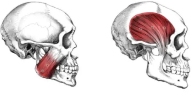
In M group, 25U of BTX-A was injected into
two points of each side (50U in total) at the lower middle third of the whole masseter muscle, which were located upper and lower side, 1 cm away from each other. In M-T group, BTX-A was injected into the masseter muscle as we had conducted in M group and 25U of BTX-A was injected into the three points of each side of the temporalis muscle at the bulkiest point which located horizontally, 1cm away from each other. Consequently, 100U of BTX-A was injected into the masseter and temporalis muscle, in total (Fig. 1).
2) Visual Analogue Scale (VAS) and Food Intake Ability (FIA)
The subjective masticatory efficiency was assessed by the Visual Analogue Scale (VAS) and Food Intake Ability (FIA). Subjective cognition on overall masticatory ability was evaluated through the VAS. The FIA questionnaire was prepared using 30 food items to quantify the subjective masticatory ability.
(1) Visual analogue scale (VAS)
The VAS was checked to assess the overall masticatory ability of subjects themselves in a 10-cm segment. The left end means ‘the worst imaginable’ and the right end means ‘the best imaginable’. The VAS score is determined by measuring from the left hand end of the line to the point that the patient marks
(2) Food Intake Ability (FIA)
The FIA questionnaire asked whether the subjects could chew the 30 food items. Thirty food items were selected from earlier study, and categorized into three groups according to the principle component factor analysis (varimax rotation) which is done earlier study.15)As a result, thirty foods divided into three groups according to similar properties; hard and tough food group (15 Items, Dried filefish, Raw carrot, Dried cuttlefish, Dried peanut, Pickled radish, Glutinous rice cake, French baguette, Hard persimmon, Caramel, Hard rice cracker, Steamed short ribs, Hard boiled
Fig. 1. Injection point of BTX-A in masseter and temporalis muscle
burdock, Cubed white radish kimchi, Apple, Roast beef), moderate hardness food group (8 Items, Yellow melon, Stuffed cucumber pickle, Watermelon, Noodles, Mandarin, Cabbage kimchi, Steamed potato, Rib of pork) and soft and runny food group (7 Items, Ham, Soybean curd, Boiled fish, Boiled chicken meat, Boiled fish paste, Boiled rice, Sweet jelly of red beans).
The FIA questionnaire consisted of five-point Likert scales: ‘cannot chew at all (1 point)’, ‘difficult to chew (2 points)’, ‘cannot say either way (3 points)’, ‘can chew some (4 points)’ and ‘can chew well (5 point)’, ‘never eaten (missing)’(Table 1).
2) Mixing Ability Index (MAI)
To measure the objective masticatory efficiency, the Mixing Ability Test reported by Sato et al. was employed. This method evaluates the masticatory efficiency by analyzing the degree of color mixed and wideness of chewed wax specimen.
(1) Fabrication of wax specimen
For this study, wax specimen was fabricated with the same design as Satos study. It was fabricated using red and green colored utility wax (Daedong Industry, Daegu, Korea). First, the sized of 2ⅹ2ⅹ 12mm3wax was sticked together parallelly to form a wax sheet. Six sheets were stacked up in lattice structure, and formed a 12ⅹ12ⅹ12mm3 sized wax cube (Fig. 2). Fabricated wax specimen was refrigerated prior to use for prevent the mixing of color and it was used immediately after taken from the refrigerator.
Foods list Cannot
chew at all Difficult
to chew Cannot say
either way Can chew
some Can chew
well Never
eaten 1. Ham
2. Boiled rice 3. Boiled fish 4. Boiled fish paste 5. Sweet jelly of red beans 6. Boiled chicken meat 7. Apple
8. Soybean curd 9. Hard rice cracker 10. Dried peanut 11. Raw carrot 12. Pickled radish 13. Dried cuttlefish 14. French baguette 15. Caramel
16. Glutinous rice cake 17. Hard boiled burdock 18. Watermelon 19. Noodles 20. Mandarin 21. Yellow melon
22. Stuffed cucumber pickle 23. Cabbage kimchi 24. Rib of pork 25. Hard persimmon
26. Cubed white radish kimchi 27. Roast beef
28. Steamed short ribs 29. Steamed potato 30. Dried filefish
Table 1. Questionnaire for the evaluation of food Intake Ability (FIA)
Fig. 2. Wax specimen Fig. 3. Chewed wax specimen
(2) Chewing test
Chewing study was done before injection and at 4, 8, and 12 weeks after injection. Subjects were instructed to chew the wax cube ten times with two seconds interval in a normal chewing pattern just using one’s own habitual masticatory side.
This procedure was repeated three times. Because the chewing speed can modify the masticatory efficiency, examiner directed to subject chew the wax specimen. The subject who does not have habitual masticatory side was instructed to chew the wax only one side throughout the study.
All measurements were made with the subject seated with the head upright, looking forward, and in an unsupported natural position.
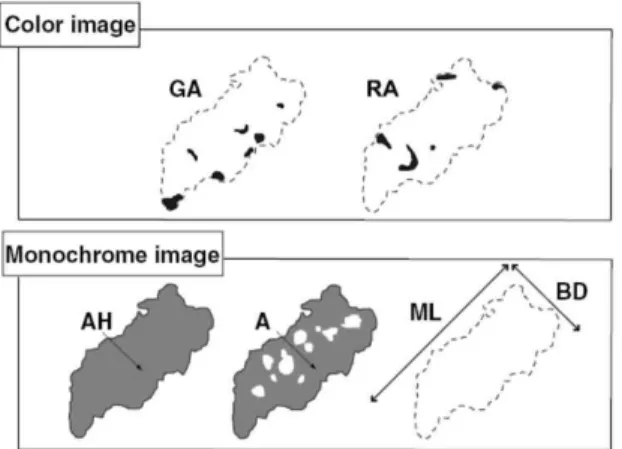
(3) Photographing and digital image analysis Digital images of the chewed wax specimen (Fig.
3) were taken using a digital camera (NIKON COOLPIC 4500, Japan) with a 30 cm distance between the camera and subject under standardized fluorescent lighting conditions. Both sides of wax specimen was photographed, because the differences in degree of color mixing between front and back side, although they are same in size and shape. The digital images were saved as JPEG files.
The digital images were analyzed using a digital image analyzer (Image pro plus 6.0, Medic Cybernetics corp., USA) to obtain information on the color and configuration. In program, we obtained two kinds of digital images: a monochrome image and a color image. On the monochrome image, the total projection area (AH), the projection area above 50 ㎛ in thickness (A), the maximum length (ML) and the maximum breadth (BD) were measured. On the color images, the green area (GA) and the red area (RA) were measured (Fig.
4).
The following four parameters were calculated from the measured items as Sato et al. suggested.
1. MIX: The ratio of color mixed area MIX = 100 - (GA + RA)/A × 100
2. TR: The ratio of area above 50㎛ in thickness to total projection area
TR = 100 - A/AH × 100
3. LB: The ratio of maximum length to maximum breadth
LB = ML/BD
4. FF: The shape factor shows how flat the sample is
FF = ML² × π/4 × AH × 100
All procedures for image analysis were performed by a single independent examiner to reduce the error.
(4) Calculation of Mixing Ability Index (MAI) MAI was obtained by applying the four parameters to discriminant function which is suggested by Jeong et al.16)
MAI = 0.100 × MIX - 0.015 × TR + 0.298 × LB + 0.000 × FF - 0.001 × AH - 7.336 4) Statistical analysis
Linear mixed models for longitudinal data was carried out to analysis the VAS, FIA and MAI in process of time and compare M group and M-T group. The interaction between times and groups was evaluated by compound symmetry covariance structure or banded toeplitz covariance structure. A
Fig. 4. Schematic image from the chewed wax specimen (from Sato et al)
Bonferroni’s correction was used for post hoc analysis. A p-value of < 0.05 was considered statistically significant. All statistical analyses were performed using SASⓇ Version 9.2 Windows Statistics Program (SAS Institute Inc., Cary, NC, USA).
Ⅲ. RESULTS
40 subjects distributed between the ages of 21 and 42 years (16 males, 24 females, mean age: 28.0
± 9.19). In M group, of the 20 subjects, 8 were male and 12 were female, with a mean age of 28.3 (± 5.1) years. In M-T group, of the 20 subjects, 8were male and 12 were female, with a mean age of 27.7 (±4.9) (Table 2.).
No subjects withdrew from this study. There were no adverse events during this study associated with BTX-A injection.
1. The VAS, FIA and MAI following BTX-A injection
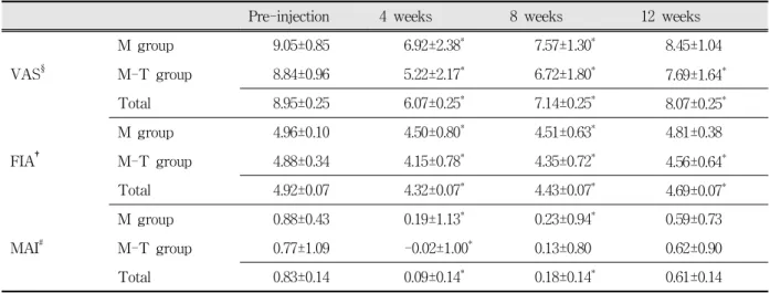
Table 3 presents the changes of the VAS, FIA and MAI during 12 weeks after BTX-A injection.
All the VAS, FIA and MAI were declined substantially at 4 weeks and it gradually recovered with time.
In M group, the VAS was significantly lower at 4 and 8 weeks compared to baseline (p-value:
<.0001, 0.0037, respectively), but it was not significant at 12 weeks (p-value: 1.0000). In M-T group, the VAS was significantly lower at 4, 8 and 12 weeks (p-value: <.0001, <.0001, 0.0476, respectively). Totally, compared to pre-injection, the VAS was significantly lower at 4, 8 and 12 weeks (p-value: <.0001, <.0001, 0.01, respectively).
M group (n=20) M-T group (n=20)
Male/Female (n) 8/12 8/12
Age (years) 28.3±5.1 27.7±4.9 Table 2. Demographic characteristics of subjects
As for the FIA, in M group, it was significantly lower at 4 and 8weeks compared to baseline (p-value: <.0001, <.0001, respectively), but not at 12 weeks (p-value: 0.2916). In M-T group, the FIA was significantly lower at 4, 8 and 12 weeks (p-value: <.0001, <.0001, 0.0003, respectively).
Totally, compared to pre-injection, the FIA was significantly lower at 4, 8 and 12 weeks (p-value:
<.0001, <.0001, <.0001, respectively).
The MAI was significantly lower than pre-injection at 4 and 8 weeks (p-value: 0.0006, 0.0164, respectively), but not at 12 weeks (p-value:
1.0000) in M group. In M-T group, the MAI was significantly lower only at 4 weeks (p-value:
0.0010). In 8 and 12 weeks, the MAI was lower than pre-injection, but it was not significant (p-value:
0.0812, 1.0000, respectively). Totally, at 4 and 8 weeks, the MAI was significantly lower than pre-injection (p-value: <.0001, 0.0004, respectively).
At 12 weeks, the MAI was lower than pre-injection but it was not statistically significant (p-value:
0.54).
2) Comparison between M group and M-T group (1) Visual Analogue Scale (VAS)
Fig. 5 presents the VAS results of both groups with time. In M group, compared with pre-injection state (9.05±0.85), a large decline of the VAS was detected at 4weeks (6.92±2.38), and it gradually recovered as time goes to 8 weeks (7.57±1.30) and 12 weeks (8.45±1.04). In the same manner, in M-T group, the VAS showed a significant decrease at 4weeks (5.22±2.17) compared with the baseline (8.84±0.96), and it gradually recovered as time goes to 8weeks (6.72±1.80) and 12 weeks (7.69±
1.64).
The VAS of M-T group was much lower than M group in 4weeks, and it was statistically significant (mean difference: 1.70, p-value: 0.0011). In 8 and 12 weeks, the VAS of M-T group was lower than M group, but it was not significant (mean difference:
0.85, 0.77, p-value: 0.0975, 0.1355, respectively).
Pre-injection 4 weeks 8 weeks 12 weeks
M group 9.05±0.85 6.92±2.38* 7.57±1.30* 8.45±1.04
VAS§ M-T group 8.84±0.96 5.22±2.17* 6.72±1.80* 7.69±1.64*
Total 8.95±0.25 6.07±0.25* 7.14±0.25* 8.07±0.25*
M group 4.96±0.10 4.50±0.80* 4.51±0.63* 4.81±0.38
FIA† M-T group 4.88±0.34 4.15±0.78* 4.35±0.72* 4.56±0.64*
Total 4.92±0.07 4.32±0.07* 4.43±0.07* 4.69±0.07*
M group 0.88±0.43 0.19±1.13* 0.23±0.94* 0.59±0.73
MAI# M-T group 0.77±1.09 -0.02±1.00* 0.13±0.80 0.62±0.90
Total 0.83±0.14 0.09±0.14* 0.18±0.14* 0.61±0.14
Values are given as mean ± standard deviation
§: Visual Analogue Scale
†: Food Intake Ability
#: Mixing Ability Index
*: p-value < 0.05 (compared to pre-injection)
Table 3. The changes of the VAS, FIA and MAI with time
Fig. 5. The VAS of two groups following BTX-A injection
Values are given as mean and standard error
†: M group: Injection into the both masseter muscles
‡: M-T group: Injection into the both masseter and temporalis muscles
*: p-value < 0.05 (compared to M group)
**:p-value < 0.05(compared to pre-injection) (n = 20 in each group)
Fig. 6. The FIA of two groups following BTX-A injection
Values are given as mean and standard error
†: M group: Injection into the both masseter muscles
‡: M-T group: Injection into the both masseter and temporalis muscles
*: p-value < 0.05 (compared to M group)
**:p-value < 0.05(compared to pre-injection) (n=20 in each group)
Fig. 7. The differences between two groups with time according to the food properties Values are given as mean
†: M: Injection into the both masseter muscles
‡: M-T: Injection into the both masseter and temporalis muscles
#: Sp1: hard and tough food group
$: Sp2: moderate hardness food group
^: Sp3: Soft and runny food group
*: p-value < 0.05 (compared to M group)
**:p-value < 0.05 (compared to pre-injection) (n=20 in each group)
(2) Food Intake ability (FIA)
Fig. 6 presents the FIA results of both groups with time. In M group, compared with pre-injection state (4.96±0.10), a large decline of the FIA was detected at 4 weeks (4.50±0.80), and it gradually recovered as time goes to 8 weeks (4.51±0.63) and 12 weeks (4.81±0.38). In the same manner, in M-T group, the FIA showed a significant decrease at 4weeks (4.15±0.78) compared with the baseline (4.88
±0.34), and it also gradually recovered as time goes to 8 weeks (4.35±0.72) and 12 weeks (4.56±0.64).
In 4 weeks, the FIA of M-T group was significantly lower than M group (mean difference:
0.35, p-value: 0.0112). In 8 and 12 weeks, M-T group was lower than M group, but it was not significant (mean difference: 0.16, 0.25, p-value:
0.2445, 0.0670, respectively).
Fig. 7 presents the changes of the FIA accordingto food properties with time and differences of two
Fig. 8. The MAI of two groups following BTX-A injection
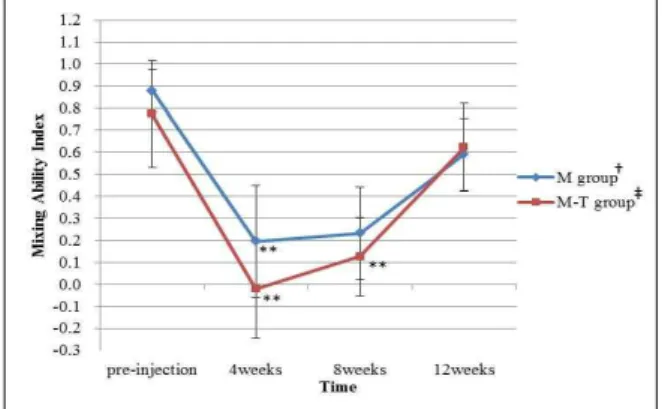
Values are given as mean and standard error
†: M group: Injection into the both masseter muscles
‡: M-T group: Injection into the both masseter and temporalis muscles
**:p-value<0.05 (compared to pre-injection) (n=20 in each group)
groups. In all food groups, the FIA was sharply decreased at 4 weeks and as time goes, it showed a pattern of gradual recovering. In all groups, it was statistically significant in 4 and 8 weeks.
Especially, the pattern was remarkable at hard and tough food group. Only the hard and tough food group in M-T group, the FIA was also significantly lower at 12 weeks (p-value: 0.0118) compared with pre-injection state.
Compared the M-T group with M group, the FIA of M-T group was lower in all food groups. There was significant difference between two groups was noted in hard and tough food group and moderate hardness group at 4 weeks (Mean difference: 0.57, 0.31, p-value: 0.0186, 0.0325, respectively). In 8 and 12 weeks, it was not significant in hard and tough food group (Mean difference: 0.28, 0.41, p-value:
0.2449, 0.0917, respectively) and moderate hardness food group (Mean difference: 0.16, 0.24, p-value:
0.2644, 0.0935, respectively). In soft and runny food group, it was not significant at 4, 8 and 12 weeks (Mean difference: 0.16, 0.03, 0.09, p-value: 0.1251,
(3) Mixing Ability Index (MAI)
Fig. 8 presents the MAI results of both groups as time. In M group, compared with pre-injection state (0.88±0.43), a Sharp drop of the MAI was detected at 4 weeks (0.19±1.13). It gradually recovered as time goes to 8 weeks (0.23±0.94) and 12 weeks (0.59±0.73). As with the M group, in M-T group, the MAI showed a significant decrease at 4weeks (-0.02±1.00) compared with the baseline (0.77±
1.09), and it gradually recovered as time goes to 8 weeks (0.13±0.80) and 12 weeks (0.62±0.90).
Compared with the M group, there was a remarkable decrease and rapid recovery of the MAI in M-T group. However, no significant difference was detected between two groups at 4, 8 and 12 weeks (Mean difference: 0.22, 0.11, -0.03, p-value:
0.4490, 0.7108, 0.9121, respectively).
Ⅳ. DISCUSSION
Botulinum toxin is a potent biological toxin produced by clostridium botulinum, which is exists in 7 different serotypes named A, B, C, D, E, F and G. BTX-A is known to be the most potent and lasting the longest among these seven serotypes, and it has proven to be a most valuable treatment for focal muscle hyperactivity disorders.17) BTX-A injected into the skeletal muscle belly prevents the release of acetylcholine by blocking the Ca2+ from the presynaptic axon of the motor endplate and blocks signal transmission at the neuromuscular junction. This process reduces muscle contraction, inducing a weakness condition.18) In orofacial region, BTX-A injected into the masticatory muscle induces masticatory muscle weakness necessarily and subsequently atrophy of masticatory muscle. So patients feel discomfort on mastication.
In relation to the BTX-A injection on masticatory muscle, most of the reports are concentrated on esthetics such as masseter hypertrophy19) or temporalis hypertrophy.20) Studies on masticatory functional changes are extremely rare, and only biting force change after botulinum toxin injection had studied.21) But evaluation of
biting force or electromyographic activity has a limitation that it is just a static analysis on mastication.
Previously mentioned, because the masticatory system is a very complex and organic system, if functional or structural changes happen in any one of the component of the masticatory system, it may be reflected by functional or structural disorder in any other compotent. The masticatory system has ability to wide range of adaptive modalities respond to transient and/or permanent demands. So this system, like any other biological system, cannot be viewed as a rigid and immutable, and dynamic analysis is crucial in study of this system.
Among the various objective methods for evaluating masticatory function, masticatory efficiency is a dynamic analysis which reflects the ones real masticatory state unlike others. It was first introduced in 1950.10) It analyzed the degree of breakdown of a food by sieving the comminuted peanuts. From then on, many studies using various test foods such as peanuts, almonds, carrots and synthetic materials have been done.22-24) Sieving method was used in many clinical fields and it is still used today, but it has limitations that the data results are not consistent. To get the reliable data results, several trials should be performed. Also, in clinical field, it is complex to use. Unlike comminution methods, a wide variety of methods have been developed and used to analyze the masticatory efficiency such as measuring color change in chewing gum,25,26) sugar loss form chewing gum27) and photometric methods to quantify changes in color.28) Since then, mixing and kneading of two colored gum29) or wax30) methods was developed and now most commonly used. It was evaluated the masticatory efficiency by the degree of mixing of two colors.
Mixing Ability Index (MAI) using two colored wax cube was developed by Sato et al. This method is easy to utilize in clinical fields and showed a reliability, validity and responsiveness for measuring the masticatory performance.12) It has been used in many dental fields.31,32)
For subjective methods, the FIA questionnaire composed of 30 types of Japanese foods was used as an effective tool for evaluation of masticatory function.33) Based on the Sugihara et al., Kim et al.
developed new FIA questionnaire composed of 30 types of Korean foods and applied it on Korean adults.15,16) But, there’s no study on relation to the botulinum toxin injection.
Therefore, this study aimed to evaluate the change of masticatory efficiency after BTX-A injection by the VAS, FIA adopted from earlier study15)and the MAI which is developed by Sato et al. Furthermore, we evaluated the differences of masticatory efficiency between masster muscle injection group and masseter and temporalis muscle injection group.
In terms of this study, the MAI was decreased after botulinum toxin injection in both M group and M-T group. Especially, it was markedly reduced at 4weeks. Since then, it was recovered gradually.
This result is similar as previous study in that biting force change after botulinum toxin injection.21) In that study, the maximum bite force decreased remarkably at 2 weeks and it gradually recovered as time goes to 4, 8 weeks by 12 weeks, and in 12 weeks, it was still lower than pre-injection state, but it was not significant.
Similar to the decrease of biting force, the masticatory efficiency also decreased. Also, the efficiency was recovered by the 12 weeks as similar to the pattern of biting force. Although mastication is affected by many factors, it seems that the change of major masticatory muscle have a greater impact on masticatory efficiency.
Molecular biologically, these results coincide with our results. Reversal of local paralysis occurs initially by neural sprouting with re-innervation of the muscle and ultimately by regeneration of the acetylcholine vesicle docking proteins, which restores function in 1 to 4 months. Other study showed that new neuromuscular junctions are formed by a process of presynaptic axonal sprouting. The duration of muscle paralysis is usually between 2 and 4 months with a gradual
recovery of full function thereafter 34)
The FIA and VAS was also showed a similar pattern as the MAI. But, unlike the MAI, the FIA and VAS was significantly decreased still at 12 weeks. Subjective discomfort on mastication was lasted longer than actual problem in masticatory efficiency. Correlation test between the VAS, FIA and MAI was not conducted in this study, but other studies on masticatory function have shown the moderate correlation of VAS and MAI, FIA and MAI.15,35)
Compare the M-T group with the M group, the VAS, FIA and MAI decreased much more in M-T group. The difference between two groups was evident at 4 weeks, and it was statistically significant in the VAS and FIA, but not in the MAI.
The reason for the difference was noted only in subjective methods is can be suspected that when subjects check the VAS and FIA, they evaluated their masticatory efficiency by just their biting force, not by overall masticatory efficiency. In 8 and 12 weeks, there was no significant difference between two groups in the VAS, FIA and MAI.
There was a rapid recovery of biting force after 4weeks in M-T group.
It can be explained that masseter muscle has more responsibility in mastication than temporalis muscle. So there was no significant difference of the MAI between two groups in this study. In earlier study, masseter muscle showed higher EMG than temporalis muscle in masticatory situation 36) In clinical field, botulinum toxin is injected into not only the masseter muscle but also temporalis muscle for treatment of sleep bruxism or myofascial pain of masticatory muscle, etc. There have been worried about worsen of masticatory function when injection is done into the masster muscle, besides the temporalis. But, it seems that the disturbance of masticatory function is not severely noticeable than injection is done only into the masseter muscle in dose of this study.
But, as mentioned above, the values in M-T group is generally lower than M group although it was not significant. These results can be explained
that the botulinum toxin was injected into two major masticaroty muscles in M-T group, while only one major muscle was injected in M group. So, if study design is modified such as more units of botulinum toxin are injected into the temporalis muscle and/or more subjects are participated, the result might be significant and more definitive.
In evaluation of the FIA according to the food properties, it was noted that the FIA of all food groups was remarkably decreased at 4 weeks and it gradually recovered as time goes. There were variances among food properties. Hard and tough food group showed marked change during 12 weeks. Disturbance of food intake ability was much more affected when we intake a type of hard and tough foods, and it lasted longer than any other food type. The FIA was not fully recovered at 12 weeks in M-T group. As expected, subjects felt more discomfort on mastication of hard foods than soft foods. The total amount of EMG activity has been shown to depend on the hardness of the food:
more EMG activity is observed for harder foods.37,
38)The character of the food influences the chewing pattern39) and the opening length depends on the size and hardness of the food bolus.40) The harder the food, the more chewing strokes are needed.41)It seems that people evaluate their masticatory ability by hard foods rather than soft foods. This result is similar as hard foods showed higher correlation with the bite force than soft foods15)and breakdown of hard foods is more strongly correlated with the maximum bite force.42)Also, there was a significant change of the FIA in soft foods group. It can be explained that not only hard and tough foods, but also soft and runny foods are affected by the BTX-A injection. In comparison of both two groups according to the food properties, general value of the FIA was lower in M-T group. There was marked difference was finding in hard and tough food group and moderate hardness group in 4 weeks and, not in 8 and 12 weeks. As mentioned above, it seems that there is a rapid recovery after 4 weeks.
There’s an opinion that the subjective methods
related to the FIA can more reflect the one’s real state better than objective methods.33,43)To evaluate the precise masticatory function, subjective methods as well as objective methods should be considered together.
A several limitations of this study were detected as the study progress. First, for blind test, as for the M group, it would be better that botulinum toxin injection is done into the masseter muscle and saline injection is done into the temporalis muscle.
It might be influence the subjective evaluation.
Seconds, the subjects were not familiar with the wax specimen. It would be better that wax was chewed by subjects preliminarily before the entry of the study. Third, if the subject enrollment was done through the pilot study within a certain limited range with similar MAI, the results would be more precise. Forth, as for the FIA, it would be needed to control the food, and subject must eat the food throughout the study. Also, the individual recognition on food might be different. Therefore, more detailed explanation of the properties and hardness of each food in the questionnaire might help decrease the individual variation. Finally, a longer follow-up will be needed to determine the definitive duration of recover the masticatory efficiency.
V. CONCLUSSION
We may conclude that masticatory efficiency was significantly lowered after BTX-A injection into the masticatory muscle, but it gradually recovered in a predictable pattern by the 12 weeks. The disturbance of subjective masticatory efficiency was lasted longer than objective masticatory efficiency. When BTX-A was injected into the masseter and temporalis muscle, the masticatory efficiency was lower than when injection was done only into the masseter muscle. It was statistically significant in the VAS and FIA at 4 weeks, but the MAI showed no significancy. After 4weeks, there was rapid recovery of muscle function in M-T group, and the difference between two groups was
not significant. It could be concluded that there will be no serious disturbance of mastication compared to injection is done only into the masseter muscle, even if injection is done into the masseter and temporalis muscle in dose of this study. According to the food properties, it was confirmed that people feel more discomfort on taking hard and tough foods after BTX-A injection and not only hard foods, but also intake of soft and runny foods was influenced by botulinum toxin injection.
REFERENCES
1. Clark GT. The management of oromandibular motor disorders and facial spasms with injections of botulinum toxin. Phys Med Rehabil Clin N Am 2003;14: 727-748.
2. Okeson JP. Management of temporomandibular disorders and occlusion. 1993, Mosby
3. van der Bilt A, Engelen L, Pereira LJ, van der Glas HW, Abbink JH. Oral physiology and mastication.
Physiol Behav 2006;89:22-27.
4. Fueki K, Kimoto K, Ogawa T, Garrett NR. Effect of implant-supported or retained dentures on masticatory performance: a systematic review. J Prosthet Dent 2007;98:470-477.
5. Zarrinkelk HM, Throckmorton GS, Ellis E 3rd, Sinn DP. A longitudinal study of changes in masticatory performance of patients undergoing orthognathic surgery. J Oral Maxillofac Surg 1995;53:777-782;
discussion 782-783.
6. Okiyama S, Ikebe K, Nokubi T. Association between masticatory performance and maximal occlusal force in young men. J Oral Rehabil 2003;30:278-282.
7. Tsuga K, Carlsson GE, Osterberg T, Karlsson S.
Self-assessed masticatory ability in relation to maximal bite force and dental state in 80-year-old subjects. J Oral Rehabil 1998;25:117-124.
8. Lepley CR, Throckmorton GS, Ceen RF, Buschang PH. Relative contributions of occlusion, maximum bite force, and chewing cycle kinematics to masticatory performance. Am J Orthod Dentofacial Orthop 2011;139:606-613.
9. Gonzalez Y, Iwasaki LR, McCall WD Jr, Ohrbach R, Lozier E, Nickel JC. Reliability of electromyographic activity vs. bite-force from human masticatory muscles. Eur J Oral Sci 2011;119:219-224.
10. Manly RS, Braley LC: Masticatory performance and
efficiency. J Dent Res 29: 448-462, 1950.
11. Edlund J, Lamm CJ. Masticatory efficiency. J Oral Rehabil 1980;7:123-130.
12. Sato H, Fueki K, Sueda S, Sato S, Shiozaki T, Kato M et al. A new and simple method for evaluating masticatory function using newly developed artificial test food. Journal of Oral Rehabilitation 2003;30:68-73.
13. Owens S, Buschang PH, Throckmorton GS, Palmer L, English J. Masticatory performance and areas of occlusal contact and near contact in subjects with normal occlusion and malocclusion. Am J Orthod Dentofacial Orthop 2002;121:602-609.
14. Magalhães IB, Pereira LJ, Marques LS, Gameiro GH.
The influence of malocclusion on masticatory performance. A systematic review. Angle Orthod 2010;80:981-987.
15. Kim BI, Jeong SH, Chung KH, Cho YK, Kwon HK, Choi CH. Subjective food intake ability in relation to maximal bite force among Korean adults. J Oral Rehabil 2009;36:168-175.
16. Jeong SH, Kang SM, Ryu JH, Kwon HK, Kim BI.
Subjective food intake ability in relation to the Mixing Ability Index in Korean adults. J Oral Rehabil 2010;37:242-247.
17. Clark GT, Stiles A, Lockerman LZ, Gross SG. A critical review of the use of botulinum toxin in orofacial pain disorders. Dent Clin North Am 2007;51:
245-261.
18. Melling J, Hambleton P, Shone CC. Clostridium botulinum toxins: nature and preparation for clinical use. 1988;Eye 2 (Pt 1):16-23.
19. Moore AP, Wood GD. The medical management of masseteric hypertrophy with botulinum toxin type A.
Br J Oral Maxillofac Surg 1994;32:26-28.
20. Isaac AM. Unilateral temporalis muscle hypertrophy managed with botulinum toxin type A. Br J Oral Maxillofac Surg 2000;38:571-572.
21. Ahn KY, Kim ST. The change of maximum bite force after botulinum toxin type a injection for treating masseteric hypertrophy. Plast Reconstr Surg 2007;
120:1662-1666.
22. Imaoka S, Ozawa T, Saitoh M, Tanaka S, Hibino K, Hirukawa T, et al. Study of a particle counting method for index of masticatory efficiency. Nihon Hotetsu Shika Gakkai Zasshi 1989;33:791-803.
23. Olthoff LW, van der Bilt A, Bosman F, Kleizen HH.
Distribution of particle sizes in food comminuted by human mastication. Arch Oral Biol 1984;29:899-903.
24. Goiato MC, Garcia AR, Dos Santos DM, Zuim PRJ.
Analysis of masticatory cycle efficiency in complete denture wearers. J Prosthodont 2010;19:10-13.
25. Matsui Y, Ohno K, Michi K, Hata H, Yamagata K, Ohtsuka S. The evaluation of masticatory function with low adhesive colour-developing chewing gum. J Oral Rehabil 1996;23:251-256.
26. Ishikawa Y, Watanabe I, Hayakawa I, Minakuchi S, Uchida T. Evaluations of masticatory performance of complete denture wearers using color-changeable chewing gum and other evaluating methods. J Med Dent Sci 2007;54:65-70.
27. Heath MR. The effect of maximum biting force and bone loss upon masticatory function and dietary selection of the elderly. Int Dent J 1982;32:345-356.
28. Gunne HS. Masticatory efficiency. A new method for determination of the breakdown of masticated test material. Acta Odontol Scand 1983;41:271-276.
29. Prinz JF. Quantitative evaluation of the effect of bolus size and number of chewing strokes on the intra-oral mixing of a two-colour chewing gum. J Oral Rehabil 1999;26:243-247.
30. Sugiura T, Fueki K, Igarashi Y. Comparisons between a mixing ability test and masticatory performance tests using a brittle or an elastic test food. J Oral Rehabil 2009;36:159-167.
31. Otomaru T, Sumita YI, Chang Q, Fueki K, Igarashi Y, Taniguchi H. Investigation of predictors affecting food mixing ability in mandibulectomy and/or glossectomy patients. J Prosthodont Res 2009;53:111- 115.
32. Tumrasvin W, Fueki K, Ohyama T. Factors associated with masticatory performance in unilateral distal extension removable partial denture patients. J Prosthodont 2006;15:25-31.
33. Sugihara et al. Multivariate analysis of food acceptance with regard to missing teeth in the elderly. Shikwa Gakuho 1989;89:1275-1280.
34. Duchen LW. An electron microscopic study of the changes induced by botulinum toxin in the motor end-plates of slow and fast skeletal muscle fibres of the mouse. J Neurol Sci 1971;14: 47-60.
35. Ahn HJ, Lee YS, Jeong SH, Kang SM, Byun YS, Kim BI. Objective and subjective assessment of masticatory function for patients with temporoman- dibular disorder in Korea. J Oral Rehabil 2011;38:
475-481.
36. Adhikari H, Kapoor A, Prakash U, Srivastava A.
Electromyographic pattern of masticatory muscles in altered dentition Part II. J Conserv Dent 2011;14:
120-127.
37. Agrawal KR, Lucas PW, Bruce IC, Prinz JF. Food properties that influence neuromuscular activity during human mastication. J Dent Res 1998;77:
1931-1938.
38. Mioche L, Bourdiol P, Martin JF, Noël Y. Variations in human masseter and temporalis muscle activity related to food texture during free and side-imposed mastication. Arch Oral Biol 1999;44:1005-1012.
39. Beyron H. Occlusal relations and mastication in Australian aborigines. Acta Odontol Scand 1964;22:
597-678.
40. Lucas PW, Ow RK, Ritchie GM, Chew CL, Keng SB.
Relationship between jaw movement and food breakdown in human mastication. J Dent Res 1986;65:400-404.
41. Horio T, Kawamura Y. Effects of texture of food on chewing patterns in the human subject. J Oral Rehabil 1989;16:177-183.
42. Salleh NM, Fueki K, Garrett NR, Ohyama T.
Objective and subjective hardness of a test item used for evaluating food mixing ability. J Oral Rehabil 2007;34:174-183.
43. Takata Y, Ansai T, Awano S, Fukuhara M, Sonoki K, Wakisaka Met al.Chewing ability and quality of life in an 80-year-old population. J Oral Rehabil 2006;
33:330-334.
국문초록
보툴리눔 A형 독소가 저작효율에 미치는 영향
연세대학교 치과대학 구강내과학교실 박형욱․권정승․김성택․최종훈․안형준
보툴리눔 독소는 Clostridium botulinum이라는 박테리아가 만들어내는 독소를 의료용으로 희석, 정제한 약제로서 이 독소 가 시냅스 전 신경말단에 부착되어 신경근접합부에서 아세틸콜린의 분비를 억제함으로써 근육의 수축을 차단하는 역할을 한다. 보툴리눔 독소 주사 요법은 구강안면 분야에서 저작근의 근경축, 근경련, 운동이상증, 측두하악관절질환, 근막통증, 이 갈이, 특발성 교근비대증 또는 만성편두통을 비롯한 두통 등 다양한 질환에서 가역적, 보존적 치료법으로 활발히 사용되고 있다. 사용 범위가 넓어지면서 교근이나 측두근 같은 저작근 부위에 주사를 하는 경우가 많아지고 있으며 그러한 경우 약제 자체의 약리학적 작용에 의해 일시적인 근마비 및 근위축에 따라 저작기능에 변화가 생기게 된다. 하지만 지금까지 보툴리눔 독소 주사치료의 효과나 활용에 대한 연구만이 주를 이루었고 부작용 중의 하나인 저작기능 변화에 대한 연구는 거의 이루어 지지 않았다. 몇몇 저작기능과 관련한 연구 또한 근전도 등을 이용한 정적인 상태의 저작력 감소에 대한 연구로, 실제로 동적 인 저작 환경을 반영하는 저작 효율에 대해서는 현재까지 전혀 연구가 이루어지지 않은 실정이다.
이에 본 연구에서는 총 40명의 건강한 성인환자를 대상으로 보툴리눔 A형 독소 주사 요법을 교근 부위에만 시술 받은 사람 과 교근과 측두근 부위 모두에 시술 받은 사람 각각 20명씩 두 군으로 나누어 주사를 시행하고, 주사 전과 주사 후 4주, 8주, 12주째의 저작 효율의 변화를 주관적 평가방법인 식품섭취능 (Food Intake Ability, 이하 FIA)과 저작 능력에 대한 주관적 인식도 (Visual Analogue Scale, 이하 VAS), 객관적 평가 방법인 Mixing Ability Test를 시행하여 알아보았다.
그 결과 보툴리눔 A형 독소 주사 후 4주 째에 저작효율의 뚜렷한 감소가 나타났으며 이후 서서히 회복되는 경향을 보였다.
주사부위를 달리하여 본 결과 교근과 측두근에 모두 주사를 시행하는 경우, 교근에만 주사를 시행하는 경우에 비해 주관적, 객관적인 값 모두 낮게 나타났다. 주관적인 값에 있어서 4주 째 유의하였으나 이후에는 유의하지 않았으며 객관적인 값은 전 기간에 걸쳐 유의하지 않았다. 실험에서 정한 주사 용량에 대해서는 측두근까지 주사가 이루어지더라도 교근만 주사를 시행한 사람과 비교하여 저작효율의 큰 차이가 없는 것으로 보인다. 또한 식품성상에 따라 저작 효율을 살펴본 결과 보툴리눔 A형 독소 주사가 단단하고 질긴 음식 섭취에 더 큰 영향을 미치는 것으로 보이며 무르고 약한 음식의 섭취 시에도 유의할만 한 영향을 받는다는 사실을 확인 할 수 있었다.
주제어: 보툴리눔 A형 독소, 저작 효율, 교근, 측두근