Introduction
Lacrimal canaliculitis is an infectious disease of the eye’s lacrimal duct system. In most cases of primary canaliculitis, no predisposing factors can be detected.
1)Studies involving dacryocystography in patients with canaliculitis have reported dilatation, irregularity, and prominent filling defects of the affected canaliculi in almost all patients.
2)Stasis secondary to diverticulum or obstruction of the canaliculus has been theorized to promote anaerobic bacterial growth, which may be a predisposing factor in canaliculitis.
1)Actinomyces spp. are most frequently implicated in this disease; however, several different organisms have been shown to cause canaliculitis.
3)The classic clinical features of lacrimal canaliculitis include unilateral conjunctivitis, mild to severe canalicular swelling, mucopurulent discharge from
the punctum, inflammation of the medial canthus, epiphora, and a red, pouting punctum.
3)Despite this classic presentation, lacrimal canaliculitis is often misdiagnosed.
3)In this report, we describe a rare case of lacrimal canaliculitis induced by Aspergillus flavus this is a fairly uncommon etiology, and no such cases have been previously reported.
Case Report
A 59-year-old man presented to our department with a 3-month history of chronic conjunctivitis in his right eye.
The patient’s conjunctivitis was diagnosed as bacterial in origin, but the patient exhibited poor response to a 4-week course of topical chloramphenicol ophthalmic drops. He developed swelling and tenderness of the medial canthal portion of the right lower eyelid. The patient’s visual acuity was 20/25 in the right eye and 20/25 in the left eye.
Upon examination of the patient’s right eye, the conjunctiva near the lesion and the right lower punctum
고신대학교 의과대학 학술지 제 권 제 호25 1 Kosin Medical Journal
Vol. 25. No. 1, pp. 106 108, 2010∼
Jung-Joo Lee
1․ Ji-Eun Lee
1․ Sang-Joon Lee
1,2Department of Ophthalmology
1, Institute for Medicine
2, Kosin University College of Medicine, Busan, Korea
――― Abstract ――――――――――――――――――――――――――――――――――――――――
Lacrimal canaliculitis is a condition normally caused by an infection of the canaliculi the condition is frequently associated with Actinomyces species. Rarely, however, bacteria, fungi, and viruses are also implicated in the infection leading to lacrimal canaliculitis. Lacrimal canaliculitis is also occasionally misdiagnosed as mucoceles, dacryocystitis, conjunctivitis, blepharitis, or chalazion.
In this report, we describe a rare case of lacrimal canaliculitis caused by Aspergillus flavus.
A 59-year-old man presented to our department with a 3-month history of chronic conjunctivitis in his right eye. The patient was initially diagnosed with bacterial conjunctivitis, but did not respond to topical antibiotics. Our examination revealed signs of canaliculitis, and Aspergillus flavus was cultured from the discharge. Based on the culture results, the patient was treated with 0.1% itraconazole ophthalmic drops administered every 2 hours and 200 mg of oral itraconazole once daily, for a total of 3 weeks. At the time of examination 3 weeks after this treatment, repeated microbiological cultures of the discharge detected no further growth of any organisms, and the patient’s symptoms were profoundly improved.
All symptoms and signs were completely resolved, and the patient exhibited no signs indicative of disease in the 2-week follow-up examination.
―――――――――――――――――――――――――――――――――――――――――――――――――
Key words : Aspergillus flavus, Canaliculitis, Chronic conjunctivitis
교신저자:Address: reprint requests to Sang Joon Lee, MD Gospel Hospital, Kosin University
#34 Amnam-dong, Seo-ku, Busan 602-702, Korea.
Tel: 82-051-990-6140, Fax. 82-051-990-3026 E-mail: hiatus@ns.kosinmed.co.kr
Misdiagnosis of Aspergillus Canaliculitis as Chronic Conjunctivitis
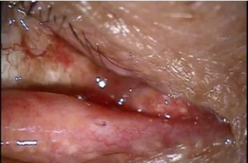
were inflamed, and the punctum was prominent and pouting (Fig.1). No erythematous swelling or tenderness was noted in the lacrimal sac region. Via the application of counterpressure with 2 cotton-tipped applicators to the affected lower medial canthus region and squeezing medially toward the inferior punctum, a large amount of mucopurulent discharge was expressed from the inferior lacrimal canaliculus (Fig. 2). The patient’s left eye was normal. Saline irrigation through the lower punctum revealed reflux from the right lower punctum. However, no lacrimal concretions were detected in the canaliculi and lacrimal sac after the application of pressure. Based on these clinical findings, the patient was diagnosed with lacrimal canaliculitis. The punctal discharge was collected for further microbiologic investigation.
Fig. 1. Slit lamp photograph of the right eye showing an inflamed lower canaliculus with a pouting punctum with purulent discharge.
Fig. 2. Palpation over the inferior canaliculus using a cotton-tipped applicator resulted in the expression of a mucopurulent discharge from the punctum.
Until the microbiologic results became available, the patient was treated for 1 week with topical 0.3% gatifloxacine ophthalmic drops 4 times daily, and 1g of oral methylol
cephalexin lysinate 3 times daily.
However, this remitting pattern of disease persisted in spite of the administration of topical and oral antibiotics.
After 1 week, the culture results revealed the presence of Aspergillus flavus. The culture results and antifungal susceptibility testing showed that the organism was susceptible to itraconazole the patient was therefore treated with 0.1% itraconazole ophthalmic drops every 2 hours, and with 200mg of oral itraconazole once daily for 3 weeks. At the 3-week evaluation, repeated microbiological cultures of the dischargesrevealed no further growth of any organisms, coupled with vast improvements in the patient’s symptoms. Complete resolution of all symptoms and signs was noted, and the patient evidenced no signs indicative of disease on a follow-up examination conducted 2 weeks later (Fig.3). No further recurrence was noted.
Fig.3. Photography of patient 5 weeks after beginning therapy. No evidence of disease upon examination.
Discussion
Lacrimal canaliculitis is an infection of the lacrimal canalicular system. Lacrimal canaliculitis has been variously misdiagnosed as conditions such as chronic conjunctivitis, blepharitis, mucoceles, dacryocystitis, hordeolum, and chalazion.
3,4)Hussain et al.
5)noted previously that patients suffering from canaliculitis frequently remain undiagnosed for months or even years, and therefore frequently receive inappropriate treatment for their recurring symptoms.
There are several reasons for this frequent misdiagnosis.
3)First, lacrimal canaliculitis is a relatively rare disease, accounting for only 2% of all patients with lacrimal disease.
6)
Furthermore, lacrimal canaliculitis may present without
고신대학교 의과대학 학술지 제 권 호 25 1 , 2010
all of its classic features, which also renders misdiagnoses more likely.
7)In our patient’s case, the etiology was first thought to be bacterial, but the patient exhibited a poor response to long-term topical antibiotics.
A variety of microorganisms have been implicated previously as pathogens that can cause lacrimal canaliculitis
3)