ficult to standardize [1]. For development and standardization of flow cytometry, the EuroFlow Consortium was formed in 2005, and the EuroFlow project was initiated in 2006 [1]. Currently Cy- tognos, Inc. (Salamanca, Spain) provides several EuroFlow prod- ucts, such as multiple myeloma (MM) minimal residual disease (MRD) kits, lymphoid screening tube (LST), acute leukemia orien- tation tube (ALOT), and plasma cell screening tube (PCST) kits [2]. The PCST kit is a pre-mixed combination of 8 conjugated anti- bodies designed for identification and quantitation of plasma cells. Additionally, it can also discriminate between normal poly- clonal plasma cells versus aberrant monoclonal plasma cells [2].
As there are no prior reports in the literature regarding the PCST kit designed by Cytognos used with the EuroFlow panel, herein we have described our experience with the PCST kit, comparing it to a conventional 4-color, 2-tube method for analysis of plasma Flow cytometry is being used increasingly in clinical hematol-
ogy to establish diagnosis, prognostic classification, and evalua- tion of treatment effectiveness of hematologic neoplasms. How- ever, flow cytometry is highly dependent on expertise, and is dif-
반응성 형질세포와 악성 형질세포 감별을 위한 형질세포 선별튜브의 유용성 평가
Utility of Plasma Cell Screening Tube Kit to Differentiate Neoplastic Plasma Cells from Reactive Plasma Cells
방해인·최태윤
Hae In Bang, M.D., Tae Youn Choi, M.D.
순천향대학교 부속 서울병원 진단검사의학과
Department of Laboratory Medicine, Soonchunhyang University Seoul Hospital, Seoul, Korea Vol. 10, No. 4: 314-320, October 2020
https://doi.org/10.47429/lmo.2020.10.4.314 진단혈액학
Corresponding author: Tae Youn Choi, M.D., Ph.D.
https://orcid.org/0000-0002-1950-8010
Department of Laboratory Medicine, Soonchunhyang University Seoul Hospital, 59 Daesagwan-ro, Yongsan-gu, Seoul 04401, Korea Tel: +82-2-709-9425, Fax: +82-2-709-9083, E-mail: choity@schmc.ac.kr Received: November 5, 2019
Revision received: June 2, 2020 Accepted: June 9, 2020
This article is available from https://www.labmedonline.org 2020, Laboratory Medicine Online
This is an Open Access article distributed under the terms of the Creative Commons Attribution Non-Commercial License (https://creativecommons.org/licenses/by-nc/4.0/) which permits unrestricted non-commercial use, distribution, and reproduction in any medium, provided the original work is properly cited.
Flow cytometry is a powerful tool for analysis of hematologic malignancies, that provides rapid, quantitative, and multiparametric analysis of het- erogeneous cell populations, but requires standardization because of complexities in panel design and interpretation. Here, we compared the Plasma Cell Screening Tube (PCST) kit (Cytognos, Spain) in conjunction with EuroFlow antibody panels for standardization of flow cytometry to a conventional method for diagnosis of plasma cell dyscrasias. Thirty-nine bone marrow samples and one peripheral blood sample from 40 patients were tested. Thirty-three patients were diagnosed with multiple myeloma (MM), and seven were in a reactive state. In PCST implementation, eight antibodies were used for staining, including anti-CD45-Pacific Blue, anti-CD19-PECy7, anti-CD138-OC515, anti-CD38-FITC, anti-CD56-PE, anti-β2- microglobulin-PerCPCy5.5, anti-kappa-APC, and anti-lambda-APC-C750. Plasma cells were initially identified using CD38 and CD138; thereafter, CD38+, CD138+ gated cells were analyzed for CD56, CD19, CD45, cytoplasmic kappa, cytoplasmic lambda, and β2-microglobulin. Conventional flow cytometry was performed with six monoclonal antibodies, including anti-CD56-FITC, anti-kappa-FITC, anti-CD19-PE, anti-lambda-PE, anti- CD138-PECy5, and anti-CD45-PECy7 (Beckman Coulter, USA). Monoclonal plasma cells with cytoplasmic light-chain restriction were detected in 30 of 33 (90.9%) MM cases by conventional methods, and 32 of 33 (97.0%) MM cases with the PCST method. No differences were noted between PCST and the conventional method in immunophenotyping and plasma cell percentages (P=0.323). Among plasma cells, levels (%) were signifi- cantly higher by the PCST approach than those in the conventional method (97.6% vs 95.8%, P=0.010). PCST exhibited better performance for plasma cell dyscrasias diagnosis, and could improve laboratory efficiency and quality.
Key Words: Flow cytometry, Immunophenotyping, Multiple myeloma, Plasma cells, Plasma cell neoplasm, Plasma cell dyscrasias
2017-03-16 https://crossmark-cdn.crossref.org/widget/v2.0/logos/CROSSMARK_Color_square.svg
criteria. Reactive plasmacytosis was defined as a level of bone marrow (BM) plasma cells >3.0%, without any evidence of a plasma cell disorder. Thirty-nine BM samples and one peripheral blood (PB) sample were analyzed. Five hundred cells in BM aspi- rate smears and two hundred cells in PB smears were counted for differentials. This study was approved by the Institutional Review Board of Soonchunhyang University Hospital, Seoul (IRB 2018- 01-001). Conventional 4-color, 2-tube analysis was performed us- ing monoclonal antibodies against CD56, CD19, CD138, and CD45 for surface staining, and using kappa, and lambda monoclonal antibodies for cytoplasmic staining [3]. White blood cell counts were adjusted to 3×10⁶/mL. For the surface staining tube, 100 μL of the washed specimens was incubated with 10 μL of each of the monoclonal antibodies for 20 minuts at room temperature in the dark. Four monoclonal antibodies were used for flow cytometric immunophenotyping: fluorescein isothiocyanate (FITC)-conju- gated anti-CD56; phycoerythrin (PE)-conjugated anti-CD19; PE- cyanine 5.1 (PECy5)-conjugated anti-CD138; and PE-Cyanine 7 (PECy7)-conjugated anti-CD45 (Beckman Coulter, Miami, FL, USA).
VersaLyse lysing solution (Beckman Coulter) was added to the tube, vortexed immediately for 10 sec, and then incubated for 10 min at room temperature in the dark. For cytoplasmic staining tube, 50 μL of the washed samples was stained with PECy5-anti- CD138 and PECy7-anti-CD45, and then incubated for 20 minutes at room temperature in the dark. Thereafter, an IntraPrep proce- dure was performed. Then, FITC-anti-kappa and PE-anti-lambda (Beckman Coulter) were added and incubated for 20 minutes at room temperature in the dark. Acquisition of 250,000 nucleated cells was performed using a BC Navios flow cytometer (Beckman Coulter), and subsequent analyses were performed using Kaluza (v.1.3, Beckman Coulter). Plasma cells were initially identified us- ing CD138 and side scatter, and CD138+ gated cells were ana- lyzed for CD56, CD19, CD45, and cytoplasmic kappa and lambda (Fig. 1A).
In the PCST method, the Beckman Coulter Navios flow cytom- eter setup and fluorescence compensation settings were performed according to the manufacturer’s instructions [4]. Fluorochrome-
were APC-conjugated anti-kappa, and APC-C750-conjugated anti- lambda (Cytognos). Both are present in the pre-mixed antibody cocktail in the PCST kit. 50 μL of the sample and 30 μL of the sur- face staining reagent were added to each tube, and PBS with 1.0%
bovine serum albumin was added to a final volume of 100 μL per tube. Incubation for 30 minutes was conducted at room tempera- ture in the dark. After washing, Reagent A (fixative solution; Nor- dic-MUbio BV, Susteren, Netherlands) was added and incubated for 15 minutes, washed with PBS, then reagent B (permeabilizing solution; Nordic-MUbio) was added. Next, 10 μL of the pre-mixed vial intracellular antibodies was added and incubated for 15 min- utes. After washing with PBS, data acquisition for 150,000 nucle- ated cells was performed using the Navios flow cytometer; subse- quent analyses were performed using Kaluza. Plasma cells were initially identified using CD38 and CD138, following which, CD38+, CD138+ gated cells were analyzed for CD56, CD19, CD45, cytoplasmic kappa, cytoplasmic lambda, and
β
2-microglobulin (Fig. 1B). Data were entered into SPSS v.22 (SPSS Inc., Chicago, IL, USA) and analyzed. To determine the correlation of each method, generalized estimating equation (GEE) and the Spearman’s rank correlation tests were performed.Among the 33 MM cases, conventional methods could detect the abnormal immunophenotypes of plasma cells with light-chain restriction in 30 cases (90.9%). However, the PCST method was able to detect abnormal immunophenotypes in 32 cases (97.0%).
In the single remaining case, malignant plasma cells were not iden- tified by either conventional or PCST methods. Therefore, there were three cases in which malignant plasma cells could not be detected by conventional methods; these cases are shown in Fig.
2. In case A, gated plasma cells comprised 1.13% by PCST and 0.72% by conventional methods. Although the detected plasma cell (%) levels were lower by conventional methods, surface anti- gens such as CD19 and CD56 could be analyzed. However, it was difficult to analyze cytoplasmic light-chain restriction due to the poor intracellular staining observed in the conventional method.
In Case B, sufficient plasma cells were present in the sample, but cytometric gating could not be performed in the conventional
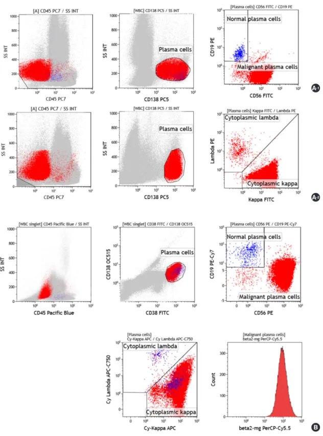
Fig. 1. Representative example of flow cytometric immunophenotyping using two methods to distinguish between neoplastic plasma cells and re- active plasma cells in a patient with multiple myeloma. (A) Conventional 4-color, 2 tube method. Plasma cells were gated with CD138+ and low side scatter, and were distinguished as neoplastic cells (CD56+/CD19-/CD138+/CD45-) and reactive plasma cells (CD56-/CD19+/CD138+/CD45+) in a first tube (A-1). Cytoplasmic light-chain restriction was analyzed in a second tube (A-2). (B) Plasma Cell Separation Tube (PCST) method. Plasma cells were gated for CD38+ and CD138+ and analyzed for CD56, CD19, CD45, cytoplasmic kappa, cytoplasmic lambda, and β2-microglobulin in one tube.
A-1
A-2
B
Fig. 2. Flow cytometry analysis of three cases in which we were not able to detect malignant plasma cells using the conventional method (A-1, B-1, C-1: Plasma Cell Separation Tube (PCST) method; A-2, B-2, C-2: Conventional method). (A) The first case was a 56-year-old female patient who was diagnosed with kidney amyloidosis, and then diagnosed with multiple myeloma (MM) because of IgM-λ type monoclonal gammopathy (M protein: 0.02 g/dL), with increased plasma cells in bone marrow aspirates (27% of all the nucleated cells, ANCs). Immunophenotyping of plasma cells was CD19-CD56+CD45-/dim+. PCST was able to identify cytoplasmic lambda light-chain restriction. However, the conventional method failed to do so. (B) The second case was a 59-year-old female patient who was admitted with left flank pain. She was diagnosed as MM because of IgD-λ type monoclonal gammopathy (M protein: 0.51 g/dL) with increased plasma cells in bone marrow aspirates (35% of ANCs). Using flow cytometry analysis, plasma cells were found to represent 7.67% of ANCs, and 99.21% of these presented CD19-CD56dim+CD45-/dim+ immunophenotypes, whereas it was difficult to gate plasma cells by the conventional method, which prevented accurate analysis.
(Continued to the next page) A-1
A-2
B-1
B-2
method without CD38 staining. In case C, malignant plasma cells were not found because they displayed normal immunopheno- types, such as CD19+, CD56-, and CD45+. Additionally, light-chain restriction could not be observed because the patient had biclonal gammopathy involving IgG-
κ
and IgG–λ
types.Among the 37 cases, with an exception of the three cases pre- senting discrepant results, there were no differences in terms of immunophenotyping results. Mean values [95% confidential inter- val, CI] of plasma cell numbers (%) detected by PCST and conven- tional methods were 14.7% [8.9-20.6%] and 16.0% [9.0-23.0%], re- spectively. The plasma cell (%) levels identified by the PCST method were 1.3% lower than by the conventional method, but there was no significant difference between the two methods (GEE, P=0.323).
The plasma cell (%) numbers detected by the two methods were significantly lower than those identified by manual differential counts (GEE, P<0.001). Compared to manual differential counts, plasma cell (%) abundance using PCST was 22.1% lower, and the conventional method was 20.9% lower. Among plasma cells, de- tection of abnormal plasma cell (%) levels was significantly higher by PCST than that by the conventional method (GEE, P=0.010).
Mean values [95% CI] of abnormal plasma cell (%) levels deter-
mined using PCST and conventional methods were 97.6% [95.7- 99.4%] and 95.8% [92.8-98.8%]. Mean fluorescent intensity (MFI) values for
β
2-microglobulin were not correlated withβ
2-microglo- bulin concentrations identified by radioimmunoassay (the Spear- man’s rank correlation coefficient=0.09, P=0.648).Among our 33 MM cases, there was only one patient in which light-chain restriction could be confirmed by the PCST method, but not by the conventional method. It could be assumed that this difference between the two methods arose due to the differing composition of assay reagents and the testing procedures involved.
This could represent an example of the importance of using vali- dated reagents and kits. Percentages of plasma cells determined by both methods were significantly lower than those determined by manual differential counts. The lower percentage of plasma cells determined by flow cytometry compared to morphology may be due to the provision of a blood-diluted sample, or a sam- ple of liquid BM [5, 6]. MFI values of
β
2-microglobulin were not correlated withβ
2-microglobulin concentrations determined by radioimmunoassay.β
2-microglobulin levels identified by flow cytometry reflect the amount ofβ
2-microglobulin on the cell sur- face. Further study will be needed to determine how the amountC-1
C-2 Fig. 2. (Continued) (C) The third case was a 72-year-old female patient who was diagnosed with MM because of biclonal gammopathy, IgG-κ and IgG-λ (M protein: 2.03 g/dL), with increased plasma cells in bone marrow aspirates (22% of ANCs). Malignant plasma cells showed normal immun- ophenotypes, CD19+ CD56- CD45+. Therefore, malignant plasma cells could not be detected with either PCST or conventional methods. Addition- ally, cytoplasmic light-chain restriction could not be observed because of biclonal gammopathy composed of two different types of light-chains, κ and λ.
sensus involving multiparametric flow cytometry in MM and re- lated disorders. The minimal test antigens for classification of ab- normal plasma cells are CD19 and CD56. Assessment of cytoplas- mic
κ
/λ
expression by flow cytometry is important to demonstrate clonality at presentation [14]. The Cytognos PCST kit contains an- tibodies against essential markers such as CD19, CD56, Igκ
, and Igλ
. The major advantage of this PCST kit is the use of multiple colors in one tube, and thus, it can recognize multiple antigens si- multaneously. For example, when abnormal plasma cells are mixed with normal plasma cells and analyzed using the PCST kit, cyto- plasmic light-chain restriction can be analyzed only in abnormal plasma cells [2]. The PCST kit contains premixed reagent combi- nations with 8 conjugated antibodies, with instructions regarding the device, and compensation settings. Therefore, a variety of lab- oratories use it for convenience and with ease. The PCST kit can be used to test PB samples, as well as BM samples [2]. Among 40 samples in our study, there was one PB sample from a patient who was diagnosed with plasma cell leukemia. Although only this single case was tested using a PB sample, there were no dif- ferences in the MFI of the target population cells compared to the results for other BM samples. The presence of clonal circulating plasma cells is an indicator of high risk of disease development in newly diagnosed MM patients [15, 16]. Therefore, prognostic eval- uation of MM patients, when needed, as well as diagnosis of plasma cell leukemia, can be undertaken using PCST.Multi-color analysis in a single tube is a powerful advantage of the PCST method for gating neoplastic plasma cells and for ana- lyzing their properties. Accelerated acquisition times and standard- ized compensation parameters may shorten total test times. Addi- tionally, standardized protocols and approaches may overcome the variability inherent in flow cytometric assays conducted by different laboratories. PCST exhibited superior performance in detection of monoclonal plasma cells than that by the conven- tional method. Therefore, PCST is suitable for immunophenotypic screening of plasma cell dyscrasias, and may improve laboratory efficiency and quality.
석의 복잡성 때문에 표준화가 필요하다. 저자들은 형질세포질환 을 진단하는 데에 있어 기존방법과 유세포분석의 표준화를 위하 여 EuroFlow와 협력하여 개발된 plasma cell screening tube (PCST) 키트(Cytognos, Spain)를 비교, 분석하였다. 40명의 환자들로부터 얻어진 39개의 골수와 한 개의 말초혈액을 검사하였다. 이 중 33명 은 형질세포골수종으로 진단받았고 7명은 염증반응으로 확인되 었다. PCST는 Pacific Blue-conjugated anti-CD45, PECy7-conju- gated anti-CD19, OC515-conjugated anti-CD138, FITC-conjugated anti-CD38, PE-conjugated anti-CD56, PerCPCy5.5-conjugated anti-
β
2-microglobulin, APC-conjugated anti-kappa, APC-C750-conju- gated anti-lambda의 8개의 항체를 사용한다. CD38과 CD138을 사용하여 형질세포를 게이팅한 후, CD56, CD19, CD45, cytoplas- mic kappa, cytoplasmic lambda,β
2-microglobulin을 분석하였다.기존 검사실 방법은 anti-CD56-FITC, anti-kappa-FITC, anti-CD19- PE, anti-lambda-PE, anti-CD138-PECy5, anti-CD45-PECy7 (Beck- man Coulter, USA)의 6개의 항체를 사용하여 검사를 시행하였다.
기존 검사실 방법에서는 형질세포골수종 33건 중 30건(90.9%), PCST 방법에서는 33건 중 32건(97.0%)에서 단클론성 형질세포 및 세포질 경쇄 제한을 확인하였다. 면역표현형과 형질세포비율에 있 어 PCST와 기존 검사의 두 방법 사이에 차이는 없었다(P=0.323).
형질세포 중, 비정상 면역표현형을 보이는 형질세포의 비율은 PCST 에서 기존 검사보다 더 높았다(97.6% vs 95.8%, P=0.010). PCST는 형질세포질환의 면역표현형을 분석하는데 더 우수한 결과를 보였 고 검사실의 효율과 질향상에 도움이 될 것으로 생각된다.
Conflicts of Interest
None declared.
Acknowledgements
The authors thank Mr. Hyung Yeol Choi, ANSE Inc., for his tech- nical assistance for this study.
REFERENCES
1. van Dongen JJ and Orfao A. EuroFlow: Resetting leukemia and lym-
phoma immunophenotyping. Basis for companion diagnostics and personalized medicine. Leukemia 2012;26:1899-907.
2. van Dongen JJ, Lhermitte L, Böttcher S, Almeida J, van der Velden VH, Flores-Montero J, et al. EuroFlow antibody panels for standardized n- dimensional flow cytometric immunophenotyping of normal, reactive and malignant leukocytes. Leukemia 2012;26:1908-75.
3. Jeong TD, Park CJ, Shim H, Jang S, Chi HS, Yoon DH, et al. Simplified flow cytometric immunophenotyping panel for multiple myeloma, CD56/CD19/CD138(CD38)/CD45, to differentiate neoplastic myeloma cells from reactive plasma cells. Korean J Hematol 2012;47:260-6.
4. EuroFlow. EuroFlow standard operating protocol (SOP) for instrument setup and compensation, BC Navios Instruments. Version 1.4. https://
www.euroflow.org/protocols (Updated on May 2014).
5. Nadav L, Katz BZ, Baron S, Yossipov L, Polliack A, Deutsch V, et al. Di- verse niches within multiple myeloma bone marrow aspirates affect plasma cell enumeration. Br J Haematol 2006;133:530-2.
6. Cogbill CH, Spears MD, Vantuinen P, Harrington AM, Olteanu H, Kroft SH. Morphologic and cytogenetic variables affect the flow cytometric recovery of plasma cell myeloma cells in bone marrow aspirates. Int J Lab Hematol 2015;37:797-808.
7. Terstappen LW, Johnsen S, Segers-Nolten IM, Loken MR. Identifica- tion and characterization of plasma cells in normal human bone mar- row by high-resolution flow cytometry. Blood 1990;76:1739-47.
8. Leo R, Boeker M, Peest D, Hein R, Bartl R, Gessner JE, et al. Multipa- rameter analyses of normal and malignant human plasma cells: CD38++, CD56+, CD54+, cIg+ is the common phenotype of myeloma cells. Ann Hematol 1992;64:132-9.
9. Harada H, Kawano MM, Huang N, Harada Y, Iwato K, Tanabe O, et al.
Phenotypic difference of normal plasma cells from mature myeloma cells. Blood 1993;81:2658-63.
10. Ruiz-Argüelles GJ and San Miguel JF. Cell surface markers in multiple myeloma. Mayo Clin Proc 1994;69:684-90.
11. Ocqueteau M, Orfao A, Almeida J, Bladé J, González M, García-Sanz R, et al. Immunophenotypic characterization of plasma cells from mono- clonal gammopathy of undetermined significance patients. Implica- tions for the differential diagnosis between MGUS and multiple my- eloma. Am J Pathol 1998;152:1655-65.
12. Lin P, Owens R, Tricot G, Wilson CS. Flow cytometric immunopheno- typic analysis of 306 cases of multiple myeloma. Am J Clin Pathol 2004;
121:482-8.
13. Seegmiller AC, Xu Y, McKenna RW, Karandikar NJ. Immunopheno- typic differentiation between neoplastic plasma cells in mature B-cell lymphoma vs plasma cell myeloma. Am J Clin Pathol 2007;127:176-81.
14. Rawstron AC, Orfao A, Beksac M, Bezdickova L, Brooimans RA, Bum- bea H, et al. Report of the European Myeloma Network on multipara- metric flow cytometry in multiple myeloma and related disorders.
Haematologica 2008;93:431-8.
15. Nowakowski GS, Witzig TE, Dingli D, Tracz MJ, Gertz MA, Lacy MQ, et al. Circulating plasma cells detected by flow cytometry as a predic- tor of survival in 302 patients with newly diagnosed multiple myeloma.
Blood 2005;106:2276-9.
16. Gonsalves WI, Morice WG, Rajkumar V, Gupta V, Timm MM, Dispen- zieri A, et al. Quantification of clonal circulating plasma cells in relapsed multiple myeloma. Br J Haematol 2014;167:500-5.