뇌에서 처음 분리했고[3], 면역세포화학(immunocytochemical) 검 사를 통해 신경세포에 특이적으로 존재함이 밝혀졌다[4]. 이어서 신경세포뿐만 아니라 말초신경내분비세포에도 NSE가 존재함이 확인되었다[5]. 신경내분비세포란 신경자극을 받아 호르몬을 분비 하는 세포로서 뇌하수체의 코르티코트로프(corticotrophs), 부갑 상선의 C 세포, 부신수질의 크롬친화세포, 췌장의 베타세포, 폐의 신경상피세포 등이 대표적이다[4, 6].
1980년대에 소세포폐암 환자의 60-81%에서 NSE 증가가 동반되 는 것이 보고된 이후 소세포폐암의 초치료 후 재발감시 및 치료반 응모니터링에도 NSE를 유용하게 사용할 수 있음이 밝혀졌다. 현 재, NSE는 폐암의 진단 및 치료에 사용할 수 있는 유용한 종양표 지자로 각광받고 있다[7-11].
최근에는 NSE가 뇌경색이나 외상으로 인한 뇌손상, 세균성뇌수 막염 등 다양한 뇌손상질환에서 손상 용적에 비례하여 증가하며, 예후예측인자로 사용이 가능한 것으로 알려져 신경계질환에도 NSE에 대한 관심이 집중되고 있다[12-14].
NSE는 초기에 방사면역측정법(radioimmunoassay, RIA)과 효소 면역측정법(enzyme immunoassay, EIA)을 이용하여 측정하였다.
서 론
에놀라제는
α
,β
,γ
의 세 가지 소단위로 구성된 이합체로α
소단 위는 다양한 조직,β
소단위는 심장이나 가로무늬근,γ
소단위는 신경에 주로 존재한다[1]. 신경원특이에놀라제(neuron specific enolase, NSE)는 신경세포에 특이적으로 존재하는 에놀라제로γγ
또는αγ
아이소형(isoform)으로 이루어져 있다[2]. NSE는 1970년대Elecsys Neuron-Specific Enolase의 수행능 평가
Performance Evaluation of the Elecsys Neuron-Specific Enolase Assay
김수경·정태동·이우창·전사일·민원기
Soo-Kyung Kim, M.D., Tae-Dong Jeong, M.D., Woochang Lee, M.D., Sail Chun, M.D., Won-Ki Min, M.D.
울산대학교 의과대학 서울아산병원 진단검사의학과
Department of Laboratory Medicine, University of Ulsan College of Medicine and Asan Medical Center, Seoul, Korea http://dx.doi.org/10.3343/lmo.2015.5.2.63
Corresponding author: Woochang Lee
Department of Laboratory Medicine, University of Ulsan College of Medicine and Asan Medical Center, 88 Olympic-ro 43-gil, Songpa-gu, Seoul 138-736, Korea
Tel: +82-2-3010-4506, Fax: +82-2-478-0884, E-mail: wlee1@amc.seoul.kr Received: July 18, 2014
Revision received: September 29, 2014 Accepted: October 10, 2014
This article is available from http://www.labmedonline.org 2015, Laboratory Medicine Online
This is an Open Access article distributed under the terms of the Creative Commons Attribution Non-Commercial License (http://creativecommons.org/licenses/by-nc/3.0/) which permits unrestricted non-commercial use, distribution, and reproduction in any medium, provided the original work is properly cited.
Background: Neuron-specific enolase (NSE) is an enzyme specifically found in neurons and neuroendocrine tissue. It is a common marker for small cell lung cancer diagnosis and is also useful as a predictor of brain damage. This study evaluates the performance of Elecsys NSE (Roche Di- agnostics, Switzerland), an electrochemiluminescent immunoassay.
Methods: The precision, linearity, limit of detection, and reference interval of the Elecsys NSE, as well as the correlation between Elecsys NSE and ELSA-NSE (Cis-Bio International, France) were evaluated in accordance with the Clinical Laboratory Standards Institute (CLSI) guidelines. Pre- ciControl Tumor Marker (Roche Diagnostics), patient sera, and sera from healthy individuals were used for the analysis.
Results: The measured coefficient of variation for the assay was below 3%, and it demonstrated linearity from 0.20 to 234.5 ng/mL. The detec- tion limit was 0.032 ng/mL and the reference interval ranged from 0.05 to 16.3 ng/mL. Compared with the ELSA-NSE assay, the correlation coeffi- cient was 0.9128.
Conclusions: The Elecsys assay showed suitable precision, linearity, limit of detection and reference range for clinical laboratory use; however, the correlation coefficient of Elecsys NSE as compared to ELSA-NSE was below 0.975. This result may be associated with the use of different monoclonal antibodies in the two different NSE assays. Elecsys NSE demonstrated a high sensitivity without the use of radioactive reagents;
therefore, Elecsys NSE will be quite useful for NSE analysis in the clinical laboratory setting.
Key Words: Analytical performance, Elecsys NSE, Neuron-specific enolase
RIA는 상대적으로 최소검출한계가 낮은 편이지만(최소검출한계
=3.0 ng/mL), 방사성동위원소를 사용하므로 검사자의 건강과 안 전에 문제를 일으킬 수 있고, 안전관리 및 폐기에 시간과 비용이 많이 소요된다[15]. EIA는 방사성동위원소를 사용하지 않지만, RIA보다 최소검출한계가 높은 문제점이 있다(최소검출한계=12.5 ng/mL)[16]. 이후 조작이 간편하고, 방사성 동위원소를 사용하지 않으면서 최소검출한계가 낮은(최소검출한계=0.25 ng/mL) 전기 화학발광면역 측정법(electrochemiluminescence immunoassay, ECLIA)이 개발되었다[17]. 저자들은 Elecsys NSE (Roche Diagnos- tics, Basel, Switzerland)의 검사성능을 평가하고자 본 연구를 시행 하였다.
대상 및 방법
1. 검사 대상의 특성
Elecsys NSE는 마이크로파티클을 이용하여 ECLIA 원리로 NSE 를 측정한다. Elecsys NSE는
γ
-enolase에 특이한 서로 다른 에피토 프를 지닌 두 개의 단클론 항체(18E5와 DN84B10)를 사용하며, 이 는α
소단위와의 교차반응은 없는 것으로 알려져 있다[18]. 검사 소요시간은 약 18분이며, 20 μL의 혈청검체를 사용한다.Elecsys NSE는 Roche사의 Elecsys 2010, ModularAnlytics E 170, cobas e 411, cobas e 601, cobas e 602 등의 장비를 이용해 분석할 수 있다. 본 연구에서는 cobas e 601 장비를 이용하여 측정하였다.
2. 정밀도(Precision)
정밀도는 Elecsys NSE의 전용 대조 물질인 PreciControl Tumor Marker 1 (Lot 171908, Roche Diagnostics)과 PreciControl Tumor Marker 2 (Lot 171909, Roche Diagnostics)를 사용하여 Clinical and Laboratory Standards Institute (CLSI) EP5-A2 지침에 따라 평가하 였다[19]. 20일간 각 농도에 대해 1일 2회, 1회 2번씩 반복 측정하였 다. 하루 2회 검사는 2시간 이상의 간격을 두고 오전과 오후로 나 누어 실시하였다. 측정된 결과를 이용하여 각 농도별 평균, 표준편 차, 검사차례내 변이계수(within run coefficient of variation, CV), 총 변이계수(total CV)를 계산하였다.
3. 직선성(Linearity)
직선성은 CLSI EP6-A의 지침에 따라 평가하였다[20]. 기존에 측 정한 고농도와 저농도 환자 검체를 CLSI EP6-A 지침에 따라 4:0, 3:1, 2:2, 1:3, 0:4의 비율로 섞어 5가지 농도를 제조하여 검사하였다.
제조사가 제시한 분석측정범위를 고려하여 저농도에서 고농도까 지 5가지 농도를 4회 반복 측정하였다. 측정된 결과로 다항회귀분 석을 시행하여 평가하였다.
4. 검출한계(limit of detection, LoD) 설정
검출한계는 CLSI EP17-A2 지침을 참고하여 설정하였다[21]. 먼저 공백한계(limit of blank, LoB)를 설정하기 위해 검사 시 사용되는 완충액(ProCell buffer, Roche Diagnostics)을 20회 반복 측정하였 고, 측정값의 평균과 표준편차를 구하여 아래 공식에 따라 LoB를 설정하였다.
LoB=Meanblank+1.645 (SDblank)
이때, Cobas e 601 장비에서는 NSE를 제조사의 검출한계인 0.05 ng/mL까지만 표시하므로 NSE가 0.05 ng/mL로 측정된 경우에는 calibration 함수 (y=2883x+1897)를 이용하여 그 농도를 추정하였다.
환자 검체를 희석하여 제조사의 검출한계(limit of detection, LoD)와 유사한 농도의 물질을 만들어 20회 반복 측정하였고, 아 래 공식에 따라 LoD를 설정하였다.
LoD=LoB+1.645 (SDlow concentration sample)
5. 참고구간 검증
참고구간 검증은 CLSI C28-A3 지침에 따라 시행하였다[22]. 건강 검진 센터에서 수집된 정상인 검체 20개를 분석하여 제조사의 참 고치와 비교하였다. 전체 20개의 검체 중 제조사의 참고구간을 벗 어나는 검체가 2개(10%) 이하이면 제조사의 참고구간을 사용할 수 있는 것으로 판단하였다[22].
6. 상관성(Method comparison)
각 검사항목에 대하여 CLSI EP9-A2 지침을 참고하여 44명의 환 자 검체를 대상으로 평가하였다. Dream Gamma-10 counter (Sin- Jin Medics Co., Seoul, Korea) 장비를 이용하여 방사면역측정 시약 인 ELSA-NSE (Cis-Bio International, Gif/Yvette, France)로 측정한 값을 기준으로 하였다. 각 장비에서 2회 반복 측정한 값의 평균값 으로 회귀방정식과 상관계수(coefficient of correlation, r)를 산출하 였다. 상관계수가 0.975 이상인 경우 상관성이 있다고 판단하였다.
7. 통계분석
통계분석은 EP Evaluator Release 9 (David G. Rhoads Assoc., Kennett square, PA, USA)과 Microsoft Excel 2007 (Microsoft cor- poration, Redmond, WA, USA)을 사용하였다.
결 과
1. 정밀도
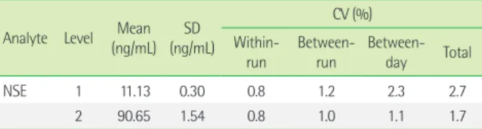
PreciControl Tumor Marker 1과 PreciControl Tumor Marker 2 두 농도에 대해 Elecsys NSE의 검사차례내 변이계수는 모두 0.8%
이었고, 총 변이계수는 각각 2.7%, 1.7%였다(Table 1).
2. 직선성
5가지 농도의 검체를 4회 반복 측정한 결과를 예상 농도와 비교 하여 선형회귀 분석을 시행하였다. 평가 결과 0.20-234.5 ng/mL 범 위에서 결정계수(coefficient of determination, R2)가 0.99 이상으로 우수한 직선성을 보였다(Fig. 1).
3. 검출한계 설정
공백검체와 저농도검체를 반복 측정하여 산출한 LoB는 0.025 ng/mL였으며, LoD는 0.032 ng/mL였다.
4. 참고구간 검증
제조사에서 제시한 참고구간은 0.05-16.3 ng/mL였다. 분석한 20 검체 중 1검체가 제조사의 참고구간을 벗어났다. 참고구간을 벗어 난 검체수가 전체 검체수의 10% 미만이었으므로 제조사의 참고구 간을 사용할 수 있는 것으로 판단하였다.
5. 상관성
44개의 검체로 Elecsys NSE와 ELSA-NSE 분석법 간의 상관성을
평가한 결과, 선형회귀모델은 Fig. 2와 같았으며, y=1.601x-21.633 수식을 따랐다. 두 장비 간의 상관계수는 0.9128로 상관성 평가 기 준인 0.975보다 낮은 값을 보였다
고 찰
NSE의 정밀도평가에서, The national academy of clinical bio- chemistry는 종양표지자의 검사내변이는 5%, 검사간변이는 10%
라는 기준을 제시하고 있다[23]. 최근 발표된 NSE의 생물학적 변이 에 관한 연구에서는 최적의 정밀도 기준을 3.4%로 제안하였다 [24]. 본 평가에서 총 변이계수는 저농도 검체에서 2.7%, 고농도 검 체에서 1.7%로 위 기준을 모두 만족하므로 우수한 정밀도를 보인 다고 판단할 수 있다.
직선성 평가 결과에서 0.20-234.5 ng/mL 범위에서 결정계수가 0.99 이상으로 우수한 직선성을 보였다. 제조사에서 제시한 분석 측정범위는 0.050-370.0 ng/mL이다. 질병과 NSE 결과를 비교한 기 존의 연구에서, 폐암환자 222명의 NSE의 농도가 0-170 ng/mL에 이 르며[25], 급성 뇌경색 환자 30명을 대상으로 했을 때 NSE 농도가 2.6-57.7 ng/mL [26], 지주막하 출혈이 있는 62명의 환자를 대상으 로 했을 때 NSE 농도가 11.2-30.2 ng/mL에 이른다는 보고가 있다 [27]. 이를 통해 볼 때 NSE가 증가하는 질병 상태라도 NSE가 Elec- sys NSE의 AMR 상한 이상으로 증가하는 경우는 드문 것으로 생 각된다.
따라서, 본 연구에서 직선성을 평가한 상기 범위가 임상적으로 의미있는 범위에 포함된다고 생각하여 평가를 진행하였다. 검출한 계는 0.032 ng/mL로 제조사가 제공한 분석측정범위의 하한치보다 Table 1. Precision profile of Elecsys NSE
Analyte Level Mean (ng/mL) SD
(ng/mL)
CV (%) Within-
run Between-
run Between- day Total
NSE 1 11.13 0.30 0.8 1.2 2.3 2.7
2 90.65 1.54 0.8 1.0 1.1 1.7
Abbreviations: CV, coefficient of variation; NSE, neuron-specific enolase; SD, stan- dard deviation.
Fig. 1. Linear regression and residual plots of Elecsys NSE. The blue line represents the linear regression and the gray line depicts a theoretical line with a slope of 1.0 and a y-intercept of 0.
250
200
150
100
50
0
15
10
5
0
-5
-10
-15
Measured (ng/mL) Residual (ng/mL)
Assigned (ng/mL) Assigned (ng/mL)
Scatter plot Residual plot
1:1 Line Fitted overall
0 50 100 150 200 250 0 50 100 150 200 250
낮았다.
Woo 등[28]은 한국 성인 844명을 대상으로 Elecsys NSE를 이용 하여 ModularAnlytics E170 장비로 분석하였을 때 NSE의 참고범 위는 7.56-15.81 ng/mL였다고 보고하였다. 제조사가 주장한 참고범 위는 16.3 ng/mL 미만으로 한국인에서의 참고범위와 유의한 차이 가 없다고 판단하여 제조사의 참고범위를 검증하였다. 분석 결과 제조사가 제시한 참고구간을 벗어난 검체는 5%로 제조사의 참고 구간인 16.3 ng/mL 미만을 사용할 수 있다고 판단하였다.
본 연구에서 RIA 원리를 사용한 기준장비와 ECLIA 방법을 사용 한 Elecsys NSE의 상관계수는 0.9128로 장비 간 상관성 평가기준 인 상관계수 0.975 미만이었다. NSE의 검사실 간 및 검사 키트 간 낮은 상관성은 이미 보고된 바 있다[29]. Štern 등[30]의 보고에 따 르면 NSE를 측정하는데 RIA 방법을 사용하는 Cis-Bio Interna- tional, Immunotech (Beckman Coulter Company, Prague, Czech Republic), Diasorin (Stillwater, Minnesota, U.S.A), PerkinElmer Life and Analytical Sciences (Wallac Oy, Turku, Finland)와 수기효 소면역측정법을 사용하는 DRG Instruments GmbH (Marburg, Germany), ECLIA를 사용하는 Roche, 면역형광법을 사용하는 B.R.A.H.M.S. AG (Hennigsdorf/Berlin, Germany)사의 키트를 비 교한 결과, RIA 원리를 사용한 키트 간의 상관계수는 모두 0.979 이상인 반면에, RIA와 그 외 원리를 이용한 키트 사이의 상관계수 는 이보다 낮은 값을 보였다(범위: 0.926-0.978). Schmitt 등 [31]은 ECLIA 방법을 이용한 Roche사와 RIA 방법을 이용한 Pharmacia 사의 NSE 검사 사이의 상관계수를 0.95로 보고한 바 있다. 이를 통 해 볼 때, 검사 원리의 차이로 인한 낮은 상관성은 불가피한 것으
로 보인다.
상관계수가 낮은 또 다른 원인으로 검사 키트마다 사용한 단일 클론항체의 차이를 생각해 볼 수 있다. 즉, 키트별로 사용한 단일 클론항체마다 NSE에 대한 친화력이 다를 수 있다[29, 30]. 또한, 단 일클론항체에 따라
γγ
아이소형에만 부착할 수도 있고,γγ
와αγ
아이소형 모두에 부착할 수도 있다[29].결론적으로 Elecsys NSE의 수행능평가 결과 정밀도와 직선성이 우수하였으며 제조사의 참고범위와 검출한계를 만족하였다. Elec- sys NSE는 방사성 동위원소를 사용하지 않으며, 민감한 검사법이 다. 본원에서는 본 연구를 통한 평가를 바탕으로 NSE를 원내 도입 할 예정이며, 이로 인해 빠른 시간 내에 임상의에게 검사 결과를 제공할 수 있을 것으로 예상한다.
요 약
배경: 신경원특이에놀라제(neuron specific enolase, NSE)는 신경 세포와 말초 신경내분비세포에 존재하며, 특히 소세포폐암의 표지 자 및 뇌손상의 예측인자로 유용한 것으로 알려져 있다. Elecsys NSE (Roche Diagnostics, Switzerland)는 NSE를 전기화학적발광면 역법으로 측정하는 검사법으로, 본 연구는 이의 수행능을 평가하 고자 하였다.
방법: CLSI 지침에 따라 Elecsys NSE 검사법의 정밀도, 직선성, 검 출한계, 참고구간, 기존 장비와의 상관성을 평가하였다. 정밀도 평 가를 위해 Elecsys NSE의 전용 대조물질인 PreciControl Tumor Marker 1과 2 (Roche Diagnostics)를 사용하였고, 직선성 평가를 Fig. 2. Method comparison between Elecsys NSE and ELSA-NSE.
250
200
150
100
50
0
100
50
0
-50
-100
e601 (ng/mL) Bias (ng/mL)
RIA (ng/mL) RIA (ng/mL)
Scatter plot Bias plot
Regular Regr 1:1 Line
0 50 100 150 200 250 0 50 100 150 200 250
위해 고농도 환자 검체와 PreciControl Tumor Marker 1을 사용하 였다. 검출한계는 완충액 및 희석한 환자검체를 사용하였다. 참고 구간검증은 정상인 검체를 사용하였다. 기존장비와의 상관성평가 는 환자 검체를 사용하여 RIA 방법인 ELSA-NSE (Cis-Bio Interna- tional, France)와 비교하였다.
결과: NSE의 총 정밀도는 저농도와 고농도에서 모두 3% 이내였고, 0.20-234.5 ng/mL 범위에서 직선성을 나타내었다. 검출한계는 0.032 ng/mL였다. 참고구간은 0.05-16.3 ng/mL로 제조사가 제시한 구간을 만족하였다. RIA와의 상관성 평가에서 상관계수는 0.9128 이었다.
결론: Elecsys NSE는 NSE 측정에 있어 정밀도, 직선성이 우수하였 고, 제조사의 검출한계와 참고구간을 만족하였다. 다만, 기존 RIA 장비와의 상관성이 낮았는데, 이는 검사키트마다 서로 다른 단일 클론항체를 사용한 것이 원인일 수 있다. Elecsys NSE는 방사성동 위원소로 인한 위험이 없으며, 민감도가 높아 일선 검사실에서의 NSE 분석장비로써 적합할 것으로 판단되었다.
REFERENCES
1. Pancholi V. Multifunctional alpha-enolase: its role in diseases. Cell Mol Life Sci 2001;58:902-20.
2. Tapia FJ, Polak JM, Barbosa AJ, Bloom SR, Marangos PJ, Dermody C, et al. Neuron-specific enolase is produced by neuroendocrine tu- mours. Lancet 1981;1:808-11.
3. Moore BW. Chemistry and biology of two proteins, S-100 and 14-3-2, specific to the nervous system. Int Rev Neurobiol 1972;15:215-25.
4. Marangos PJ, Polak JM, Pearse AGE. Neuron-specific enolase: a probe for neurons and neuroendocrine cells. Trends Neurosci 1982;5:193-6.
5. Schmechel D, Marangos PJ, Brightman M. Neurone-specific enolase is a molecular marker for peripheral and central neuroendocrine cells.
Nature 1978;276:834-6.
6. Kaltsas GA, Besser GM, Grossman AB. The diagnosis and medical management of advanced neuroendocrine tumors. Endocr Rev 2004;25:458-511.
7. Carney DN, Ihde DC, Cohen MH, Marangos PJ, Bunn PA Jr, Minna JD, et al. Serum neuron-specific enolase: a marker for disease extent and response to therapy of small-cell lung cancer. Lancet 1982;1:583-5.
8. Ariyoshi Y, Kato K, Ishiguro Y, Ota K, Sato T, Suchi T. Evaluation of serum neuron-specific enolase as a tumor marker for carcinoma of the lung. Gan 1983;74:219-25.
9. Akoun GM, Scarna HM, Milleron BJ, Bénichou MP, Herman DP. Se- rum neuron-specific enolase. A marker for disease extent and re-
sponse to therapy for small-cell lung cancer. Chest 1985;87:39-43.
10. Molina R, Holdenrieder S, Auge JM, Schalhorn A, Hatz R, Stieber P. Di- agnostic relevance of circulating biomarkers in patients with lung can- cer. Cancer Biomark 2010;6:163-78.
11. Ebert W, Hoppe M, Muley T, Drings P. Monitoring of therapy in inop- erable lung cancer patients by measurement of CYFRA 21-1, TPA- TP CEA, and NSE. Anticancer Res 1997;17:2875-8.
12. Inoue S, Takahashi H, Kaneko K. The fluctuations of neuron-specific enolase (NSE) levels of cerebrospinal fluid during bacterial meningitis:
The relationship between the fluctuations of NSE levels and neurologi- cal complications or outcome. Acta Paediatr Jpn 1994;36:485-8.
13. Cunningham RT, Watt M, Winder J, McKinstry S, Lawson JT, Johnston CF, et al. Serum neurone-specific enolase as an indicator of stroke vol- ume. Eur J Clin Invest 1996;26:298-303.
14. Ondruschka B, Pohlers D, Sommer G, Schober K, Teupser D, Franke H, et al. S100B and NSE as useful postmortem biochemical markers of traumatic brain injury in autopsy cases. J Neurotrauma 2013;30:1862-71.
15. Cunningham RT, Johnston CF, Irvine GB, McIlrath EM, McNeill A, Bu- chanan KD. Development of a radioimmunoassay for neurone spe- cific enolase (NSE) and its application in the study of patients receiv- ing intra hepatic arterial streptozotocin and floxuridine. Clin Chim Acta 1990;189:275-86.
16. Fu X, Meng M, Zhang Y, Yin Y, Zhang X, Xi R. Chemiluminescence enzyme immunoassay using magnetic nanoparticles for detection of neuron specific enolase in human serum. Anal Chim Acta 2012;722:
114-8.
17. Muley T, Ebert W, Stieber P, Raith H, Holdenrieder S, Nagel D, et al.
Technical performance and diagnostic utility of the new Elecsys neu- ron-specific enolase enzyme immunoassay. Clin Chem Lab Med 2003;41:95-103.
18. Sterk M, Oenings A, Eymann E, Roos W. Development of a new auto- mated enzyme immunoassay for the determination of neuron-specific enolase. Anticancer Res 1999;19:2759-62.
19. Clinical and Laboratory Standards Institute, eds. Evaluation of preci- sion performance of quantitative measurement methods; approved guideline. CLSI document EP5-A2. 2nd ed. Wayne, PA: Clinical and Laboratory Standards Institute, 2004.
20. Clinical and Laboratory Standards Institute, ed. Evaluation of the Lin- earity of Quantitative Measurement Procedures: a Statistical Approach;
Approved Guideline. CLSI document EP6-A. Wayne, PA: Clinical and Laboratory Standards Institute, 2003.
21. Clinical and Laboratory Standards Institute, ed. Evaluation of precision
performance of quantitative measurement methods; approved guide- line. CLSI document EP17-A2. 2nd ed. Clinical and Laboratory Stan- dards Institute, 2012.
22. Clinical and Laboratory Standards Institute, ed. Defining, Establish- ing, and verifying reference intervals in the clinical laboratory; ap- proved guideline. CLSI document C28-A3. 3rd ed. Clinical and Labora- tory Standards Institute, 2008.
23. Sturgeon CM and Diamandis EP, eds. Use of Tumor Markers in Clinical Practice: quality Requirements. Washington, DC: American Associa- tion for Clinical Chemistry, 2009.
24. Braga F, Ferraro S, Mozzi R, Dolci A, Panteghini M. Biological varia- tion of neuroendocrine tumor markers chromogranin A and neuron- specific enolase. Clin Biochem 2013;46:148-51.
25. Mumbarkar PP, Raste AS, Ghadge MS. Significance of tumor markers in lung cancer. Indian J Clin Biochem 2006;21:173-6.
26. Ku BD and Chung KC. Change of Serum Neuron Specific Enolase Level During Acute Stage of Cerebral Infarction. J Korean Neurol As- soc 2009;27:13-8.
27. Moritz S, Warnat J, Bele S, Graf BM, Woertgen C. The prognostic value of NSE and S100B from serum and cerebrospinal fluid in pa- tients with spontaneous subarachnoid hemorrhage. J Neurosurg Anes- thesiol 2010;22:21-31.
28. Woo HY, Kim YJ, Park H. Establishment of reference intervals of tu- mor markers in Korean adults. Korean J Lab Med 2008;28:179-84.
29. Stern P, Bartos V, Uhrova J, Bezdickova D, Vanickova Z, Tichy V, et al.
Performance characteristics of seven neuron-specific enolase assays.
Tumour Biol 2007;28:84-92.
30. Štern P, Bartoš V, Uhrová J, Springer D, Vaníčková Z, Tichý V, et al.
The comparability of different neuron-specific enolase immunoassays and its impact on external quality assessment system. Klin Biochem Metab 2007;15:21-6.
31. Schmitt UM, Stieber P, Hasholzner U, Pahl H, Hofmann K, Fateh- Moghadam A. Methodological and clinical evaluation of two auto- mated enzymatic immunoassays as compared with a radioimmunoas- say for neuron-specific enolase. Eur J Clin Chem Clin Biochem 1996;
34:679-82.