• 검색 결과가 없습니다.

Hepatic Arterial Complications after Liver Transplantation: A Single-Center Experience

N/A
N/A
Protected

Academic year: 2021

Share "Hepatic Arterial Complications after Liver Transplantation: A Single-Center Experience"

Copied!
8
0
0

로드 중.... (전체 텍스트 보기)

전체 글

(1)

Hepatic Arterial Complications after Liver Transplantation:

A Single-Center Experience

Department of Surgery, Gacheon University Gil Hospital, Incheon, Korea

Keun Jeong Lee, M.D., Sang Tae Choi, M.D., Chung Min, M.D., Jung Nam Lee, M.D., Woon Ki Lee, M.D., Jeong-Heum Baek, M.D., Keon Kuk Kim, M.D.,

Jin Mo Kang, M.D. and Won Suk Lee, M.D.

Background: We wanted to explore performing hepatic arterial reconstruction in living donor liver transplantation (LDLT) using right lobe liver grafts and cadaveric liver transplantation (CLT) in a single center.

Methods: Thirty five LDLTs were performed from April 2005 to August 2009. The back wall support suture without twisting was used in most cases. A single RHA was anastomosed to the RHA in 24 patients, to the proper HA in 2 patients, to the RAHA in 4 patients, to the LHA in 2 patients and to an aberrant RHA arising from the SMA in 3 patients. The diameter of the donor RHA was between 1.5 mm and 3.0 mm (mean: 2.5 mm). In the 34 patients who underwent CLT, most of the arterial anastomoses were usually performed using two cuffs at the recipient HA and the GDA bifurcation and a branching point on the donor CHA with running and intermittent stay suture.

Results: The total incidence of HA complication was 4.34% (3/69): 1 HAT (2.85%) occurred in a case of LDLT and 2 HAS (5.88%) occurred in a case of CLT. HAT occurred in 1 recipient on the 1st day following LDLT and 2 HAS occurred in CLT recipients at one and two months, respectively, following LDLT.

Conclusions: HA complications occurred as a mild type of late complication and these complications might not be fatal in CLT. A low incidence of HAT can be achieved with using non-twisting method-guided microsurgical techniques for creating hepatic arterial anastomosis in LDLT. When early HAT occurs, early surgical reconstruction is mandatory for preventing the loss of the graft. Back wall sutures with only single needle suture might be a feasible method for HA microsurgical reconstruction.

Key Words: Hepatic artery, Thrombosis, Arterial occlusive disease, Liver transplantation 중심 단어: 간동맥, 혈전증, 동맥 폐색증, 간이식

Correspondence:Sang Tae Choi, Department of Surgery, Gacheon University Gil Hospital, 1198 Guwol- dong, Namdong-gu, Incheon 405-760, Korea Tel: +82-32-460-3244, Fax: +82-32-460-3247, E-mail: bcon1218@hanmail.net

Received : July 22, 2011, Revised : August 27, 2011 Accepted : September 5, 2011

Introduction

Hepatic artery complication that occurred in liver transplantation is a potentially devastating problem.

HAT developed 3∼9% in even CLT(1,2). when it oc- curred immediately, HAT may induced the graft fail- ure, sepsis due to liver abscess, biliary stricture and leakage. Delayed, HAT presented with mild clinical symptoms such as cholangitis, altered liver function test(2,3). It was initially suggested that surgical anasto- mosis technique was the most important risk factor for

HAT. Attempting to reduce the incidence of HAT, we undertook a retrospective review of patients at our in- stitution undergoing LDLT and CLT between April 2005 and August 2009, with the hepatic artery anasto- mosis performed by a single microvascular surgeon.

In this study, we intended to explore our experi- ence in hepatic reconstruction and the management of hepatic artery complications following living donor and cadaveric liver transplantation over initial five years.

Materials and Methods 1) Recipient profile

Records were from a retrospectively collected data-

base from April 2005 to August 2009. There were 34

adult CLTs and 35 LDLTs conducted at our center dur-

(2)

Type Anastomoed artery Size (mean, mm) n (%) SD Complication LDLT

CLT

RHA-RHA RHA-PHA RHA-RAHA RHA-LHA

RHA-aberrant RHA

a

CHA-CHA

CHA-RHA

2.6 3.2 1.3 2.4 2.5 13 7

24 (68.5%) 2 (5.7%) 4 (11.4%)

2 (5.7%) 3 (8.5%)

30 4

0.26 0.1 0.07

0.1 0.08 1.57 0.07

2/24 - - - - 1/30

- Abbreviation: SD, standard deviation.

Aberrant right hepatic artery arising from superior mesenteric artery

a

. Table 1. Anastomosed arterial feature and complication

ing this period. Male/female ratio was 51/18, the me- dian age being 47 (range: 14∼69) years. The indica- tions for LT were HBV-associated cirrhosis (n=32), HBV-associated HCC (n=15), fulminant hepatic failure due to toxin (n=5), primary biliary cirrhosis (n=1), al- coholic liver cirrhosis (n=10), fulminant hepatic failure due to HAV (n=3), HCV(n=1), HAV and HBV co-in- fection (n=1). MELD score is mean 19.2 (6∼42) in LDLT, mean 28.5 (10-50) in CLT. Right lobe without middle hepatic vein was used in most cases (n=33) except 2 case using right lobe with middle hepatic vein in LDLT.

2) Surgical technique

Hepatic arterial reconstruction was performed under the magnified scope of 3.5 times or 5∼10 times zoom magnification using an operating microscope (Olym- pus, Tokyo, Japan), completed by end-to-end anasto- mosis method between donor hepatic artery and recip- ient hepatic artery with interrupted sutures. In LDLT, conventional twisting method that turns over the hep- atic artery was used in early 2 cases. After having dif- ficulty in twisting too short donor hepatic artery, the back wall first suture without twisting can be done in the other case (n=33) by using nylon microsuture with single needle (prolene 9/0, Ethicon, Voerderstedt, Ger- many). Initial sutures can be done at the deepest and most difficult points in posterior wall, located in the farthest away from operator. Another stitch was placed from the outer side of healthy donor arterial wall to the inner side of recipient arterial wall which was li- able to make the intimal dissection during dissection.

The subsequent sutures were placed forward or back- ward on either side adjacent to the previous suture.

After the posterior wall suture finished, anterior wall suture could be done with intermittent suture. The su- ture method guaranteed the good operation field and the secure intimal adjustment and prevented the in- timal dissection and further hepatic arterial compli- cation. This suture method does not require turning over the hepatic artery. It takes a less time to anasto- mosis, there is a little change to intimal damage and tension may occur during turning the short hepatic artery.

Most arterial anastomosis in CLT was usually per- formed using the cuff at the recipient hepatic artery and gastroduodenal artery bifurcation and a branching point on the donor common hepatic artery with inter- mittent stay suture and a running suture using prolene 7/0 (Ethicon, Voerderstedt, Germany) under the mag- nified scope of 3.5 times. In 2 case of young recipi- ent, donor celiac trunk anastomosis to supraceliac aor- ta was used for arterial reconstruction. Anastomosis between the recipient and graft HA was resolved by cutting both arterial ends obliquely to adjust the length in 5 cases.

3) Statistics

Statistical analysis was performed using SPSS, ver-

sion 14.0 (Statistical Package for the Social Sciences,

Chicago, U.S.A.). Survival curves were calculated us-

ing Kaplan-Meier analysis to compared survivals among

groups. The chi-square test was used to compare the

frequency of the complication.

(3)

Sex/age Primary disease Type Onset (#POD) Graft Recipient Treatment Biliary stricture Result M/46

M/30 F/53

HBV/HCC HBV/LC HBV/LC

LDLT CLT CLT

1 58 31

RHA CHA CHA

Rt. GEPA CHA CHA

GSV to supraceliac aorta Balloon/stent Balloon/stent

Yes No No

Survival Survival Survival Abbreviations: LDLT, living donor liver transplantation; CLT, cadaveric liver transplantation; POD, postoperation day; GSV, great saphe- nous vein.

Table 2. Clinical data from 3 patients with hepatic arterial complication after liver transplantation

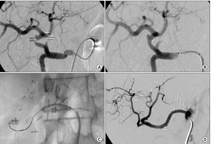
Fig. 1. Ischemic necrosis followed by hepatic artery stenosis. CT shows ischemic necrosis followed by hepatic arterial thrombosis, which occurred at segment VI at 1st postoperation day. Hepatic angiography show arterial vasospam ans thrombus occurred at the right gastroepiploic artery (A, B). Hepatic flow can be preserved after jump graft by using great saphenous vein from supraceliac aorta to right gastroepiploic artery (C). The ischemic necrosis portion show relatively good parenchymal regeneration and duct dilatation in 7 months later (D, E).

Results

In the 35 patients who underwent LDLT, all single donor right hepatic arteries were anastomosed; to right hepatic artery in 24 patients, to proper hepatic artery in 2 patients, to right anterior hepatic artery in 4 pa- tients, to left hepatic artery in 2 patients, to aberrant right hepatic artery arising from superior mesenteric artery in 3 patients. The diameter of donor right hep- atic artery is between 1.5 mm and 3.0 mm (mean 2.5 mm).

In the 34 patients who under CLT, most arterial anastomosis was usually performed using a Carrel pat-

ch at the recipient hepatic artery and gastroduodenal artery bifurcation and a branching point on the donor common hepatic artery or common hepatic artery and gastroduodenal bifurcation with intermittent stay suture and a running suture except 4 cases (between the re- cipient and graft HA after cutting both arterial end ob- liquely to adjust the length). Anastomosed arterial fea- ture and complication were summarized in Table 1.

The total incidence of HA complication was 4.34%

(3/69), 1 HAT (2.85%) occurred in only LDLT, 2 HAS

(5.88%) in CLT (1 case occurred in non-anastomosis

site). HAT occurred at 1st day following LDLT which

were revascularized with autogenous saphenous vein

between right gastroepiploic artery anastomosed to do-

(4)

Fig. 2. Hepatic angiography showing arterial kicking and stenosis. Hepatic angiography show arterial kicking and stenosis at the anas- tomosis of CHA in CLT. The kicking of anastomosis site was revised by ballooning and stenting (A, B). Intimal dissection and stenosis in non-anastomotic site was relieved by ballooning and stenting (C, D).

nor right hepatic artery and recipient abdominal aorta 1 day postoperatively, 2 HAS occurred one and two month following CLT (mean 52.6 months), no symp- tom was presented. HAT was identified by serial US, CT and angiography. HAS was found accidentally by routine CT. No death related to hepatic artery compli- cations occurred.

Hepatic arterial complication developed in 3 (4.34%) of 69 patients, as summarized in Table 2.

1) Hepatic artery thrombosis (Fig. 1)

One patient was identified with HAT at the first post-operative day. He suffered from HBV related he- patocellular carcinoma and transarterial chemoembo- lization (TACE) twice preoperatively. His hepatic artery flow in common hepatic artery proved to be minimal in operative field, because of intimal damage after

TACE. Right hepatic artery of donor was anastomosed to the right gastroepiploic artery of the recipient.

Thrombosis occurred at the site of right gastroepiploic artery, demonstrated arterial vasospasm and thrombus in angiography. Jump graft by using great saphenous vein from supraceliac aorta to right gastroepiploic ar- tery was performed in end-to-side arterial anastomosis.

2) Hepatic artery stenosis (Fig. 2)

Among the CLT patient, one patient was found

with common hepatic artery stenosis during checking

the commputed tomography, suspicious of acute rejec-

tion. Intimal dissection and stenosis in the recipient

artery (not the anastomosis) was revealed in conven-

tional angiography, balloon and coronary stent in-

sertion was done. One patient’s commputed tomog-

raphy showed 7 mm sized contrast filling around vas-

(5)

cular structure in right hepatic artery can not rule out pseudoaneurysm, proved to be kicking of common hepatic artery anastomosis site in angiography. This stenosis was revised by ballooning and stenting.

Discussion

Hepatic artery thrombosis (HAT) after liver trans- plantation is a potentially life threatening complica- tion(1). HAT presently complicates 3∼9% of all CLTs (4,5). Reconstruction of the hepatic artery is more dif- ficult and challenging issue especially in living donor liver transplantation (LDLT), compared to CLT. The in- cidence of arterial thrombosis has been reduced dra- matically from 25% without a microscope to 0∼3.8%

with a microscope(6-8). The technique of microsur- gical hepatic artery reconstruction has contributed greatly to a reduced incidence of HAT and HAS.

Technical failure of the reconstruction usually leads to re-transplantation or even death, and the procedure is complicated by anatomical variation, vascular con- sistency, and the hemodynamic situation of the recipi- ents during the operation. Hepatic artery reconstruc- tion is one of the key steps for LDLT. In a hospital launching liver transplantation, initial patient loss could make transplantation impossible to proceed with the operation if patient loss occurred because of the arte- rial complication. Initial failure will almost lead to the cessation of transplantation at launching hospitals.

Therefore, transplantation surgeons were assisted by plastic surgeons that had many micorosurgical experi- ences in the beginning of liver transplantation opera- tion. Recently transplantation surgeons themselves have performed hepatic artery anastomosis in most trans- plant centers. Proper anastomotic vessel choice via mi- crosurgical technique in hepatic arterial reconstruction would reduce significantly the incidence of hepatic ar- tery complications and provide an excellent graft sur- vival following LDLT. The recipient’s hepatic artery is located deep in the abdominal cavity and the oper- ative field is limited and movable during ventila- tion(4-6), the intimal damage may be more severe, and the usable vessel grafts are limited. Because of the short and small hepatic artery, size discrepancy

between the graft and recipient hepatic arteries, poor arterial pathologic state such as intimal dissection and reconstruction of hepatic arteries is a challenge to sur- geons in LDLT(9,10). Hepatic arterial complications in- cluding thrombosis, stenosis and aneurysm formation are life-threatening in LDLT by causing graft failure and irreversible biliary damage(9-11). So these early experiences have been accumulated, several techni- ques have attempted to improve the success rate. Re- cently many centers have designed new technique in order to reduce the arterial damage and complication which happens in operation(12-14). Other methods such as the fish-mouth method, funnelization method, or end-to-side anastomosis were not used in this study; sometimes it is dangerous to use the conven- tional twist reconstruction technique, because it may require turning over a damaged, short artery, some re- ports support a back wall support suture technique with double needle suture instead of conventional ro- tating method. There are a little literatures comparing conventional method with the back wall suture me- thod. Some studies reported the advantage of the back wall method(15,16). Using back wall support su- ture under microsurgical reconstruction, they could minimize the artificial injuries of hepatic arteries and improve the survival rate of the post liver transplan- tation by reducing hepatic artery thrombus(15-17).

In our study, all anastomoses were carried out by one transplant surgeon using atraumatic microvascular techniques in 5∼10 times zoom magnification using an operating microscope during the reconstructions in LDLT, 3.5 times under the magnified scope in CLT.

Only 1 case of HAT in recipients occurred which was

in the 35 incipient patients following LDLT. Surgical

techniques have been known as major factor for hep-

atic artery thrombosis. The key points in hepatic artery

reconstruction included selecting a reciprocal stump

location for arterial reconstruction: a thicker and heal-

thy arterial stump in the recipient was chosen for the

first anastomosis(18). When separating the arteries from

surrounding structures, it is important to preserve

plenty of soft tissue around the artery. The patient’s

liver condition such as fibrosis, ascite and coagulop-

athy influenced by primary disease could make it more

(6)

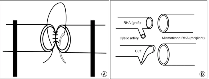
Fig. 3. Schematic drawing of the microvascular anastomosis. Schematic drawings show the microvascular anastomosis. The recipient and graft artery are clamped without twisting. The first suture is placed at upper and posterior wall at most deep portion of two arteries (A), and hepatic artery cuff by using cystic attery to overcome size discrepancy (B).

difficult.

To minimize the incidence of intimal dissection and injury, careful division and preparation of recipient ar- tery are necessary. When donor artery diameter is too small, isolation of smaller hepatic artery could be pos- sible by dividing the deep recipient liver parenchyma to overcome the size discrepancy. Intimal damage and injury of hepatic artery usually occurs during dissec- tion proximally to visualize smaller diameter of hepatic artery. In the case of intimal dissection, recipient hep- atic artery is not suitable for reconstruction, some studies have been reported various applications by us- ing splenic artery, right gastroepiploic artery and inter- position graft between common hepatic artery and do- nor hepatic artery or between CHA and abdominal aorta, instead of dissected hepatic artery. Some reports suggested graft interposition increased the risk of HAT(19). Mostly, direct end to end anastomosis was simplest way to avoid the size mismatching and make larger anastomosis decreased risk for thrombosis theoretically. From the view of this, donor’s cuff be- tween right hepatic artery and cystic artery will be good option to anastomose with larger recipient’s hepatic artery, as possible (Fig. 3).

Some reports have provided the convenience and usefulness about hepatic artery anastomosis by using microsuture with double-arm needle(17). Initially, we

must distress ourselves about the amount safe anasto- mosis of pressure hepatic arteries with previous single arm needle suture, having no idea of the new instru- ment and surgical product. While some cases of much shorter hepatic artery were difficult to use the conven- tional rotating method, we used the back wall suture with not rotation but fixed method. Because recipient artery tends to be more intimal dissection or damaged than healthy donor artery, each stitch was placed from the outer side of donor artery to the inner side of re- cipient artery with single needle, could make it dis- tinct to penetrate the full thickness of poor recipient’s arterial wall and attach the both arterial intimas.

It is more important to adjust arterial course and ax- is in hepatic arterial anastomosis, even if some tor- tuous or enough long in LDLT. While CHA in CLT has so thicker and larger diameter that approximate length detached arteries is more important compared with LDLT. Inapproximate lengthy anastomosis between do- nor and recipient artery without detaching from the recipient’s hepatoduodenal ligament enough distally causes the kicking and stenosis under the natural posi- tion of whole liver.

Hepatic arterial kicking and stenosis in CLT may be

more frequent than LDLT. Released liver may com-

press the anastomosis of length hepatic artery, the liv-

er retracted to obtain visual field during arterial

(7)

anastomosis.

In the case of early HAT, several methods, includ- ing re-transplantation, urgent revascularization, or re- vision of anastomosis, have been performed to avoid irreversible damage of the allograft(20-22). In our study, one recipients underwent urgent revascularization im- mediately after HAT was confirmed on the 1st day af- ter LDLT, and were subsequently cured with satisfac- tory liver function. We performed hepatic reconstruc- tion between RHA and RGEPA in HAT patient suspi- cious of inflow insufficiency after repeated TACE. CT show ischemic necrosis followed by hepatic arterial thrombosis, which occurred at segment VI at 1st day postoperatively day. Hepatic angiography show arterial vasospasm and thrombus occurred at the right gastro- epiploic artery. In emergency operation, we anastmo- sed celiac axis and right gastroepiploic artery by using the great saphenous vein without aorta clamping.

Celiac axis is more better inflow than abdominal aorta, no need to clamp the aorta and high blood pressure cause the GSV to tear after finishing direct anastomosis to aorta. Right gastroepiploic artery was anastomosed with donor hepatic artery is better distal conduit could be possible to anastomose in end-to-side fashion with larger GSV. It is difficult to anastomose the short and small donor hepatic artery to larger GSV directly.

Therefore we anastomosed interposed right gastroepip- loic artery to GSV by end to side fashion after check- ing anastomosis security between epiploic artery and donor hepatic artery. This patient didn’t receive throm- bolytic and anticoagulant therapy with monitoring of coagulation status, and thereafter HA inflow returned to normal. The majority of late hepatic complication occurred to only CLT, presented with no specific symptom. Patient with stenosis was treated with bal- loon angioplasty and endoluminal stent placement (4,16). The stenosis proximal part from anastomosis site was found on the other patient. However, no death was associated with vascular complications. Late HAT, which occurred after the first postoperative month, can be well tolerated, and portal flow can support the liver until the collateral circulation has de- veloped(23). Stent insertion after balloon angioplasty was performed for the patient in our study with late

HAT. Liver function test results and general condition were good. However, long-term results of late HAT patients will require further research.

Conclusion

Careful preoperative evaluation and intraoperative microsurgical technique for hepatic artery reconstruc- tions are the keys to prevent vascular complications following LDLT. Immediate surgical intervention is re- quired for acute vascular complications, whereas late complications remain asymptomatic and may be treat- ed by balloon angioplasty and endovascular stent placement.

REFERENCES

1) Stange BJ, Glanemann M, Nuessler NC, Settmacher U, Steinmüller T, Neuhaus P. Hepatic artery thrombosis af- ter adult liver transplantation. Liver Transpl 2003;9:

612-20.

2) Bhattacharjya S, Gunson BK, Mirza DF, Mayer DA, Buckels JA, McMaster P, et al. Delayed hepatic artery thrombosis in adult orthotopic liver transplantation-a 12-year experience. Transplantation 2001;71:1592-6.

3) Settmacher U, Stange B, Haase R, Heise M, Steinmüller T, Bechstein WO, et al. Arterial complications after liver transplantation. Transpl Int 2000;13:372-8.

4) Takatsuki M, Chiang YC, Lin TS, Wang CC, Concejero A, Lin CC, et al. Anatomical and technical aspects of hepatic artery reconstruction in living donor liver tran- splantation. Surgery 2006;140:824-8.

5) Uchiyama H, Hashimoto K, Hiroshige S, Harada N, Soejima Y, Nishizaki T, et al. Hepatic artery re- construction in living-donor liver transplantation: a re- view of its techniques and complications. Surgery 2002;

131(1 Suppl):S200-4.

6) Wei WI, Lam LK, Ng RW, Liu CL, Lo CM, Fan ST, et al. Microvascular reconstruction of the hepatic artery in live donor liver transplantation: experience across a decade. Arch Surg 2004;139:304-7.

7) Broelsch CE, Whitington PF, Emond JC, Heffron TG, Thistlethwaite JR, Stevens L, et al. Liver transplantation in children from living related donors. Surgical techni- ques and results. Ann Surg 1991;214:428-37.

8) Miyagi S, Enomoto Y, Sekiguchi S, Kawagishi N, Sato A, Fujimori K, et al. Microsurgical back wall support su- ture technique with double needle sutures on hepatic ar- tery reconstruction in living donor liver transplantation.

Transplant Proc 2008;40:2521-2.

9) Shirouzu Y, Kasahara M, Morioka D, Sakamoto S, Taira

(8)

K, Uryuhara K, et al. Vascular reconstruction and com- plications in living donor liver transplantation in infants weighing less than 6 kilograms: the Kyoto experience.

Liver Transpl 2006;12:1224-32.

10) Kim BW, Won JH, Lee BM, Ko BH, Wang HJ, Kim MW. Intraarterial thrombolytic treatment for hepatic ar- tery thrombosis immediately after living donor liver transplantation. Transplant Proc 2006;38:3128-31.

11) Kaneko J, Sugawara Y, Akamatsu N, Kishi Y, Niiya T, Kokudo N, et al. Prediction of hepatic artery thrombosis by protocol Doppler ultrasonography in pediatric living donor liver transplantation. Abdom Imaging 2004;29:

603-5.

12) Katz E, Fukuzawa K, Schwartz M, Mor E, Miller C. The splenic artery as the inflow in arterial revascularization of the liver graft in clinical liver transplantation. Transplan- tation 1992;53:1373-4.

13) Cherqui D, Riff Y, Rotman N, Julien M, Fagniez PL.

The recipient splenic artery for arterialization in ortho- topic liver transplantation. Am J Surg 1994;167:327-30.

14) Ikegami T, Kawasaki S, Hashikura Y, Miwa S, Kubota T, Mita A, et al. An alternative method of arterial re- construction after hepatic arterial thrombosis following living-related liver transplantation. Transplantation 2000;

69:1953-5.

15) Yang Y, Yan LN, Zhao JC, Ma YK, Huang B, Li B, et al. Microsurgical reconstruction of hepatic artery in A-A LDLT: 124 consecutive cases without HAT. World J Gastroenterol 2010;16:2682-8.

16) Yan S, Zhang QY, Yu YS, He JJ, Wang WL, Zhang M, et al. Microsurgical reconstruction of hepatic artery in

living donor liver transplantation: experiences and les- sons. Hepatobiliary Pancreat Dis Int 2009;8:575-80.

17) Okazaki M, Asato H, Takushima A, Nakatsuka T, Sarukawa S, Inoue K, et al. Hepatic artery reconstruction with double-needle microsuture in living-donor liver transplantation. Liver Transpl 2006;12:46-50.

18) Okochi M, Ueda K, Hirose T, Okochi H, Watanabe H, Suzuki Y, et al. A modified technique for hepatic artery reconstruction in living donor liver transplantation.

Microsurgery 2010;30:541-4.

19) Mazzaferro V, Esquivel CO, Makowka L, Belle S, Kahn D, Koneru B, et al. Hepatic artery thrombosis after pe- diatric liver transplantation-medical or surgical event?

Transplantation 1989;47:971-7.

20) Silva MA, Jambulingam PS, Gunson BK, Mayer D, Buckels JA, Mirza DF, et al. Hepatic artery thrombosis following orthotopic liver transplantation: a 10-year expe- rience from a single centre in the United Kingdom. Liver Transpl 2006;12:146-51.

21) Matsuda H, Yagi T, Sadamori H, Matsukawa H, Shi- noura S, Murata H, et al. Complications of arterial re- construction in living donor liver transplantation: a sin- gle-center experience. Surg Today 2006;36:245-51.

22) Jiang XZ, Yan LN, Li B, Zhao JC, Wang WT, Li FG, et al. Arterial complications after living-related liver trans- plantation: single-center experience from West China.

Transplant Proc 2008;40:1525-8.

23) Sheiner PA, Varma CV, Guarrera JV, Cooper J, Garatti

M, Emre S, et al. Selective revascularization of hepatic

artery thromboses after liver transplantation improves pa-

tient and graft survival. Transplantation 1997;64:1295-9.

수치

Table  2.  Clinical  data  from  3  patients  with  hepatic  arterial  complication  after  liver  transplantation
Fig. 2. Hepatic angiography showing arterial kicking and stenosis. Hepatic angiography show arterial kicking and stenosis at the anas- anas-tomosis of CHA in CLT
Fig. 3. Schematic drawing of the microvascular anastomosis. Schematic drawings show the microvascular anastomosis

참조

관련 문서

The associations of hepatic steatosis and fibrosis using fatty liver index and BARD score with cardiovascular outcomes and mortality in patients with new‑onset type 2

In this study, we investigated the time course of neutrophil recruitment and infiltration in the early stages of arterial throm- bosis using an FeCl 3 -induced carotid

Background: Incidence of complications related extracorporeal membrane oxygenation (ECMO) support as a bridge to lung transplantation (BTT) and its association with the

MAFLD, metabolic dysfunction-associated fatty liver disease; NAFLD, nonalcoholic fatty liver disease; CVD, cardiovascular disease; CI, confidence interval; Model 1,

cACLD = compensated advanced chronic liver disease, dACLD = decompensated advanced chronic liver disease, LS = liver stiffness, LS-SIR = liver-to-spleen signal intensity

Undiagnosed malignancy in patients with deep vein thrombosis: incidence, risk indicators, and diagnosis.. Monreal M, Fernandez-Llamazares J, Perandreu

The locations of aneurysms were middle cerebral artery in 15 patients, cerebral artery in 15 patients, cerebral artery in 15 patients, cerebral artery in

Department of Education, National Center for Educational Statistics, Adult Literacy in America, National Adult Literacy Survey, 1992, Table 1.1b, p.. In addition, the United