Vol. 20, No. 4, Demcember, 2012
□ Case Report □Headache with Dural Thickening on MRI as an Initial Presentation of Leukemia: A Case Report
Department of Pediatrics, Soonchunhyang University College of Medicine, Seoul, Korea
Sang Soo Yoo, M.D. and Eun Sook Suh, M.D.
= Abstract =
Headache can be divided into two categories: primary and secondary. Secondary headaches are associated with central nervous system or other pathology, and the underlying cause of a secondary headache can lead to death. For that reason, a thorough history and examination is key to determining the cause of headache. Other investigations including neuroimaging studies should be strongly considered in children with a history worrisome for intracranial pathology or an abnormal neurologic examination. We present a case of headache with diffuse hypertrophy of the dura mater due to lymphomatous dural infiltration. MRI revealed diffusely enhancing dural thickening. Biopsy of the dura mater found precursor B-cell lymphoblastic lymphoma. The patient was diagnosed with acute lymphoblastic leukemia and the headache was the initial presentation of acute leukemia. In this case, the rapid expansion of the dura mater resulted in elevation of intracranial pressure and caused an acute onset, progressive headache.
Key Words : Headache, Meninges, Leukemia
1)
Introduction
Headaches are a common complaint encoun- tered in the pediatric population and the pre- valence of headache increases with increasing age. Some studies indicate that 39% of children experience headache by 7 years of age and 70% by age 15
1).
Headache can be a symptom of many patho- logies. The second edition of the International Classification of Headache Disorders (ICHD- II) makes a distinction between primary and secondary headaches
2).
Received : 2 December 2012, Revised : 17 December 2012 Accepted : 28 December 2012
Corresponding author : Eun Sook Suh, M.D.
Department of Pediatrics, Soonchunhyang University, Hannam dong 657, Yongsan-gu, Seoul 140-743, Korea Tel : +82.2-709-9338, Fax : +82.2-794-5471 E-mail: [email protected]
Approximately 10% of patients presenting to the emergency department complaining of headache have a secondary headache
3). Some causes of secondary headache are potentially life-threatening and deadly. Therefore, during the course of the evaluation, consideration must be given to other disorders which may cause secondary headache in children and adole- scents.
We report a case of secondary headache with thickening of the dura mater on imaging finding as first manifestation of acute leukemia.
Case Report
A 13-year-old man suffered an acute onset,
daily, progressive, global headache. The pain
was characterized by a sense of pressure and
not affected by positional change. There was no
- 257 -
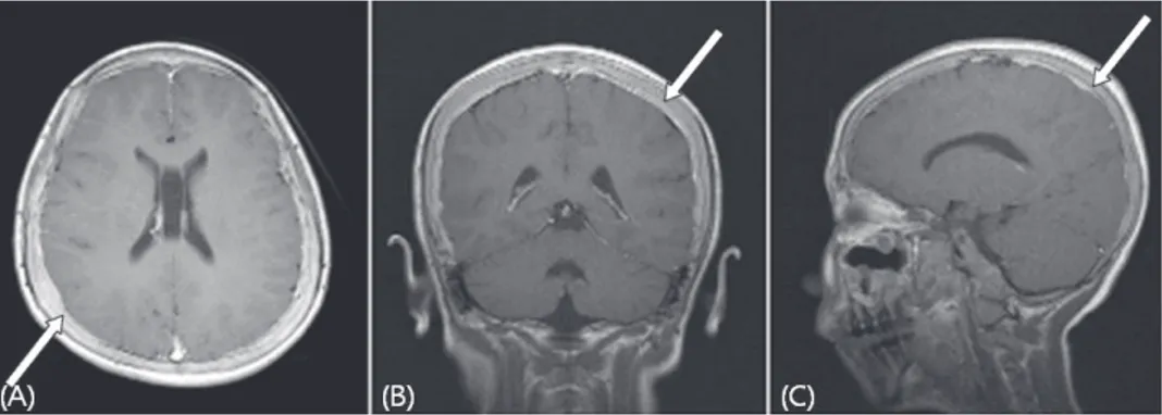
Fig. 1. Magnetic resonance imaging shows diffuse dural thickening with enhance- ment (A, B, C). T1-weighted axial and coronal and sigittal view with gadolinium enhancement. The lesion is hyperintense on T1-weighted image (white arrow).
history associated with trauma. A tension type of headache was considered and treated con- servatively but the headache persisted.
Three days prior to presentation, the patient suddenly started vomiting and showed neck stiff- ness and positive Kernig sign at the time of presentation. The patient's mental status was normal and other neurologic examination showed no focal abnormality. Fundoscopic evaluation revealed mild papilledema in both eyes at first.
Laboratory data: Routine infectious and meta- bolic laboratory findings including electrolytes, and liver enzymes were all within normal limits.
White blood cell count was 6200/uL and lym- phocytosis was present(85%). Hemoglobin and hematocrit was 13.3 g/dL and 39.3%. Platelet count was 150,000/uL. The erythrocyte sedi- mentation rate (ESR) had slightly risen to 29 mm/hr. Lumbar puncture revealed an opening pressure of 400 mmH
2O and showed 0 white blood cells, 18 red blood cells, glucose level of 66 mg/dL, protein level of 20.4 mg/dL. Cere- brospinal fluid (CSF) studies showed no organi- sms on gram stain, negative viral, fungal, bac- terial cultures. Polymerase chain reaction (PCR) did not detect enterovirus or adenovirus.
Radiologic findings: Brain magnetic resona-
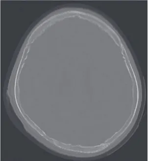
nce imaging (MRI) showed diffusely enhancing dural thickening on T1-and T2-weighted, gadolinium-enhanced coronal and axial and sagittal views (Fig. 1. A-C). Brain computeri- zed tomography (CT) showed multiple osteo- lytic bony changes in calvarium and skull base (Fig. 2). Chest and abdominal CT were per- formed to find evidence of systemic neoplastic involvement and revealed a soft tissue mass with osteolytic bony destruction in left 6th rib and hepatosplenomegaly (Fig. 3. A, B).
Pathologic data: A burr-hole biopsy of the right parietal skull bone, dura mater, and galeal tissue was done. Pathological examination of the biopsy specimen suggested precursor B- cell lymphoblastic lymphoma. Posterior supe- rior iliac bone marrow biopsy specimen showed hypercellular marrow with infiltration of mono- tonous atypical lymphoid cells, suggestive of acute lymphoblastic leukemia. Immunopheno- typing results were positive for CD10, CD19, CD34, and negative for TdT.
Finally acute B-cell lymphoblastic leukemia
was diagnosed and diffuse hypertrophy of the
pachymeninges on MRI was considered secon-
dary change of acute leukemia.
Fig. 2. Brain computerized tomography (CT) shows diffuse osteolytic bony changes in
Discussion
Headaches represent a major health concern as one of the most common medical complaints.
Although primary headaches such as migraine and tension type headache account for the vast majority of childhood headaches, physicians cannot neglect the possibility of secondary causes of headache. Secondary headaches usu- ally have identifiable etiologies including head or neck trauma, vascular disorders, intracranial neoplasms, epileptic seizures, acute substance use, and intracranial infection
2). Clearly some of these causes of secondary headache can be fatal to the patient. Therefore, a comprehensive headache evaluation is aimed at excluding secondary causes. The headache history (onset, frequency, duration, location, character, associated symptoms, aggravating factor, relie- ving factor, history of trauma and family history
of headache) plays an essential role in the exclusion of secondary causes and provides the correct diagnosis
4). The neurologic and physical examination (growth abnormalities, rapidly increasing head circumference, neck stiffness, papilledema, decreased level of consciousness, hemiparesis, or other neurologic deficits) should be performed to confirm a diagnosis.
Several studies
5-7)have shown that children with headaches related to fatal causes such as brain tumor have identifiable neurologic fin- dings.
Several studies have evaluated the value of neuroimaging study for children with headaches
8, 9)
. CT can detect most of the abnormalities that cause a headache, MRI is considered supe- rior especially in detecting small lesions, poste- rior fossa lesions and vascular malformations.
But neuroimaging studies are not indicated routinely in children with recurrent episode of headaches and a normal neurologic finding.
American Academy of Neurology and Child Neurology Society suggest that physicians should consider neuroimaging in children with an abnormal neurologic finding, associated sei- zures, a recent onset of severe headaches, a change in headache type, or associated features to suggest neurologic dysfunction
10).
Our patient had acute onset, progressive
headache and a sign of elevated intracranial
pressure. CSF study revealed high opening
pressure but there is no evidence of infectious
cause. So we tried imaging study to find out
possible secondary cause of headache and MRI
showed diffuse thickening with enhancement of
dura mater. Dura mater biopsy revealed precur-
sor B-cell lymphoblastic lymphoma. Hypertro-
phy of the dura mater by leukemic and lym-
phomatous cells elevates intracranial pressure
- 259 -
Fig. 3. Chest CT shows soft tissue mass (white arrow) with osteolytic bony destruction in left 6th anterior rib (A). Abdominal CT shows hepatosplenomegaly (B).
and we think the headache was caused by elevated intracranial pressure.
The two main anatomic features of the men- inges are the pachymeninges (dura mater) and the leptomeninges (arachnoid and pia mater).
Hematologic malignancies and other carcinomas including breast, colon, kidney, lung cancer may disseminate to the dura mater and dural meta- stases may arise by direct extension from skull metastasis or by hematogeneous spread
11).
Dural metastases are often clinically asymp- tomatic. But symptomatic dural metastases have several clinical presentations including elevated intracranial pressure (23.5%), neurologic deficit (20%), comatose mental state (10%), seizure (9%) and headache (7%)
12).
Dural metastases show thickening with en- hancement of the dura mater, which is often focal but may spread widely along the dura mater on contrast enhanced MRI
13).
In our case, there were no malignant cells in the CSF and moderate rate (55%) of positive CSF cytologic results was reported in a subset of 11 patients with a dural enhancement on MRI
14). The soft tissue mass on chest and abdominal CT was considered granulocytic sar- coma, but pathologic study has not been per-
formed.
Acknowledgement
A careful history should be obtained and a thorough physical examination should be per- formed in patients with headaches. If necessary, neuroimaging studies should be considered to find out possible secondary cause of headache.
국 문 요 약
두통과 MRI 영상에서의 경막 비후가 백혈병의 첫 증상으로 발현된 1례
순천향대학교 의과대학 소아과학교실
유 상 수ㆍ서 은 숙
두통은 일차성과 이차성의 두 가지로 분류할 수
있다. 이차성 두통은 중추신경계 또는 다른 원인에
의한 두통을 의미하며, 이차성 두통의 원인에 따라
환아를 사망에 이르게 할 수 있다. 저자들은 급성,
지속성 두통으로 내원한 환아의 MRI 영상에서 경막
비후를 보인 증례를 소개한다. 경막의 생검 결과 전
구 B세포성 림프모세포 림프종이 확인되었다. 환아
는 급성 림프모구성 백혈병을 진단받았고 두통은 급
성 백혈병의 첫 증상이었다. 이 증례에서 보는 바와
같이 이차성 두통에 대한 가능성을 염두에 두고 원
인을 밝혀내려는 노력이 중요하며, 두개 내 병변이 의심되는 병력이나 비정상적인 신경학적 진찰을 보 이는 환아에게는 영상학적 검사가 고려되어야 한다.
References
1) Bille BS. Migraine in school children. A study of the incidence and short-term prognosis, and a clinical, psychological and electroencephalo- graphic comparison between children with mig- raine and matched controls. Acta Paediatr Suppl. 1962;136:1-51.
2) Headache Classification Subcommittee of the International Headache Society. The Interna- tional Classification of Headache Disorders: 2nd edition. Cephalalgia. 2004;24(Suppl 1):9-160.
3) Peters KS. Secondary headache and head pain emergencies. Prim Care. 2004;31:381-93, vii.
4) Dooley JM, Gordon KE, Wood EP, Camfield CS, Camfield PR. The utility of the physical exami- nation and investigations in the pediatricneu- rology consultation. Pediatr Neurol. 2003;28:
96-9.
5) Lewis DW, Qureshi F. Acute headache in child- ren and adolescents presenting to the emer- gency department. Headache. 2000;40:200-3.
6) Lobera Gutierrez de Pando E, Lopez Navarro JA, Youssef Fasheh W, Vernet Bori A, Luaces Cubells C. Headache in a short-stay unit. A retrospective study of 140 cases. An Esp De Pediatr. 1999;50:562-5.
7) Burton LJ, Quinn B, Pratt-Cheney JL, Pourani M. Headache etiology in a pediatric emergency department. Pediatr Emerg Care. 1997;13:1-4.
8) Maytal J, Bienkowski RS, Patel M, Eviatar L.
The value of brain imaging in children with headaches. Pediatrics. 1995;96:413-6.
9) Dooley JM, Camfield PR, O'Neill M, Vohra A.
The value of CT scans for children with head- aches. Can J Neurol Sci. 1990;17:309-10.
10. Lewis DW, Ashwal S, Dahl G, Dorbad D, Hirtz D, Prensky A, et al. Practice parameter: eval- uation of children and adolescents with recur- rent headaches: report of the Quality Standards Subcommittee of the American Academy of Neurology and the Practice Committee of the Child Neurology Society. Neurology. 2002;59:
490-8.
11. Gavrilovic IT, Posner JB. Brain metastases:
epidemiology and pathophysiology. J Neuroon- col. 2005;75:5-14.
12. Laigle-Donadey F, Taillibert S, Mokhtari K, Hildebrand J, Delattre JY. Dural metastases. J Neurooncol. 2005;75:57-61.
13. Fukui MB, Meltzer CC, Kanal E, Smirnioto- poulos JG. MR imaging of the meninges. Part II. Neoplastic disease. Radiology. 1996;201:
605-12.
14. Freilich RJ, Krol G, DeAngelis LM. Neuroima- ging and cerebrospinal fluid cytology in the diagnosis of leptomeningeal metastasis. Ann Neurol. 1995;38:51-7.