© 2018 Korean Breast Cancer Society. All rights reserved. http://ejbc.kr | pISSN 1738-6756 eISSN 2092-9900
This is an Open Access article distributed under the terms of the Creative Commons Attribution Non-Commercial License (http://creativecommons.org/
licenses/by-nc/4.0) which permits unrestricted non-commercial use, distribution, and reproduction in any medium, provided the original work is properly cited.
Breast cancer has been divided into five subtypes based on their intrinsic molecular entities: normal-like, luminal A, lu- minal B, human epidermal growth factor receptor 2-enriched, and basal-like [1]. Among them, basal-like breast cancer, commonly known as triple-negative breast cancer (TNBC), has attracted much interest due to its poor prognosis and lack of effective targeted therapies [2]. Intensive studies are, there- fore, currently underway to identify the underlying molecular mechanism that drives the growth and metastasis of TNBC [3]. In addition, as early diagnosis enables early treatment and
helps significantly delay disease progression and expand the overall lifetime of patients, the discovery of specific or selec- tive biomarkers for early stage TNBC has become an attrac- tive research area [4]. Nonetheless, there is still no specific biomarker that can provide a definitive diagnosis of TNBC.
Current cancer screening is based on a combination of mam- mography and self-examination, but their prognostic value remains controversial [5]. No laboratory tests utilizing serum, saliva, or urine samples have been established to be effective in the primary diagnosis of breast cancer [5]. Thus, early stage biomarkers of TNBC to establish or develop precise early di- agnostic methods that meet the criteria for an accurate, rela- tively noninvasive, reliable, and low-cost test remain to be dis- covered [6,7]. Recently, disease-specific autoantibodies de- rived from patient serum have become useful and cost-effec- tive biomarkers, providing a potential gateway for diagnosing cancer [8]. Compared with other biomarkers such as DNA, RNA, or proteins, autoantibodies have several advantages in that they are structurally stable, specific, and present in large amounts in serum [7]. Thus, disease-specific autoantibodies are currently being investigated in a range of cancers to over- come the obstacles encountered for their early detection [8- 10]. In an effort to identify a useful early stage autoantibody biomarker in the sera from TNBC samples, we utilized a hu-
Identification of the Thioredoxin-Like 2 Autoantibody as a Specific Biomarker for Triple-Negative Breast Cancer
Jee Min Chung1,2,3,*, Yongsik Jung4,*, Young Pil Kim5, Jinsue Song5, Soyeon Kim1,2,3, Ji Young Kim4, Mira Kwon1,2,3,
Jung Hyun Yoon1,2,3, Myo-Deok Kim5, Jun-kyoung Lee5, Da-Yoon Chung5, Seo Yun Lee1,2, Jooseong Kang5, Ho Chul Kang1,2,3
1Genomic Instability Research Center and Departments of 2Physiology, 3Biomedical Sciences, and 4Surgery, Ajou University School of Medicine, Suwon;
5Department of Bio-Engineering, Life Science RD Center, Sinil Pharmaceutical Co., Seongnam, Korea BRIEF COMMUNICATION
J Breast Cancer 2018 March; 21(1): 87-90 https://doi.org/10.4048/jbc.2018.21.1.87
Triple-negative breast cancer (TNBC) has a higher risk of death within 5 years of being diagnosed than the other forms of breast cancer. It is the second leading cause of death due to cancer among women. Currently, however, no diagnostic blood-based biomarker exists to identify the early stages of TNBC. To address this point, we utilized a human protein microarray system to identify serum autoantibodies that showed different expression patterns between TNBC and normal serum samples, and identi- fied five autoantibodies showing TNBC-specific expression.
Among them, we selected the thioredoxin-like 2 (TXNL2) auto- antibody and evaluated its diagnostic relevance by dot blot anal- ysis with the recombinant TXNL2 protein. We demonstrated that the TXNL2 autoantibody showed 2- to 6-fold higher expression in TNBC samples than in normal samples suggesting that serum TXNL2 autoantibodies are potential biomarkers for TNBC.
Key Words: Autoantibodies, Biomarkers, Breast neoplasms, Protein array analysis
Correspondence to: Ho Chul Kang
Department of Physiology, Ajou University School of Medicine, 206 World cup-ro, Yeongtong-gu, Suwon 16499, Korea
Tel: +82-31-219-5044, Fax: +82-31-219-5049 E-mail: hckang@ajou.ac.kr
*These authors contributed equally to this work.
This work was supported by the Technology Innovation Program (R0003804) funded by the Ministry of Trade, Industry & Energy, Republic of Korea and the Korea Health Technology R&D Project through the Korea Health Industry Development Institute, funded by the Ministry of Health & Welfare, Republic of Korea (HI16C0992). This work was also supported in part by the Brain Research Program through the National Research Foundation of Korea funded by the Ministry of Science, ICT & Future Planning (NRF-
2015M3C7A1029038).
Received: January 4, 2018 Accepted: March 4, 2018
Journal of
Breast
Cancer
88 Jee Min Chung, et al.
http://ejbc.kr https://doi.org/10.4048/jbc.2018.21.1.87
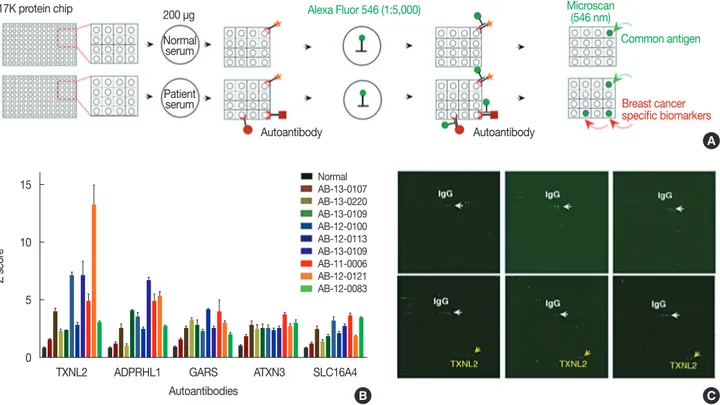
man protein microarray system containing 16,368 human re- combinant proteins. Human serum proteome analysis dem- onstrated that approximately 3,700 separate proteins exist in a physiological state. One issue with using human serum to identify biomarkers is the high background protein content, which causes noise signals under certain experimental condi- tions [11]. To minimize background signals in the protein mi- croarray, the amount of serum was adjusted so that up to 200 µg of serum proteins was used, which greatly reduced the in- terference by noise signals (data not shown). To identify auto- antibodies in TNBC samples, we used the HuProtTM human proteome microarray v2.0 (CDI Laboratories Inc., Mayaguez, USA), each containing 16,368 unique full-length human pro- tein antigens (Figure 1A). In total, 20 human serum samples (10 TNBC and 10 normal) were subjected to protein micro- array analysis. This study was approved by the Institutional Re- view Board of Ajou University Hospital (IRB No. AJIRB-BMR- KSP-15-362) and written informed consent was obtained from all participants. The result of protein microarray was analyzed
by GenePix analysis software (Molecular Devices, San Jose, USA) (Figure 1B). Briefly, microarray slides were blocked with filtrated dry milk and were then incubated with serum samples. After washing, the arrays were incubated with anti- human IgG (H+L) conjugated to AlexaFluor 546 (Cat. No.
A-21445; Invitrogen, Carlsbad, USA). The arrays were then washed, dried, and immediately scanned with a GenePix 4000B Fluorescence Scanner (Molecular Devices). The GenePix analy- sis software determined that five autoantibodies, thioredoxin- like 2 (TXNL2), ADP-ribosylhydrolase like 1 (ADPRHL1), glycyl-TRNA synthetase (GARS), spinocerebellar ataxia type 3 protein (ATXN3) and solute carrier family 16 member 4 (SLC16A4), had a significantly higher expression in the TNBC samples than in the normal samples. The differential expression of these five autoantibody biomarkers is shown in Figure 1B. Among them, we selected the TXNL2 autoanti- body biomarker not only because it showed the largest differ- ence between the TNBC and normal samples but also because the TXNL2 autoantigen is known as a potential diagnostic in-
Figure 1. Identification of five autoantibodies for triple-negative breast cancer (TNBC) biomarkers using a 17K human protein microarray system. (A) To identify novel TNBC-specific biomarkers, normal or TNBC patient sera containing autoantibodies were applied on 17K protein microarray. TNBC- specific autoantibodies were detected by anti-human Alexa Flour 546 (Invitrogen) and scanned by GenePix 4000B (Molecular Devices). Signal intensi- ty for each spot was obtained as the ratio of foreground to background signals and was normalized with the glutathione s-transferase signal intensity.
The mean signal intensity of each protein on the chip was calculated. (B) Five autoantibodies, thioredoxin-like 2 (TXNL2), ADP-ribosylhydrolase like 1 (ADPRHL1), glycyl-TRNA synthetase (GARS), spinocerebellar ataxia type 3 protein (ATXN3), and solute carrier family 16 member 4 (SLC16A4), were determined by the GenePix analysis. The differential expression of these five autoantibody biomarkers were calculated by statistical analysis. (C) High- power image of 17K protein microarray. Yellow arrow indicated TXNL2 autoantibody and white arrow was shown IgG as a positive control. TXNL2 autoantibody specifically and strongly interacted with the TXNL2 antigen.
15
10
5
0
Z score
B C
A
17K protein chip Alexa Fluor 546 (1:5,000)
Common antigen
Breast cancer specific biomarkers Microscan
(546 nm)
Autoantibody Autoantibody
200 µg Normal
serum
Patient serum
Normal AB-13-0107 AB-13-0220 AB-13-0109 AB-12-0100 AB-12-0113 AB-13-0109 AB-11-0006 AB-12-0121 AB-12-0083
TXNL2 ADPRHL1 GARS ATXN3 SLC16A4
Autoantibodies
Thioredoxin-Like 2 Autoantibody as a Biomarker for Triple-Negative Breast Cancer 89
https://doi.org/10.4048/jbc.2018.21.1.87 http://ejbc.kr
dicator of breast cancer [12]. Figure 1C shows that the TXNL2 autoantibody specifically and strongly interacted with the TXNL2 antigen, suggesting that it is a highly specific bio- marker for TNBC.
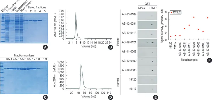
For independently verifying the TXNL2 autoantibody as a TNBC-specific biomarker, we attempted to purify the recom- binant TXNL2 antigen from BL21-Gold(DE3)pLysS Compe- tent Cells (Agilent, Santa Clara, USA). The purity of the TXNL2 protein is directly associated with the reliability of the TXNL2 autoantibody as a TNBC-specific biomarker. To ob- tain high-purity and sufficiently large yields of recombinant TXNL2, the full-length cDNA of TXNL2 was optimized for codon usage. Fortunately, we found that glutathione s-trans- ferase (GST)-TXNL2 protein was successfully expressed and purified using a Glutathione Sepharose 4B (GE Healthcare, Little Chalfont, UK). Most GST-TXNL2 proteins were ob- served in the soluble fraction after sonication and migrated to
~63 kDa on an sodium dodecyl sulfate polyacrylamide gel electrophoresis (SDS-PAGE) as a single band (Figure 2A).
Densitometric analysis by the ImageJ program (https://imagej.
net/) indicated that GST-TXNL2 comprised 42% of the total proteins. As shown in Figure 2B, following affinity chroma- tography, the fractions containing GST-TXNL2 were further separated by using gel filtration with Superdex 75 prep grade resin (GE Healthcare) that displays a high resolution for pro- teins in the molecular weight range of 3,000 to 70,000 Da. Fi- nally, we found that the final eluted fractions contained highly pure GST-TXNL2 (Figure 2C). The molecular weight and ho- mogeneity of the sample were checked using a TSKgel- S3000SWXL (Tosoh Bioscience, Tokyo, Japan) (Figure 2D).
Taken together, these data suggest that GST-TXNL2 protein might be a usable antigen for testing of the TXNL2 autoanti- body as a TNBC-specific biomarker.
To validate the specificity of the TXNL2 autoantibody for TNBC, we employed a quantitative dot blot system. The dot blot method is the best technique for rapidly assessing the quantity of a target antigen across many samples at once. It is also a popular method for screening antibodies for target specificity. The feasibility of the dot blot analysis was verified by analyzing the GST protein and the specificity of a rabbit
Figure 2. Thioredoxin-like 2 (TXNL2) autoantibody is a potential biomarker for diagnosing triple-negative breast cancer (TNBC). (A) Sodium dodecyl sulfate polyacrylamide gel electrophoresis (SDS-PAGE) analysis of the recombinant glutathione s-transferase (GST)-tagged TXNL2 protein. Total cell lysate and each subcellular fractions of the isopropyl β-D-1-thiogalactopyranoside-induced Escherichia coli BL21 (DE3) expressing GST-tagged TXNL2 protein was loaded in SDS-PAGE. The soluble fraction containing GST-tagged TXNL2 protein was applied in a Glutathione Sepharose 4B res- in (GE Healthcare). Subsequently resin conjugated TXNL2 was detached by elution buffer containing reduced glutathione. (B) Superdex 75 (GE Healthcare) gel-filtration fractionation profile for the further purification of the target protein. (C) SDS-PAGE profile of GST-tagged TXNL2 protein eluted from gel filtration. (D) A high-performance liquid chromatography chromatogram of recombinant GST-tagged TXNL2 protein. (E) Dot blot analysis of TXNL2 for detection of TXNL2 autoantibody in TNBC serum sample. Purified GST or GST tagged TXNL2 protein was spotted on nitrocellulose mem- brane and applied with normal or TNBC patient sera. GST was used as a negative control. Autoantibody against TXNL2 was detected by anti-human horseradish peroxidase conjugated antibody and developed using an enhanced chemiluminescent substrate. (F) Graph shows that amount of TXNL2 autoantibody derived from human sera. TXNL2 autoantibody was expressed 2- to 6-fold higher in TNBC serum than in normal. Densitometric analy- sis was performed using the ImageJ program (https://imagej.net/).
MarkerTotal cell lysatePelletSuppematantFlow thr ough
AB-13-0109
AB-12-0034
AB-12-0113
AB-12-0121
AB-11-0006
AB-12-0100
AB-12-0083
19120
19117 1,600
1,400 1,200 1,000 800 600400 2000 0.090.08 0.070.06 0.050.04 0.030.02 0.010
8 6 4 2 0
20 40 60 80 100 120 140 2 4 6 8 10 12 14 16 18 20 22 24 1 2 3 4 5
Eluted fractions
Volume (mL) Volume (mL)
Blood samples
Abs 280 nm (mA.U.)Abs 280 nm (mA.U.) Signal intensity (arbitrary unit)
B
F A
D E
C 3 3.5 4 4.5 5 5.5 6 6.5 7 7.5 8 8.5 9
Fraction numbers
Mock TXNL2 GST
PatientNormal 19120 19117 AB-13-0109 AB-12-0034 AB-12-0113 AB-12-0121 AB-11-0006 AB-12-0100 AB-12-0083
TXNL2
90 Jee Min Chung, et al.
http://ejbc.kr https://doi.org/10.4048/jbc.2018.21.1.87
anti-GST antibody was evaluated (data not shown). For the dose–response curve, we serially diluted GST proteins from 0.1 μg to 2 μg under nondenaturing condition. Dot blot analy- sis yielded a linear curve between 0.2 μg and 1.8 μg of protein with a coefficient of determination (R2) of 0.999 when simple linear regression analysis was performed (data not shown).
Beyond 2 μg, the signal intensity started to plateau. Based on the control experiment, we loaded 1.5 μg GST or GST-TXNL2 proteins onto the nitrocellulose membrane; we then incubated each membrane with normal or TNBC sera, which were used in the protein microarray experiment, as shown in Figure 2E.
Dot blot data revealed that the amount of the TXNL2 autoan- tibody derived from each serum sample showed similar ex- pression levels to those obtained using the protein microarray, indicating that the protein microarray data are reliable for de- tecting autoantibodies from sera (Figure 2F). In addition, we found that the TXNL2 autoantibody was more highly ex- pressed in TNBC samples than in normal samples. Taken to- gether, these results suggest that the TXNL2 autoantibody is a strong serum biomarker to detect TNBC.
Clinically, autoantibodies are currently being used to diag- nose various immune diseases. However, they are also found in various cancers and neurological diseases, indicating that a diagnostic method using autoantibodies can be applied to a wide range of diseases beyond immunological diseases. That said, autoantibodies against cancer-related antigens may be de- tected in the sera of patients with various types of cancer, and there are still few ways to identify disease-specific autoantibod- ies from patient blood. Of these methods, protein microarrays have the advantage of being able to identify autoantibodies ef- ficiently by using only a small amount of blood. For this rea- son, many research groups are now trying to identify autoanti- bodies as specific biomarkers for a number of diseases [13].
Recently, it has been reported that TXNL2 is also a TNBC- specific marker that is highly expressed in breast cancer pa- tients [12]. Moreover, the same study determined that TXNL2 protein is directly linked to the growth and metastasis of breast cancer cells and is associated with decreased overall pa- tient survival, suggesting that abnormal expression of TXNL2 is directly or indirectly linked to TNBC initiation or progres- sion [12]. TXNL2 has a unique protein structure consisting of a Trx homology region in the N terminus, followed by two tandem repeats of the Grx domains, which play a role in reac- tive oxygen species (ROS) detoxification and cell viability [12].
Remarkably, the deletion of TXNL2 gene in mice leads to em- bryonic lethality [14], indicating its role in protecting cells against oxidative stress during embryogenesis. In addition, TXNL2 is a critical factor in regulating ROS levels and nuclear factor κB activity for controlling tumor development and me-
tastasis in primary human breast cancers, demonstrating that it has essential roles in cancer cells [12]. In the current study, we identified the TXNL2 autoantibody as a TNBC-specific biomarker. Although the disease-specific linkage between au- toantibodies and autoantigens is not clear, our data suggests that abnormal expression or dysfunctions of TXNL2 might be closely related to the generation of the TXNL2 autoantibody in TNBC.
CONFLICT OF INTEREST
The authors declare that they have no competing interests.
REFERENCES
1. Perou CM, Sørlie T, Eisen MB, van de Rijn M, Jeffrey SS, Rees CA, et al.
Molecular portraits of human breast tumours. Nature 2000;406:747-52.
2. Kalimutho M, Parsons K, Mittal D, López JA, Srihari S, Khanna KK.
Targeted therapies for triple-negative breast cancer: combating a stub- born disease. Trends Pharmacol Sci 2015;36:822-46.
3. Collignon J, Lousberg L, Schroeder H, Jerusalem G. Triple-negative breast cancer: treatment challenges and solutions. Breast Cancer (Dove Med Press) 2016;8:93-107.
4. Fleisher B, Clarke C, Ait-Oudhia S. Current advances in biomarkers for targeted therapy in triple-negative breast cancer. Breast Cancer (Dove Med Press). 2016;8:183-97.
5. Wahba HA, El-Hadaad HA. Current approaches in treatment of triple- negative breast cancer. Cancer Biol Med 2015;12:106-16.
6. Ramachandran N, Srivastava S, Labaer J. Applications of protein micro- arrays for biomarker discovery. Proteomics Clin Appl 2008;2:1444-59.
7. Conrad K, Roggenbuck D, Reinhold D, Sack U. Autoantibody diagnos- tics in clinical practice. Autoimmun Rev 2012;11:207-11.
8. Xu YW, Peng YH, Chen B, Wu ZY, Wu JY, Shen JH, et al. Autoantibod- ies as potential biomarkers for the early detection of esophageal squa- mous cell carcinoma. Am J Gastroenterol 2014;109:36-45.
9. Zaenker P, Ziman MR. Serologic autoantibodies as diagnostic cancer biomarkers: a review. Cancer Epidemiol Biomarkers Prev 2013;22:
2161-81.
10. Macdonald IK, Parsy-Kowalska CB, Chapman CJ. Autoantibodies: op- portunities for early cancer detection. Trends Cancer 2017;3:198-213.
11. Pieper R, Gatlin CL, Makusky AJ, Russo PS, Schatz CR, Miller SS, et al.
The human serum proteome: display of nearly 3700 chromatographi- cally separated protein spots on two-dimensional electrophoresis gels and identification of 325 distinct proteins. Proteomics 2003;3:1345-64.
12. Qu Y, Wang J, Ray PS, Guo H, Huang J, Shin-Sim M, et al. Thioredoxin- like 2 regulates human cancer cell growth and metastasis via redox ho- meostasis and NF-kappaB signaling. J Clin Invest 2011;121:212-25.
13. Zhu H, Cox E, Qian J. Functional protein microarray as molecular de- cathlete: a versatile player in clinical proteomics. Proteomics Clin Appl 2012;6:548-62.
14. Cha H, Kim JM, Oh JG, Jeong MH, Park CS, Park J, et al. PICOT is a critical regulator of cardiac hypertrophy and cardiomyocyte contractili- ty. J Mol Cell Cardiol 2008;45:796-803.