Analysis of Protrusio Acetabuli Using a CT-based Diagnostic Method in Korean Patients with Marfan Syndrome: Prevalence and Association with Other Manifestations
A new CT-based diagnostic method of protrusio acetabuli (PA) was introduced. However, prevalence of PA by this method and correlation between PA and other manifestations of Marfan syndrome (MFS) is unknown in Korean MFS patients. This study aimed to investigate the prevalence of PA diagnosed by a CT-based method in Korean patients with MFS, the association of PA with other manifestations of MFS, and the contribution of PA to MFS diagnosis. We retrospectively reviewed the records of 146 MFS patients with the presence of a causative FBN1 mutation and 146 age- and sex-matched controls from a single tertiary care center. All MFS patients underwent a complete assessment of criteria based on the revised Ghent nosology. PA was assessed quantitatively using a CT-based circle-wall distance (CWD) method. PA was diagnosed in 77.4% of patients in the MFS group and in 11.0% of the control group. CWD was significantly different between the two groups (1.50 mm vs. -0.64 mm, P < 0.001). The presence of PA did not correlate with the presence of ectopia lentis, aortic root diameter, or history of aortic dissection. The presence of PA did not have a significant impact on the final diagnosis of MFS. Even though the presence of PA does not related to the cardinal clinical features of MFS or influence MFS diagnosis, its presence may be helpful for the suspicion of MFS when aortic dissection or aneurysm is found on CT angiography of the aorta because of the high frequency of PA in MFS patients.
Keywords: Protrusio Acetabuli; Marfan Syndrome; FBN1 Mutation; Computed Tomography
Kwang Jin Chun,1 Jeong Hoon Yang,1,2 Shin Yi Jang,1 Seung Hwa Lee,1 Hye Bin Gwag,1 Tae-Young Chung,3 June Huh,4 Chang-Seok Ki,5 Kiick Sung,6 Seung-Hyuk Choi,1 Sung Mok Kim,7 Yeon Hyeon Choe,7 and Duk-Kyung Kim1
1Division of Cardiology, Department of Medicine, Heart Vascular Stroke Institute, 2Department of Critical Care Medicine, 3Department of Ophthalmology, 4Department of Pediatrics,
5Department of Laboratory Medicine and Genetics,
6Department of Thoracic and Cardiovascular Surgery, 7Department of Radiology, Cardiovascular and Stroke Imaging Center, Samsung Medical Center, Sungkyunkwan University School of Medicine, Seoul, Korea
Received: 14 November 2014 Accepted: 13 May 2015 Address for Correspondence:
Duk-Kyung Kim, MD
Division of Cardiology, Department of Medicine, Heart Vascular Stroke Institute, Samsung Medical Center, Sungkyunkwan University School of Medicine, 81 Irwon-ro, Gangnam-gu, Seoul 135-710, Korea
Tel: +82.2-3410-3419, Fax: +82.2-3410-3849 E-mail: [email protected]
Funding: This work was supported by the Samsung Biomedical Research Institute (grant C-A9-204-3).
http://dx.doi.org/10.3346/jkms.2015.30.9.1260 • J Korean Med Sci 2015; 30: 1260-1265
INTRODUCTION
Marfan syndrome (MFS) is an autosomal dominant connective tissue disorder with cardiovascular, skeletal and ocular mani- festations (1). The diagnosis of MFS relies on the revised Ghent nosology, which weights cardiovascular manifestations more heavily and in which aortic root aneurysm and ectopia lentis are the cardinal clinical features (2, 3). In the revised Ghent no- sology, the most selective systemic features were included in the systemic score and each element was assigned one, two, or three points based on its importance to diagnosis (2). Protrusio acetabuli (PA) was assigned two points, as was dural ectasia, spontaneous pneumothorax, anterior chest deformity, and hind- foot deformity (2).
PA has various etiologies, including infectious, neoplastic, in- flammatory, metabolic, traumatic, and genetic causes (4). There are a few methods to diagnose PA using plain anteroposterior
radiographs of the pelvis. The prevalence of PA, which is differ- ent according to the diagnostic method, is 23%-46% in Korean MFS patients and 16%-100% in Western countries (5-9). The three most widely used criteria are the Steel method (using a center-edge angle of Wiberg > 50°), the Armbuster method (an acetabular-ilioischial distance of ≥ 3 mm in male patients or
≥ 6 mm in female patients), and crossing of the teardrop figure (Fig. 1) (5, 10, 11). Some consider the Armbuster method the most reliable measurement of PA, but other studies have report- ed that plain radiographic measurements of PA become unreli- able as pelvic tilt increases (11, 12).
In MFS, computed tomography (CT) angiography is com- monly performed to investigate the abnormality of aorta. In the axial view, the loss of the normal oval shape of the pelvic inlet at the level of the acetabulum can be easily identified and is con- sidered a sign of PA. A new CT-based diagnostic criterion was recently introduced and the degree of PA could be measured
using this method (13).
Some skeletal manifestations of MFS have been known to be associated with other skeletal manifestations or the severity of aortic root dilatation (14, 15). One study reported that dural ec- tasia was related to skeletal manifestations and aortic Z-scores (14). However, PA has not been evaluated to correlate with oth- er skeletal manifestations of MFS or aortic root dilatation.
Therefore, we investigated the prevalence of PA using the CT- based diagnostic method in Korean patients with MFS, the as- sociation of PA with other manifestations of MFS, and contribu- tion of PA to MFS diagnosis.
MATERIALS AND METHODS Study population
We reviewed data that was entered into our Marfan database between March 2006 and September 2013. Total 154 patients
≥ 18 yr of age with a FBN1 mutation were identified. Eight pa- tients who did not undergo CT examination due to renal dys- function and contrast allergy were excluded. All 146 patients fulfilled MFS diagnostic criteria by the revised Ghent nosology.
A
C
B
D CWD
a b
c d
Fig. 1. Diagnostic methods for protrusio acetabuli using plain anteroposterior radiographs of the pelvis and CT images. (A) The center-edge angle (CEA) is formed by a vertical line drawn through the center of the femoral head and a line drawn from the center through the lateral edge of the acetabular roof. A CEA of >50° is considered indicative of protrusio acetabuli. (B) The acetabular-ilioischial distance represents the transverse distance between the ilioischial line (a) and the acetabular line (b). Crossing of the ilioischial line by the acetabular line by > 3 mm medially in men or > 6 mm in women is considered indicative of protrusio acetabuli. (C) Radiographic changes of the teardrop figure in protrusio acetabuli. a, opened; b, closed; c, crossed; d, reversed. (D) CT-based circle-wall distance (CWD) method. A 10 cm radius circle is adapted to the inner acetabular wall of the pelvis. The distance between the line of the circle and the medial most point of the inner pelvic wall of the acetabular fossa was measured. Measurement of CWD is indi- cated. CT, computed tomography.
b a CEA
Revised Ghent nosology
In the revised Ghent nosology, cardiovascular manifestations are more heavily weighted and aortic root aneurysm and ecto- pia lentis are the cardinal clinical features. In the absence of any family history, the presence of these two manifestations is suffi- cient for diagnosis of MFS. In the absence of either of the cardi- nal clinical features, the presence of a FBN1 mutation or a com- bination of systemic score is required (Table 1).
Control population
The control cases were selected from patients who visited the emergency room between July 2007 and December 2013 and underwent abdominal and/or pelvic CT, and were age- and sex-matched to Marfan patients. Control candidates with hip joint problems or any known connective tissue disease were excluded from this study.
CT image
Multidetector computed tomography (MDCT) was performed using a dual-source CT scanner (Definition Flash; Siemens Health- care, Forchheim, Germany) with 2 × 64 × 0.6 mm collimation, section acquisition of 2 × 64 × 0.6 mm, and z-flying focal spot technique in both the study and control populations. The scan- ning parameters were 100 kV, 200 mA with automatic tube cur- rent modulation (Caredose 4D; Siemens Healthcare), a pitch of 1.0-1.2 and a 0.28-sec tube rotation time.
Measurements and definitions
PA was assessed quantitatively by the CT-based circle-wall dis-
tance (CWD) method (Fig. 1). This method uses axial CT imag- es and was introduced by Lundby et al. (13) in 2011. In this meth- od, PA was quantified by using a circle with a 10 cm radius. The circle was fitted to the inner pelvic wall at the level of the ace- tabulum where the cranial border of the superior pubic ramus fuses with the anterior column of the acetabulum. This level usually includes the fovea of the femoral head. The circle was best fitted to the concavities of the inner walls of the anterior and posterior columns of the acetabulum. We then measured the distance between the line of the circle and the medial most point of the inner pelvic wall of the acetabular fossa (CWD). A positive distance indicates that this medial most point is medial to the circle. Increasing degrees of PA have increasing positive values of CWD. A cut-off value of 1.25 mm was used for diagno- sis of PA via the CWD method (13).
We divided the systemic scores into a skeletal score, which included all skeletal manifestations, and non-skeletal scores which included pneumothorax, skin striae, myopia > 3 diop- ters, and mitral valve prolapse.
Statistics
Continuous data are expressed as the mean ± standard devia- tion or median and interquartile range. Categorical data are ex- pressed as frequency and percentage. To evaluate differences between the study groups, we used the Student’s unpaired t-test for normally distributed data and the Mann-Whitney test for skewed data. Categorical data was analyzed using the chi-square test or Fisher’s exact tests by SPSS software (SPSS for Windows, version 20.0, IBM Corp., Armonk, NY, USA).
Table 1. Diagnostic criteria for Marfan syndrome (MFS) according to the revised Ghent nosology In the absence of family history (FH)
(1) Ao (Z ≥ 2) AND EL = MFS (2) Ao (Z ≥ 2) AND FBN1 = MFS
(3) Ao (Z ≥ 2) AND Systemic score (≥ 7 pts) = MFS (4) EL AND FBN1 with known Ao = MFS In the presence of FH
(5) EL AND FH of MFS (as defined above) = MFS
(6) Systemic score ( ≥ 7 pts) AND FH of MFS (as defined above) = MFS
(7) Ao (Z ≥ 2 above 20 yr old, ≥ 3 below 20 yr) AND FH of MFS (as defined above) = MFS Scoring of systemic feature (maximum total 20 points)
Wrist AND thumb sign - 3 (wrist OR thumb sign - 1)
Pectus carinatum deformity - 2 (pectus excavatum or chest asymmetry - 1) Hind foot deformity - 2 (plain pes planus - 1)
Pneumothorax - 2 Dural ectasia - 2 Protrusio acetabuli - 2
Reduced US/LS AND increased arm/height AND no severe scoliosis - 1 Scoliosis or thoracolumbar kyphosis - 1
Reduced elbow extension - 1
Facial features (3/5) - 1 (dolichocephaly, enophthalmos, downslanting palpebral fissures, malar hypoplasia, retrognathia) Skin striae - 1
Myopia > 3 diopters - 1 Mitral valve prolapse (all types) - 1
Ao, aortic diameter at the sinuses of Valsalva above indicated Z-score or aortic root dissection; EL, ectopia lentis; FBN1, fibrillin-1 mutation; US/LS, upper segment/lower seg- ment ratio.
Ethics statement
This study received institutional review board approval (IRB File No. 2015-03-081). Informed consent was waived due to ret- rospective study.
RESULTS
PA was diagnosed in 77.4% of the MFS group and in 11.0% of controls (Table 2). The median CWD was 1.50 mm (range, -5.90 - 9.40 mm) in MFS group and -0.64 mm (range, -4.00 - 3.20 mm) in controls (P < 0.001, Fig. 2).
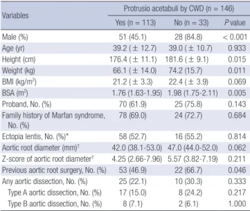
Clinical characteristics of MFS patients according to presence of PA were summarized in Table 3. Of patients with MFS and PA, 45.1% were male; in patients with MFS but without PA, 84.8%
were male (P < 0.001). The mean height of patients with PA ver- sus those without PA was 176.4 vs. 181.6 cm (P = 0.015), mean weight was 66.1 vs. 74.2 kg (P = 0.011), mean body mass index (BMI) was 21.2 vs. 22.4 kg/m2 (P = 0.069), and median body sur- face area (BSA) was 1.76 vs. 1.98 m2 (P = 0.005), respectively.
Hence, anthropometric data showed that patients with PA gen- erally had a smaller body habitus. Important diagnostic criteria,
such as a family history of MFS, ectopia lentis, and aortic root diameter, were not significantly different between the two groups (P = 0.684, P = 0.814, and P = 0.062, respectively). Rates of aor- tic dissection did not differ between the two groups (P = 0.333).
The prevalence of systemic score points ≥ 7 was more com- mon in the PA-positive group than in the PA-negative group (78.8 vs. 60.6%, P = 0.035, Table 4). Median systemic score points were
Table 2. Prevalence of protrusio acetabuli in Marfan syndrome and normal controls
Variables MFS (n = 146) Controls (n = 146) P value
No. of hips (persons) 292 (146) 292 (146)
Age (yr) 39.1 (± 12.2) 39.1 (± 12.2) 0.970
Males, No. (%) 79 (54.1) 79 (54.1) 1.000
Protrusio acetabuli, No. (%) 113 (77.4) 16 (11.0) < 0.001 CWD (mm)
Median (min, max) (interquartile range)
1.50 (-5.90, 9.40) (0.65−2.60)
-0.64 (-4.00, 3.20) (-1.40–0.63)
< 0.001
MFS, Marfan syndrome; CWD, circle-wall distance.
Table 3. Clinical characteristics of patients with vs. without protrusio acetabuli in Marfan syndrome
Variables Protrusio acetabuli by CWD (n = 146)
Yes (n = 113) No (n = 33) P value
Male (%) 51 (45.1) 28 (84.8) < 0.001
Age (yr) 39.2 ( ± 12.7) 39.0 ( ± 10.7) 0.933
Height (cm) 176.4 ( ± 11.1) 181.6 ( ± 9.1) 0.015
Weight (kg) 66.1 ( ± 14.0) 74.2 (15.7) 0.011
BMI (kg/m2) 21.2 ( ± 3.3) 22.4 ( ± 3.9) 0.069
BSA (m2) 1.76 (1.63-1.95) 1.98 (1.75-2.11) 0.005
Proband, No. (%) 70 (61.9) 25 (75.8) 0.143
Family history of Marfan syndrome, No. (%)
78 (69.0) 24 (72.7) 0.684
Ectopia lentis, No. (%)* 58 (52.7) 16 (55.2) 0.814
Aortic root diameter (mm)† 42.0 (38.1-53.0) 47.0 (44.0-52.0) 0.062 Z-score of aortic root diameter† 4.25 (2.66-7.96) 5.57 (3.82-7.19) 0.211 Previous aortic root surgery, No. (%) 53 (46.9) 22 (66.7) 0.046 Any aortic dissection, No. (%) 25 (22.1) 10 (30.3) 0.333 Type A aortic dissection, No. (%) 17 (15.0) 8 (24.2) 0.217 Type B aortic dissection, No. (%) 8 (7.1) 2 (6.1) 1.000
*7 patients were not assessed for ectopia lentis; †22 patients who previously under- went aortic root surgery at an outside hospital could not be assessed for aortic root diameter. CWD, circle-wall distance; BMI, body mass index; BSA, body surface area.
Fig. 2. Scattergram of CWD in patients with MFS and normal controls. CWD, circle- wall distance; MFS, Marfan syndrome.
MFS 10
5
0
-5
-10
Control
CWD (mm)
Table 4. Analysis of systemic scores of patients with vs. without protrusio acetabuli in Marfan syndrome
Variables Protrusio acetabuli by CWD (n = 146)
Yes (n = 113) No (n = 33) P value Systemic score ≥ 7, No. (%) 89 (78.8) 20 (60.6) 0.035 Systemic score (point) 10.0 (7.0-12.0) 7.0 (4.0-8.0) < 0.001 Skeletal score (point) 8.0 (6.0-10.0) 6.0 (3.5-7.5) < 0.001 Wrist and/or thumb sign, No. (%) 84 (74.3) 19 (57.6) 0.063 Pectus carinatum or excavatum
deformity, No. (%)
12 (10.6) 5 (15.2) 0.538
Hindfoot deformity, No. (%) 68 (60.2) 23 (69.7) 0.321
Dural ectasia, No. (%) 68 (60.2) 20 (60.6) 0.965
Reduced US/LS and increased arm/height ratio, No. (%)
26 (23.0) 5 (15.2) 0.332
Scoliosis or thoracolumbar
kyphosis, No. (%) 23 (20.4) 2 (6.1) 0.055
Reduced elbow extension, No. (%) 22 (19.5) 6 (18.2) 0.869 Facial features, No. (%) 81 (71.7) 21 (63.6) 0.376 PA (-) skeletal score (point) 6.0 (4.0-8.0) 6.0 (3.5-7.5) 0.322 Non-skeletal score (point) 1.0 (1.0-2.0) 1.0 (0.5-2.0) 0.493
Pneumothorax, No. (%) 14 (12.4) 3 (9.1) 0.763
Skin striae, No. (%) 74 (65.5) 23 (69.7) 0.652
Myopia > 3 diopters, No. (%) 32 (28.3) 9 (27.3) 0.906
MVP, No. (%) 39 (34.5) 6 (18.2) 0.074
CWD, circle-wall distance; PA, protrusio acetabuli; US/LS, upper segment/lower seg- ment ratio; MVP, mitral valve prolapse.
Table 5. Fulfilled by revised Ghent nosology with or without FBN1 mutation or protru- sio acetabuli
Not considering PA point
Considering PA point Fulfilled revised Ghent nosology, No. (%) 145 (99.3) 146 (100.0) Fulfilled revised Ghent nosology before FBN1
mutation was confirmed, No. (%) 141 (96.6) 143 (97.9) FBN1, fibrillin-1 mutation; PA, protrusio acetabuli.
higher in the PA-positive group (10.0 vs. 7.0 points, P < 0.001) and median skeletal score points were also higher in the PA-pos- itive group (8.0 vs. 6.0 points, P < 0.001). Because the presence of PA is worth 2 points in the calculation of the systemic score, we compared skeletal score points other than PA points. The median skeletal score points excluding PA did not differ between the two groups (6.0 vs. 6.0 points, P = 0.322). We analyzed each element of the skeletal score. There was also no association be- tween PA and the elements comprising the skeletal score. How- ever, scoliosis was numerically but not statistically significantly more frequent in the PA-positive group (20.4 vs. 6.1%, P = 0.055).
The median non-skeletal score points were not significantly dif- ferent (1 vs. 1 point, P = 0.493). CWD was weakly correlated with skeletal score points (P < 0.001, correlation coefficient = 0.391).
Among 146 patients with FBN1 mutation, only one patient did not fulfill the revised Ghent nosology if the two points at- tributable to PA were not included (Table 5). In real world clini- cal practice, genetic diagnosis of FBN1 mutation takes several weeks or months. Therefore, if we assume that we do not know the FBN1 mutation status, three patients would not have met the revised Ghent nosology. If both FBN1 mutation and pres- ence of PA were not included, five patients did not meet the di- agnostic criteria for MFS.
DISCUSSION
In the revised Ghent nosology, two points are assigned to the presence of PA in systemic score criteria (2). There are various methods for diagnosing PA, usually using plain anteroposterior radiographs of the pelvis. Of this, the Steel and Armbuster meth- ods were representative. The Steel method uses the center-edge angle of Wiberg, and the Armbuster method uses the acetabu- lar-ilioischial distance. In one study, the prevalence of PA in pa- tients with MFS differed depending on the method used, with 27% of patients considered to have PA using the Steel method and 16% using the Armbuster method (9). Other studies have reported that CT or magnetic resonance imaging (MRI) are go- ing to be more reliable (11, 12).
In patients with MFS, CT is commonly used to evaluate dila- tation, aneurysm, or dissection of the aorta. In PA, a loss of the normal oval shape of the pelvic inlet at the level of the acetabu- lum is typically noted on axial CT images. Lundby et al. (13) re- cently introduced a new method to diagnose PA using axial CT
images. They measured the distance between the line of the imaginary 10 cm radius circle and the medial most point of the inner pelvic wall of the acetabular fossa. In their study, the exact prevalence of PA by the CWD method was not reported, but was calculated to be 74.7% in Ghent nosology positive patients and 3.7% in controls (13). In our study, the prevalence of PA us- ing the CWD method was similar in the MFS group and more frequent in controls compared to Lundby’s study. PA developed more frequently in females and patients with a smaller body habitus.
The CWD method uses a circle with a 10 cm radius to quan- tify and cut-off value for diagnosis of PA was 1.25 mm. Some study reported that there might be racial and ethnic differences in clinical manifestations between Asian and Western MFS pa- tients (16, 17). Akutsu et al. (16) reported that major skeletal cri- teria were less frequent in the Japanese population than the West- ern population, especially arm span to height ratio > 1.05, sco- liosis, reduced extension at elbows, and joint hypermobility. Yoo et al. (17) reported that Korean MFS patients less frequently ful- filled major skeletal criteria than Western MFS patients. There- fore, further study is needed to investigate the prevalence of PA between Asian and Western MFS patients, and establish appro- priate cut-off value when using the CWD method for diagnosis of PA in Korean MFS patients. However, the absence of a gold standard for PA diagnosis prevents the establishment of an ap- propriate cut-off value.
MFS has multiple manifestations that may be related to each other. Indeed, PA was known to associate with scoliosis (15).
Sheikhzadeh et al. (14) reported that dural ectasia, which adds two points to the systemic score like PA, correlates with aortic Z-scores and skeletal involvement. In our study, aortic root di- ameter, aortic Z-scores, family history of MFS, and ectopia len- tis was not significantly different between the PA-positive and PA-negative groups. Furthermore, PA was neither associated with a history of aortic dissection nor a particular type of aortic dissection. Patients with PA had higher total systemic and skel- etal scores than those without PA, but this difference was lost when PA was not included in systemic and skeletal scores. Con- sidering each element of the skeletal score, there was no rela- tionship between each element of the skeletal score and PA, ex- cept the higher tendency for scoliosis observed in the MFS group.
In the study of Sheikhzadeh et al. (14), they included dural ecta- sia points in their skeletal score calculation. Therefore, if dural ectasia was not considered in their skeletal score calculation, it might be possible that there was no association between dural ectasia and skeletal score. An other study reported that no as- sociation between aortic dilatation and dural ectasia (18).
CT angiography is useful in the diagnosis of other manifesta- tions of MFS in addition to cardiovascular findings. We can eval- uate the presence of pectus carinatum or excavatum, scoliosis or thoracolumbar kyphosis, apical blebs, and dural ectasia as
well as the presence of PA. Sohn et al. (7) reported that 24% of the study population could be diagnosed with MFS using only CT images. Among our 146 patients, only one patient would not fit criteria for MFS if we excluded the presence of PA. Even though PA does not greatly impact MFS diagnosis, the presence of PA may be helpful for the suspicion of MFS when aortic dissection or aneurysm is found on CT angiography of the aorta.
Our study has several limitations. First, the symptoms and signs of PA were missing in our study. Because of retrospective design of the study, we could not assess how many MFS patients with PA had the symptoms. Second, our MFS patients were un- likely to represent individuals from the general population. Our hospital is a major hospital in Korea, and most patients were referred from other hospitals for surgery for aortic root dilata- tion or dissection. Accordingly, most of our MFS patients satisfy the aortic Z-score or aortic root dissection criteria, which are essential for the diagnosis of MFS under the revised Ghent no- sology. Third, our study did not include patients less than 18 yr of age, because they usually do not undergo CT due to concerns regarding radiation exposure.
In conclusion, even though the presence of PA does not re- lated to the cardinal clinical features of MFS or influence MFS diagnosis, its presence may be helpful for the suspicion of MFS when aortic dissection or aneurysm is found on CT angiogra- phy of the aorta because of the high frequency of PA in MFS pa- tients.
AUTHOR CONTRIBUTION
Conception and design of this study: Chun KJ, Choi SH, Kim DK.
Data collection: all authors. Analysis and interpretation of data:
Chun KJ, Choi SH, Kim DK. Manuscript preparation: Chun KJ, Kim DK. Manuscript approval: all authors.
ORCID
Kwang Jin Chun http://orcid.org/0000-0001-7683-1343 Shin Yi Jang http://orcid.org/0000-0003-4319-7029 Seung-Hyuk Choi http://orcid.org/0000-0002-0304-6317 Duk-Kyung Kim http://orcid.org/0000-0001-9227-4196 REFERENCES
1. Koh KK, Hyon MS, Lim HJ, Kim CH, Oh BH, Park YB, Choi YS, Seo JD, Lee YW. Cardiovascular manifestations of marfan syndrome. Korean Circ J 1987; 17: 777-82.
2. Loeys BL, Dietz HC, Braverman AC, Callewaert BL, De Backer J, De- vereux RB, Hilhorst-Hofstee Y, Jondeau G, Faivre L, Milewicz DM, et al.
The revised Ghent nosology for the Marfan syndrome. J Med Genet 2010;
47: 476-85.
3. De Paepe A, Devereux RB, Dietz HC, Hennekam RC, Pyeritz RE. Revised
diagnostic criteria for the Marfan syndrome. Am J Med Genet 1996; 62:
417-26.
4. McBride MT, Muldoon MP, Santore RF, Trousdale RT, Wenger DR. Pro- trusio acetabuli: diagnosis and treatment. J Am Acad Orthop Surg 2001;
9: 79-88.
5. Steel HH. Protrusio acetabuli: its occurrence in the completely expressed Marfan syndrome and its musculoskeletal component and a procedure to arrest the course of protrusion in the growing pelvis. J Pediatr Orthop 1996; 16: 704-18.
6. Do T, Giampietro PF, Burke SW, Davis JG, Raggio C, Schneider R, Boachie- Adjei O, Brill P. The incidence of protrusio acetabuli in Marfan’s syndrome and its relationship to bone mineral density. J Pediatr Orthop 2000; 20:
718-21.
7. Sohn GH, Jang SY, Moon JR, Yang JH, Sung K, Ki CS, Oh JK, Choe YH, Kim DK. The usefulness of multidetector computed tomographic angiog- raphy for the diagnosis of Marfan syndrome by Ghent criteria. Int J Car- diovasc Imaging 2011; 27: 679-88.
8. Yang JH, Han H, Jang SY, Moon JR, Sung K, Chung TY, Lee HJ, Ki CS, Kim DK. A comparison of the Ghent and revised Ghent nosologies for the diagnosis of Marfan syndrome in an adult Korean population. Am J Med Genet A 2012; 158A: 989-95.
9. Sponseller PD, Jones KB, Ahn NU, Erkula G, Foran JR, Dietz HC 3rd. Pro- trusio acetabuli in Marfan syndrome: age-related prevalence and associ- ated hip function. J Bone Joint Surg Am 2006; 88: 486-95.
10. Armbuster TG, Guerra J Jr, Resnick D, Goergen TG, Feingold ML, Ni- wayama G, Danzig LA. The adult hip: an anatomic study. Part I: the bony landmarks. Radiology 1978; 128: 1-10.
11. Van de Velde S, Fillman R, Yandow S. Protrusio acetabuli in Marfan syn- drome. History, diagnosis, and treatment. J Bone Joint Surg Am 2006; 88:
639-46.
12. Richards PJ, Pattison JM, Belcher J, Decann RW, Anderson S, Wynn- Jones C. A new tilt on pelvic radiographs: a pilot study. Skeletal Radiol 2009; 38: 113-22.
13. Lundby R, Kirkhus E, Rand-Hendriksen S, Hald J, Pripp AH, Smith HJ.
CT of the hips in the investigation of protrusio acetabuli in Marfan syn- drome. A case control study. Eur Radiol 2011; 21: 1485-91.
14. Sheikhzadeh S, Sondermann C, Rybczynski M, Habermann CR, Brock- staedt L, Keyser B, Kaemmerer H, Mir T, Staebler A, Robinson PN, et al.
Comprehensive analysis of dural ectasia in 150 patients with a causative FBN1 mutation. Clin Genet 2014; 86: 238-45.
15. Wenger DR, Ditkoff TJ, Herring JA, Mauldin DM. Protrusio acetabuli in Marfan’s syndrome. Clin Orthop Relat Res 1980: 147; 134-8.
16. Akutsu K, Morisaki H, Takeshita S, Ogino H, Higashi M, Okajima T, Yo- shimuta T, Tsutsumi Y, Nonogi H, Morisaki T. Characteristics in pheno- typic manifestations of genetically proved Marfan syndrome in a Japa- nese population. Am J Cardiol 2009; 103: 1146-8.
17. Yoo EH, Woo H, Ki CS, Lee HJ, Kim DK, Kang IS, Park P, Sung K, Lee CS, Chung TY, et al. Clinical and genetic analysis of Korean patients with Marfan syndrome: possible ethnic differences in clinical manifestation.
Clin Genet 2010; 77: 177-82.
18. Fattori R, Nienaber CA, Descovich B, Ambrosetto P, Reggiani LB, Pepe G, Kaufmann U, Negrini E, von Kodolitsch Y, Gensini GF. Importance of dural ectasia in phenotypic assessment of Marfan’s syndrome. Lancet 1999; 354: 910-3.