Received: May 13, 2018 Revised: June 2, 2018 Accepted: June 7, 2018 CliniCAl
neurophysiology
Correspondence to Ju-Hun Lee
Department of Neurology, Kangdong Sacred Heart Hospital, Hallym University College of Medicine, 150 Seongan-ro, Gangdong-gu, Seoul 05355, Korea Tel: +82-2-2224-2206
Fax: +82-2-2224-2114 E-mail: leejuhun@hallym.or.kr
http://www.e-acn.org copyright © 2018 the Korean Society of clinical neurophysiology
Pulsatility of middle cerebral arteries is better correlated with white matter hy- perintensities than aortic stiffening
Sang-Hwa Lee1, Yerim Kim2, Yeongbae Lee3, and Ju-Hun Lee2
1Department of Neurology, Chuncheon Sacred Heart Hospital, Hallym University College of Medicine, Chuncheon, Korea
2Department of Neurology, Kangdong Sacred Heart Hospital, Hallym University College of Medicine, Seoul, Korea
3Department of Neurology, Gachon University Gil Medical Center, Incheon, Korea
Background: Pulsatility of cerebral arteries and aortic stiffness have been associated with white matter hyperintensities (WMH). We explored which is better correlated with the severity of WMH in a population with acute lacunar infarct.
Methods: We included patients with acute small subcortical infarcts who underwent transcranial Doppler (TCD) and brachial ankle pulse wave velocity (baPWV). Exclusion criteria were any stenosis or occlusion on major cerebral arteries on magnetic resonance angiography; poor temporal inson- ation windows; ankle brachial index < 0.9; and atrial fibrillation. We assessed the performance of the pulsatility index of bilateral middle cerebral arteries (PI-MCA) and baPWV for predicting moder- ate-to-severe WMH, defined as an Age Related White Matter Changes score > 5, and then sought to find independent predictors using binary logistic regression analysis.
Results: Eighty-three patients (56 males, mean age 61.5 ± 11.4) participated in the study. Uni- variate analysis showed old age and high PI-MCA were significantly correlated with moder- ate-to-severe WMH. However, baPWV was not associated with the severity of WMH. Multivar- iate analysis revealed old age (odds ratio per 1-year increase, 1.068; p = 0.044) and upper tertile of PI-MCA (odds ratio, 5.138; p = 0.049) were independently associated with moderate-to-se- vere WMH. Receiver-operating characteristics showed PI-MCA differentiated those with and without moderate-to-severe WMH with an area under the curve of 0.719.
Conclusions: PI-MCA derived from TCD was better correlated with the severity of WMH than baPWV in a population with lacunar infarction. Pulsatility of cerebral arteries may better pre- dict cerebral small vessel disease than the aortic stiffness index.
Key words: Pulsatility index; Aortic stiffness; Transcranial Doppler ultrasonography
INTRODUCTION
Pulse wave velocity (PWV) is known as an index to quantify arterial stiffness.1 More specifically, aortic PWV, calculated on the basis of pulse transit time and the distance between carotid and femoral arteries, is a well-known predictor of car- diovascular mortality and morbidity.2,3 However, aortic PWV measurement may not be practical in the clinical setting due to the difficulty in applying pressure transducers on target arteries and the reluctance of some patients in exposing the inguinal area. Brachial-ankle PWV (baPWV) may provide qual- itatively similar information to aortic PWV, although some portions of baPWV may be influenced by peripheral arterial stiffness.4 Therefore, baPWV is widely used in clinical practice for the assessment of central arterial stiffness.
Transcranial Doppler (TCD) is a useful and safe way to ob- tain valuable hemodynamic data in cerebrovascular disease.
The pulsatility index (PI), derived from TCD, has long been proposed to reflect small vessel vascular resistance.5,6
Both pulsatility of cerebral arteries obtained from TCD7-10 and central arterial stiffness derived from PWV8,11,12 are known to be associated with cerebral small vessel disease (SVD). However, it remains unclear whether PI obtained from TCD or baPWV better correlates with cerebral SVD.
We explored whether PI derived from TCD or baPWV better correlates with the severity of white matter hyperintensities (WMH) in a population with lacunar infarction.
MATERIALS AND METHODS
Subjects
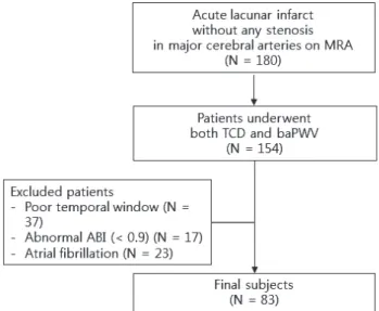
This is a retrospective hospital-based study. We reviewed consecutive patients with acute cerebral infarction who were admitted to two training hospitals in Korea between January 2014 and June 2015. We found 180 patients with lacunar infarct without any stenotic lesions in major cerebral arteries (anterior cerebral arteries, middle cerebral arteries, posterior cerebral arteries, basilar artery, vertebral arteries, and internal cerebral arteries) on magnetic resonance angi- ography (MRA). We defined lacunar infarct as a small infarct (less than 2 cm on diffusion weighted image) in the territory of perforating arteries. Among them, we included 154 pa- tients who underwent both TCD and baPWV examinations
(Fig. 1). We excluded the patients with any of the following:
1) poor temporal insonation windows (n = 37); 2) ankle-bra- chial index < 0.9 (n = 17); 3) atrial fibrillation documented on electrocardiography (n = 23).
TCD and baPWV examination
TCD examination was performed by experienced ultraso- nographers in each hospital. Middle cerebral arteries (MCAs) were examined at a depth of 50-60 mm through both tem- poral windows. Pulsatility index (PI) was automatically cal- culated for each vessel using the formula: PI = (peak systolic velocity-end-diastolic velocity) / mean velocity. We mea- sured PI at least two points in each MCA (proximal and distal points of MCA stem). We defined the PI of bilateral middle cerebral arteries (PI-MCA) as the mean of all measured PI val- ues in both MCAs.
BaPWV was measured using the Form PWV/ABI device (VP 2000; Colin Medical Technology, Komaki, Japan). Patients were examined in the supine position after being at rest for at least 10 minutes. Waveform data and time intervals be- tween the waves were automatically obtained from volume plethysmography cuff sensors on the right brachium and both ankles. The distance between the brachium and the ankle for baPWV was calculated according to patient height.
We used the average value of the baPWVs obtained on both sides.
Fig. 1. Flow diagram for study subjects. MRA, magnetic resonance an- giography; TCD, transcranial Doppler; baPWV, brachial ankle pulse wave velocity; ABI, ankle-brachial index.
Rating of white matter hyperintensities
We scored the severity of WMH using the Age Related White Matter Changes (ARWMC) scale according to operational definitions suggested by Xiong et al.13 for improving reliabil- ity of the ARWMC scale. We classified the severity of WMH as follows: absent (score 0), mild (1-5), moderate (6-10), and severe (11-15). The scoring was done independently by one neurologist blinded to TCD and baPWV findings as well as clinical features of patients.
Risk factors and confounding variables
We collected information on the patients’ age, sex, cardio- vascular risk factors, and National Institutes of Health Stroke Scale (NIHSS) at the time of hospitalization. History of stroke included all hemorrhagic or ischemic strokes. Hypertensive patients were defined as individuals previously diagnosed
with hypertension or individuals with a blood pressure of
> 140/90 mmHg in repeated measurements at rest after acute phase. Diabetes patients were defined as individuals previously diagnosed with diabetes or individuals with fast- ing blood glucose > 126 mg/dL or glycated hemoglobin (HbA1c) > 6.5%. Hyperlipidemic patients were defined as in- dividuals diagnosed with hyperlipidemia or individuals with fasting low-density lipoprotein > 130 mg/dL or total choles- terol > 200 mg/dL, which is the cutoff value of borderline high cholesterol according to NCEP III guidelines.14 Smokers were defined as current smokers.
Statistical analysis
We investigated the association between the severity of WMH and PI-MCA, and between the severity of WMH and baPWV. Values of PI-MCA and baPWV were split into tertiles.
We used chi-square tests to compare categorical variables and t-test (or Mann-Whitney test as appropriate) to analyze factors associated with moderate-to-severe WMH. To search for independent predictors of moderate-to-severe WMH, bi- nary logistic regression analysis was used, with age, sex, and cardiovascular risk factors (hypertension, previous stroke, diabetes, hyperlipidemia, and smoking) as covariates. Signif- icance level was set at p < 0.05 for all statistical analyses. The moderate-to-severe WMH predicting capability of PI-MCA and baPWV was evaluated through a receiver-operating characteristic curve.
RESULTS
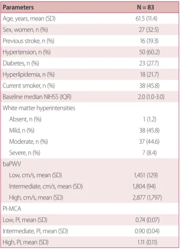
Eighty-three patients (56 male, 67.5%, mean age 61.5 ± 11.4) participated in the study. Patients’ baseline median NIHSS score was 2.0 (interquartile range 1.0-3.0). Detailed baseline characteristics are described in Table 1.
In univariate analysis (Table 2), old age was significantly associated with moderate-to-severe WMH (p < 0.001). PI- MCA was also significantly associated with moderate-to-se- vere WMH (p < 0.001, linear-by-linear association Chi-square tests). baPWV tended to associate with the severity of WMH, but this association was not statistically significant (p = 0.082, linear-by-linear association tests).
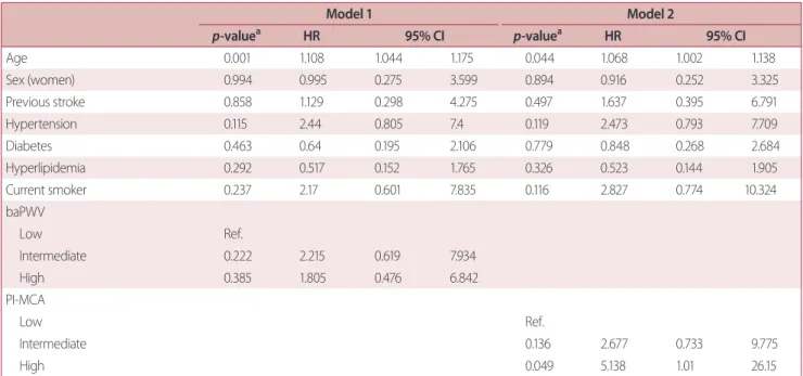
We conducted multivariate binary logistic analysis using baPWV (Model 1, Table 3) and using PI-MCA (Model 2, Table 3).
Table 1. Baseline characteristics
Parameters N = 83
Age, years, mean (SD) 61.5 (11.4)
Sex, women, n (%) 27 (32.5)
Previous stroke, n (%) 16 (19.3)
Hypertension, n (%) 50 (60.2)
Diabetes, n (%) 23 (27.7)
Hyperlipidemia, n (%) 18 (21.7)
Current smoker, n (%) 38 (45.8)
Baseline median NIHSS (IQR) 2.0 (1.0-3.0)
White matter hyperintensities
Absent, n (%) 1 (1.2)
Mild, n (%) 38 (45.8)
Moderate, n (%) 37 (44.6)
Severe, n (%) 7 (8.4)
baPWV
Low, cm/s, mean (SD) 1,451 (129)
Intermediate, cm/s, mean (SD) 1,804 (94) High, cm/s, mean (SD) 2,877 (1,797) PI-MCA
Low, PI, mean (SD) 0.74 (0.07)
Intermediate, PI, mean (SD) 0.90 (0.04)
High, PI, mean (SD) 1.11 (0.11)
NIHSS, National Institutes of Health Stroke Scale; IQR, interquartile range;
baPWV, brachial ankle pulse wave velocity; PI-MCA, pulsatility index of bilateral middle cerebral arteries; PI, pulsatility index.
Table 2. Factors associated with white matter hyperintensities in univariate analysis White matter hyperintensity
p-valuea Absent or mild (N = 39) Moderate or severe (N = 44)
Age, years, mean (SD) 56.5 (11.4) 65.9 (9.5) < 0.001
Sex, women, n (%) 10 (37.0) 17 (63.0) 0.207
Previous stroke, n (%) 34 (50.7) 33 (49.3) 0.16
Hypertension, n (%) 18 (54.5) 15 (45.5) 0.262
Diabetes, n (%) 28 (46.7) 32 (53.3) 0.925
Hyperlipidemia, n (%) 29 (44.6) 36 (55.4) 0.411
Current smoker, n (%) 20 (44.4) 25 (55.6) 0.613
Baseline mean NIHSS (SD) 2.1 (1.5) 2.7 (2.1) 0.175
baPWV 0.082
Low, n (%) 16 (59.3) 11 (40.7)
Intermediate, n (%) 13 (46.4) 15 (53.6)
High, n (%) 10 (35.7) 18 (64.3)
PI-MCA < 0.001
Low, n (%) 20 (74.1) 7 (25.9)
Intermediate, n (%) 12 (42.9) 16 (57.1)
High, n (%) 7 (25.0) 21 (75.0)
NIHSS, National Institutes of Health Stroke Scale; WMH, white matter hyperintensities; baPWV, brachial ankle pulse wave velocity; PI-MCA, pulsatility index of bilateral middle cerebral arteries.
aChi-square test, t-test, Mann-Whitney test as appropriate.
Table 3. Predicting facotrs for moderate-to-severe white matter hyperintensities in multivariate analysis
Model 1 Model 2
p-valuea HR 95% CI p-valuea HR 95% CI
Age 0.001 1.108 1.044 1.175 0.044 1.068 1.002 1.138
Sex (women) 0.994 0.995 0.275 3.599 0.894 0.916 0.252 3.325
Previous stroke 0.858 1.129 0.298 4.275 0.497 1.637 0.395 6.791
Hypertension 0.115 2.44 0.805 7.4 0.119 2.473 0.793 7.709
Diabetes 0.463 0.64 0.195 2.106 0.779 0.848 0.268 2.684
Hyperlipidemia 0.292 0.517 0.152 1.765 0.326 0.523 0.144 1.905
Current smoker 0.237 2.17 0.601 7.835 0.116 2.827 0.774 10.324
baPWV
Low Ref.
Intermediate 0.222 2.215 0.619 7.934
High 0.385 1.805 0.476 6.842
PI-MCA
Low Ref.
Intermediate 0.136 2.677 0.733 9.775
High 0.049 5.138 1.01 26.15
HR, heart rate; CI, confidence interval; baPWV, brachial ankle pulse wave velocity; Ref., reference; PI-MCA, pulsatility index of bilateral middle cerebral arter- ies; PI, pulsatility index.
aBinary logistic regression analysis.
In multivariate analysis, old age and upper tertile of PI-MCA were independently associated with moderate-to-severe WMH (p = 0.044 and 0.049, respectively).
In the receiver-operating characteristics (Fig. 2), PI-MCA differentiated those with and without moderate-to-severe WMH with an area under the curve of 0.719 (95% confi- dence interval [CI], 0.65-0.832). However, the performance of baPWV for predicting moderate-to-severe WMH was low, with an area under the curve of 0.625 (95% CI, 0.505-0.745).
DISCUSSION
Our data showed that PI-MCA derived from TCD was sig- nificantly associated with the severity of WMH. The ability to predict moderate-to-severe WMH was better for PI-MCA than for baPWV.
Aortic stiffness derived from PWV is known to be associ- ated with cerebral SVD.8,11,12 Our data showed that baPWV tended to be associated with the severity of WMH in univar- iate analysis although this association was not statistically significant. Aortic stiffening with advancing age increases the transfer of excessive, potentially harmful pulsatile energy
into the peripheral vessels. As a result, increased transmis- sion of pulsatile energy into smaller arteries may lead to reactive changes in vessel resistance, increasing peripheral resistance. In the long term, persistent reactive changes in vessel resistance may lead to remodeling of wall compo- nents and permanent vessel changes, which may be the un- derlying mechanism of the association between aortic PWV and cerebral small vessel disease.15
Like aortic stiffness, PI-MCA is known to be well correlated with cerebral small vessel disease.5,6 These two measure- ments may be associated with cerebral small vessel disease through a complex function of various hemodynamic fac- tors. For example, PI derived from TCD may be not depen- dent solely on cerebrovascular resistance, but may describe cerebral perfusion pressure in a more accurate manner.16 High PI was significantly associated with low cerebral perfu- sion pressure. Cerebral blood flow is determined by cerebral perfusion pressure as well as cerebral vascular resistance.
Therefore, low cerebral perfusion pressure (CPP) and high cerebrovascular resistance (CVR) in patients with high PI may cause chronic ischemic damage to the brain. The mecha- nism behind the better predictive ability of PI-MCA when compared to baPWV is not easily understandable and war- rants further research. A previous study also showed a sig- nificantly stronger association of leukoaraiosis with PI-MCA than with any other physiological measure.17 We think PI- MCA reflects cerebral blood flow more accurately through interplay with CPP and CVR than other physiological mea- sures such as aortic stiffness.
Our study has some limitations. First, it was a small cross-sectional and observational study and therefore there may be unmeasured confounders potentially associated with WMH. The association between baPWV and WMH was not statistically significant in our study. The statistical signif- icance between PI-MCA and WMH also diminished in the multivariate analysis. We think that age may be relatively a strong predictor compared with baPWV or PI-MCA. Relative- ly weak predictors might be obscured probably due to small sample size. Second, we did not assess other hemodynamic measures such as heart rate and blood pressure, and blood viscosity parameters such as hematocrit, which influence PI. We could not adjust the time of examination after acute ischemic stroke. In addition, we did not adjust drugs such as cilostazol or statin which can influence on PI. However, Fig. 2. In the receiver-operator characteristics, PI-MCA differentiated
those with and without moderate-to-severe WMH with area under curve of 0.719 (95% CI, 0.65-0.832). However, the performance of baP- WV for predicting moderate-to-severe WMH was low, with area under curve of 0.625 (95% CI, 0.505-0.745). PI-MCA, pulsatility index of bilateral middle cerebral arteries; WMH, white matter hyperintensities; baPWV, brachial ankle pulse wave velocity; CI, confidence interval.
we minimized the influence of irregular heart rate on PI through exclusion of patients with atrial fibrillation. Further- more, it was a strength of this study that we excluded any stenosis of proximal cerebral arteries on MRA. When there is a significant stenosis on the internal cerebral artery, PI-MCA becomes low and therefore inaccurately reflects cerebral small vessel disease. Therefore, exclusion of any stenosis in proximal cerebral arteries is a strong point of this study as it allows for the accurate assessment of small vessel disease.
PI-MCA derived from TCD was better correlated with the severity of WMH than baPWV in this study’s lacunar infarc- tion population. Pulsatility of cerebral arteries may be more predictive of cerebral small vessel disease than the aortic stiffness index.
Acknowledgements
This research was performed without any sources of funding.
REFERENCES
1. O’Rourke MF, Staessen JA, Vlachopoulos C, Duprez D, Plante GE.
Clinical applications of arterial stiffness; definitions and reference values. Am J Hypertens 2002;15:426-444.
2. Blacher J, Guerin AP, Pannier B, Marchais SJ, Safar ME, London GM. Impact of aortic stiffness on survival in end-stage renal dis- ease. Circulation 1999;99:2434-2439.
3. Laurent S, Boutouyrie P, Asmar R, Gautier I, Laloux B, Guize L, et al. Aortic stiffness is an independent predictor of all-cause and cardiovascular mortality in hypertensive patients. Hypertension 2001;37:1236-1241.
4. Sugawara J, Hayashi K, Yokoi T, Cortez-Cooper MY, DeVan AE, Anton MA, et al. Brachial-ankle pulse wave velocity: an index of central arterial stiffness? J Hum Hypertens 2005;19:401-406.
5. Legarth J, Thorup E. Characteristics of doppler blood-velocity waveforms in a cardiovascular in vitro model. i. the model and the influence of pulse rate. Scand J Clin Lab Invest 1989;49:451-457.
6. Giller CA, Hodges K, Batjer HH. Transcranial doppler pulsatility in vasodilation and stenosis. J Neurosurg 1990;72:901-906.
7. Heliopoulos I, Artemis D, Vadikolias K, Tripsianis G, Piperidou C, Tsivgoulis G. Association of ultrasonographic parameters with
subclinical white-matter hyperintensities in hypertensive pa- tients. Cardiovasc Psychiatry Neurol 2012;2012:616572.
8. Mitchell GF, van Buchem MA, Sigurdsson S, Gotal JD, Jonsdottir MK, Kjartansson Ó, et al. Arterial stiffness, pressure and flow pul- satility and brain structure and function: the age, gene/environ- ment susceptibility--Reykjavik study. Brain 2011;134(Pt 11):3398- 3407.
9. Kidwell CS, el-Saden S, Livshits Z, Martin NA, Glenn TC, Saver JL.
Transcranial doppler pulsatility indices as a measure of diffuse small-vessel disease. J Neuroimaging 2001;11:229-235.
10. Mok V, Ding D, Fu J, Xiong Y, Chu WW, Wang D, et al. Transcranial doppler ultrasound for screening cerebral small vessel disease: a community study. Stroke 2012;43:2791-2793.
11. Henskens LH, Kroon AA, van Oostenbrugge RJ, Gronenschild EH, Fuss-Lejeune MM, Hofman PA, et al. Increased aortic pulse wave velocity is associated with silent cerebral small-vessel disease in hypertensive patients. Hypertension 2008;52:1120-1126.
12. Kim DH, Choi JH, Moon JS, Kim HJ, Cha JK. Association between the severity of cerebral small vessel disease, pulsatility of cerebral arteries, and brachial ankle pulse wave velocity in patients with lacunar infarction. Eur Neurol 2010;64:247-252.
13. Xiong Y, Yang J, Wong A, Wong CH, Chan SS, Li HH, et al. Oper- ational definitions improve reliability of the age-related white matter changes scale. Eur J Neurol 2011;18:744-749.
14. National Cholesterol Education Program (NCEP) Expert Panel on Detection, Evaluation, and Treatment of High Blood Cho- lesterol in Adults (Adult Treatment Panel III). Third Report of the National Cholesterol Education Program (NCEP) Expert Panel on Detection, Evaluation, and Treatment of High Blood Choles- terol in Adults (Adult Treatment Panel III) final report. Circulation 2002;106:3143-3421.
15. Mitchell GF. Effects of central arterial aging on the structure and function of the peripheral vasculature: implications for end-or- gan damage. J Appl Physiol (1985) 2008;105:1652-1660.
16. de Riva N, Budohoski KP, Smielewski P, Kasprowicz M, Zweifel C, Steiner LA, et al. Transcranial doppler pulsatility index: what it is and what it isn’t. Neurocrit Care 2012;17:58-66.
17. Webb AJ, Simoni M, Mazzucco S, Kuker W, Schulz U, Rothwell PM.
Increased cerebral arterial pulsatility in patients with leukoarai- osis: arterial stiffness enhances transmission of aortic pulsatility.
Stroke 2012;43:2631-2636.