Secondary Amyloidosis and Mycosis Fungoides
Vol. 29, No. 1, 2017 79
Received March 21, 2016, Revised June 16, 2016, Accepted for publication June 25, 2016
Corresponding author: Myung Hwa Kim, Department of Dermatology, Dankook University Hospital, 201 Manghyang-ro, Dongnam-gu, Cheonan 31116, Korea. Tel: 82-41-550-6485, Fax: 82-41-552-7541, E-mail:
This is an Open Access article distributed under the terms of the Creative Commons Attribution Non-Commercial License (http://creativecommons.
org/licenses/by-nc/4.0) which permits unrestricted non-commercial use, distribution, and reproduction in any medium, provided the original work is properly cited.
Copyright © The Korean Dermatological Association and The Korean Society for Investigative Dermatology
pISSN 1013-9087ㆍeISSN 2005-3894
Ann Dermatol Vol. 29, No. 1, 2017 https://doi.org/10.5021/ad.2017.29.1.79
CASE REPORT
Secondary Cutaneous Amyloidosis in a Patient with Mycosis Fungoides
Chan Hee Nam, Min Kee Park, Mi Soo Choi, Seung Phil Hong, Byung Cheol Park, Myung Hwa Kim
Department of Dermatology, Dankook University Medical College, Cheonan, Korea
Secondary cutaneous amyloidosis refers to clinically un- apparent amyloid deposits within the skin in association with a pre-existing skin condition or skin tumors, such as basal cell carcinoma, porokeratosis, solar elastosis, Bowen’s disease, and mycosis fungoides. A 70-year-old woman presented with a 6-month history of asymptomatic multiple yellowish plaques on both legs. She had been diagnosed with mycosis fungoides 7 years ago and was treated with psoralen and ul- traviolet A radiation (PUVA) therapy, narrow-band ultra- violet B (UVB) therapy, and acitretin for 5 years. Finally, she reached complete remission of mycosis fungoides. However, new yellowish lesions started to appear 1 year after dis- continuing the phototherapy. A physical examination re- vealed multiple yellowish plaques on both extremities. The plaques were well circumscribed and slightly elevated. All laboratory tests were normal. A biopsy specimen showed multiple nodular deposits of eosinophilic amorphous materi- al in papillary dermis and upper reticular dermis. The depos- its represented apple green birefringence on Congo red stain viewed under polarized light. Acellular small nodules in the upper dermis consisted of randomly oriented, non-branch- ing, 6.67∼12.7 nm thick amyloid fibrils on electron microscopy. We report an interesting and rare case of secon- dary cutaneous amyloidosis after narrow-band UVB therapy
and PUVA therapy in a patient with mycosis fungoides. (Ann Dermatol 29(1) 79∼82, 2017)
-Keywords-
Amyloidosis, Electron microscopy, Mycosis fungoides, PUVA therapy, Ultraviolet therapy
INTRODUCTION
Amyloidosis can be subdivided into cutaneous amyloi- dosis and systemic amyloidosis with cutaneous involve- ment1,2. Amyloid precipitates are seen in the extracellular space of the dermis in both groups. Amyloid accumulates in the papillary dermis in patients with cutaneous amyloi- dosis, whereas it accumulates in subpapillary layers, der- mal appendages, and blood vessels in patients with sys- temic amyloidosis and cutaneous involvement3,4. Secondary cutaneous amyloidosis refers to clinically unapparent amy- loid deposits within the skin in association with preexist- ing skin conditions or skin tumors, such as basal cell carci- noma, porokeratosis, seborrheic keratosis, solar elastosis, Bowen’s disease, and mycosis fungoides5-9. It has also been reported following psoralen and ultraviolet A radia- tion (PUVA) therapy10,11. We report an interesting and rare case of secondary cutaneous amyloidosis that occurred in patient with mycosis fungoides who was treated with PUVA and narrow band ultraviolet B (UVB) therapy.
CASE REPORT
A 70-year-old woman presented with a 6-month history of asymptomatic multiple yellowish plaques on both lower extremities, including thighs and legs. She had been diag- nosed with mycosis fungoides 7 years ago and had been treated with PUVA therapy, narrow-band UVB therapy,
CH Nam, et al
80 Ann Dermatol
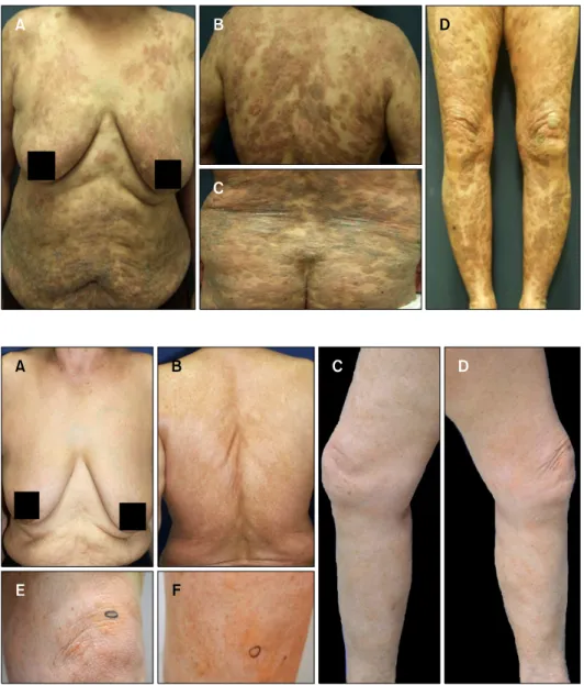
Fig. 1. (A∼D) The patient was diagnosed with mycosis fungoides (MF) 7 years ago. Erythematous and brownish scaly oozing plaques on the entire body. MF stage: T3b- N0M0.
Fig. 2. (A, B) Complete remission of mycosis fungoides. The patient had been treated with psoralen and ultraviolet A radiation therapy, nar- row-band phototherapy, and acitretin for 5 years and had reached com- plete remission of mycosis fun- goides. (C∼F) Multiple yellowish plaques on the both lower extre- mities including thigh, knee and lower leg. New yellowish lesions started to appear 1 year after dis- continuing phototherapy. The pla- ques were well circumscribed and slightly elevated.
and acitretin medication for 5 years (Fig. 1). She had been treated with PUVA therapy firstly for 25 months and then treated with UVB therapy for 21 months. The photo- therapy was done once a week or twice a week. The total number of exposure of PUVA was 72 and the cumulative PUVA radiation dose was 216.9 J/cm2. The total number of exposure of UVB was 98 and the cumulative UVB radi- ation dose was 110.35 J/cm2. Finally, she reached com- plete remission of mycosis fungoides (Fig. 2A, B). However, new yellowish lesions started to appear 1 year after dis- continuing the phototherapy. A physical examination re- vealed multiple yellowish plaques on both lower ex- tremities (Fig. 2C, D). The plaques were well circumscribed and slightly elevated (Fig. 2 E, F). Laboratory tests, includ- ing a complete blood cell count, differential leukocyte count, erythrocyte sedimentation rate, and blood chem- istry studies were all normal. A biopsy specimen showed
multiple nodular deposits of eosinophilic amorphous ma- terial in papillary dermis and upper reticular dermis (Fig.
3A, B). The deposits represented apple green birefringence on Congo red stain viewed under polarized light micro- scopy (Fig. 3C). The acellular small nodules in the upper dermis consisted of randomly oriented, non-branching, non-anastomosing 6.67∼12.7 nm thick amyloid fibrils on electron microscopy (Fig. 3D). Therefore, we confirmed the diagnosis of secondary cutaneous amyloidosis. We re- viewed past biopsy slides to determine when the amyloid had formed. Dense atypical lymphocyte infiltration in the dermis and epidermotrophism without evidence of amy- loidosis were observed at the mycosis fungoides diagnosis 7 years ago. We found small multifocal deposits of faintly eosinophilic amorphous material confined to the papillary dermis but no evidence of mycosis fungoides 2 years ago during complete remission of the mycosis fungoides.
Secondary Amyloidosis and Mycosis Fungoides
Vol. 29, No. 1, 2017 81 Fig. 3. (A) Multiple nodular deposits of fissured faintly eosinophilic amorphous material in the papillary dermis and upper reticular dermis (H&E, ×100). (B) Large, fissured, homogenous, eosinophilic masses in the papillary dermis (H&E, ×400). (C) The deposits were birefringence positive on Congo red staining viewed under polarized light microscopy (Congo red, ×100). (D) Electron micrographs show acellular small nodules consisting of randomly oriented, non-branching, non-anastomosing, 6.67∼12.7 nm thick (mean, 9.3 nm) amyloid fibrils.
DISCUSSION
Localized amyloidosis refers to single organ-limited amy- loid deposition. Localized cutaneous amyloidosis can oc- cur as a primary phenomenon as in lichen or macular amyloidosis, or as a secondary phenomenon associated with another cutaneous pathology. Although amyloid deposits in different clinicopathological types of amyloidosis have histologically and morphologically identical properties, it has become apparent that there are several mechanisms of amyloid accumulation1,12.
In secondary amyloidosis associated with chronic in- flammatory conditions, such as hidradenitis suppurativa, the amyloid protein precursors are acute phase reactants.
However the deposits in localized amyloidosis are likely proteins synthesized within the affected tissue. Secondary localized cutaneous amyloidosis is usually associated with tumors of epidermal origin, and the amyloid is thought to be derived from keratinocytes1,12,13. Although the pre- cursor proteins of cutaneous amyloidosis have not been fully characterized, they are predominantly derived from keratinocytes in cutaneous amyloidosis. Converting a pre- cursor to amyloid requires a conformational switch from α-pleated sheet arrangement to a β-pleated sheet structure.
It is postulated that keratin tonofilaments undergo “fila- mentous degeneration” and keratinocytes “drop off” into the dermis forming amyloid13. A commonly accepted pathogenic theory of amyloidosis is that apoptotic basal
CH Nam, et al
82 Ann Dermatol
keratinocytes (colloid bodies) release cytokeratins, which are covered with autoantibodies, phagocytized by macro- phages, and enzymatically degraded into amyloid K (keratin-associated amyloid), which is a key feature of or- gan-limited cutaneous amyloidosis13,14.
In our case, the clinical and histological features of cuta- neous amyloidosis were observed without any clinical or histological features of mycosis fungoides. Secondary amyloid deposits can originate in association with mycosis fungoides. Several cases of secondary cutaneous amyloi- dosis associated with mycosis fungoides have been re- ported without any history of phototherapy8,9,15. Izumi et al.15 reported that CD8+ T cells in poikilodermatous my- cosis fungoides might be attributable to the formation of amyloid material from attacks on epidermal keratinocytes.
The other theory associated with mycosis fungoides is that amyloid forms through prolonged scratching and rubbing, such as in friction amyloidosis. This theory is also sup- ported by the fact that this patient complained of severe pruritus at the diagnosis of mycosis fungoides. A possible alternative mechanism of this case is amyloid formation through a cytotoxic effect of PUVA therapy. Secondary cu- taneous amyloidosis was detected in five of 61 patients with mycosis fungoides simultaneously after PUVA by Zemheri et al.14. The mechanism of PUVA is phototoxic reactions resulting from direct cellular damage caused by an inflammatory, non-immunological mechanism. PUVA primarily targets DNA, and other important targets of psor- alens are specific receptors, such as epidermal growth fac- tor receptor. More recently, PUVA therapy can induce programmed cell death (apoptosis) in skin infiltrating T-helper lymphocytes and keratinocytes, so it cause inter- face changes10,11,14. Lastly, we can also consider that amy- loidosis might develop de novo without relation with my- cosis fungoides or PUVA therapy. However, clinical fea- tures of amyloidosis in this patient did not correspond to primary amyloidosis such as lichen or macular amyloidosis.
In addition, amyloidosis occurred at multiple sites along the both lower extremities, including thighs, knees, and lower legs. Accordingly, we think that amyloidosis in this patient could develop secondary to mycosis fungoides or PUVA therapy rather than de novo. We report an interest- ing and rare case of secondary cutaneous amyloidosis that developed during PUVA therapy and progressed for more than 1 year even though PUVA therapy was discontinued and mycosis fungoides was in complete remission.
REFERENCES
1. Breathnach SM. Amyloid and amyloidosis. J Am Acad Dermatol 1988;18:1-16.
2. Lee DY, Kim YJ, Lee JY, Kim MK, Yoon TY. Primary localized cutaneous nodular amyloidosis following local trauma. Ann Dermatol 2011;23:515-518.
3. Kumakiri M, Hashimoto K. Histogenesis of primary localized cutaneous amyloidosis: sequential change of epidermal keratinocytes to amyloid via filamentous degeneration. J Invest Dermatol 1979;73:150-162.
4. Li WM. Histopathology of primary cutaneous amyloidoses and systemic amyloidosis. Clin Dermatol 1990;8:30-35.
5. Hashimoto K, King LE Jr. Secondary localized cutaneous amyloidosis associated with actinic keratosis. J Invest Dermatol 1973;61:293-299.
6. Tsuji T, Asai Y, Hamada T. Secondary localized cutaneous amyloidosis in solar elastosis. Br J Dermatol 1982;106:
469-475.
7. Aso M, Hagari Y, Nakamura K, Mihara M, Shimao S. A case of secondary cutaneous amyloidosis: epidermal keratinocytes produce amyloid in the cytoplasm. J Cutan Pathol 1990;
17:176-181.
8. Holzmann H, Schott HJ. Amyloid demonstration in the skin in mycosis fungoides. Klin Wochenschr 1965;43:1061- 1062.
9. Schott HJ, Holzmann H. Detection of amyloid deposits in mycosis fungoides. Arch Klin Exp Dermatol 1965;222:
632-641.
10. Hashimoto K, Kumakiri M. Colloid-amyloid bodies in PUVA-treated human psoriatic patients. J Invest Dermatol 1979;72:70-80.
11. Greene I, Cox AJ. Amyloid deposition after psoriasis therapy with psoralen and long-wave ultraviolet light. Arch Dermatol 1979;115:1200-1202.
12. Powell AM, Albert S, Bhogal B, Black MM. Discoid lupus erythematosus with secondary amyloidosis. Br J Dermatol 2005;153:746-749.
13. Eto H, Hashimoto K, Kobayashi H, Fukaya T, Matsumoto M, Sun TT. Differential staining of cytoid bodies and skin- limited amyloids with monoclonal anti-keratin antibodies.
Am J Pathol 1984;116:473-481.
14. Zemheri IE, Ozkanli SS, Zindanci I, Senol S, Akbulak O, Topaloğlu Demir F. PUVA phototherapy-induced secondary amyloidosis in patients with mycosis fungoides: a rare adverse effect of phototherapy. Turk J Med Sci 2014;44:
89-94.
15. Izumi K, Arita K, Horie K, Hoshina D, Shimizu H. Localized cutaneous amyloidosis associated with poikilodermatous mycosis fungoides. Acta Derm Venereol 2014;94:225-226.