大훌훌放射線햄學會誌 第 24 卷 第 l 廳 pp. 20 - 24, 1988 Journal of Korean Radiological Society, 24(1) 20-24, 1988
뇌수막종의 비전형적 전산화단층촬영 소견
고려대학교 의파대학 방사선과학교실
임진우·박정국·전혜정·김정혁·차인호·서원혁
- Abstract -
The Atypical CT Findings of Meningioma
)in Woo Lim, M.D., )eong Kook Park, M.D., Hae )eong )eon, M.D.
)ung Hyuk Kim, M.D., In Ho Cha, M.D., Won Hyuck Suh, M.D.
Department of Radiology
,
College of Medicine,
Korea UniversityThe intracranial meningioma represent 15% of all intracranial tumors in adult and CT is the most usef비 for identifying the meningiomas‘ Most intracranial meningiomas have typical CT findings of round, sharply delineated, isodence or hyperdense tumor in a juxtadural location with intense homogenous enhancement and mild degree of peritumoral edema.
The authors reviewed 88 cases of meningioma confirmed pathologically in Korea University Medical Center for recent 4 years. Among them, 14 cases had unusual CT findings. We described and analized atypical CT findings of these 14 patients. Atypical findings consisted of irregular eccentric low density in 4 cases, peripheral cyst formation in 3 cases, marked edema in 3 cases, low density mass with thick rim enhancement in 2 cases
There is one case of multiple lesions‘
1.
서 료르 」뇌수막종은 두개강내종양의 약 15% 를 차지하는 비 교석 흔한 종양이여, 전산화단층촬영이 정확한 진단 을 하는데 큰 비좋을 차지하고 있마. 대부분의 뇌수박 종은 특징적인 전산화단층촬영 소견을 보이고 있는 데, 즉 조영천 전산화단층촬영 스캔에서 경계가 명확 하고 뇌실질과 동일 음영。l거나 보다 더 증가된 음영 을 보이고 종괴의 일부가 골이냐 뇌수막에 붙어 있으 며 조영증강후스캔에서 종괴 전체의 균등한조영증강 을 보이여 또한 주밴 뇌살잘에 대한 종괴효과와 경도 의 주밴부종을 대부분에서 보여주고 있마 ]-4)
이 논문은 1987 년 12월 15 일에 접 수하여 1988년 2월 17일 에 채택되었음
저자들은 최근 4 년간 고려대학교 부속병원에서 수 술파 영리학석으로확인된 88예의 뇌수악종중중싱부 괴사, 낭포형성, 윤상증강과 중심성 저밀도, 광뱀위 한 주밴부종 동으로 >1]천형적 전산화단층촬영 소견을 보인 14예를 경험하였기에 분석 검토하여 보았마.
1I.
대상 및 방법최근 4 년간 고려대학교 의과대학 부속영원에서 천 산화단층 촬영 후 수술과 영 리 학적 으로 확선된 88예 중 비전형적인 전산화단층촬영 소견을 보인 14예를 그 대 상으로 하였마. 88예중 소위 천형석인 소견을 보인 74 예는 모두 종괴의 경계가 명확하고 조영증강 후 종괴 전체의 균등한 조영증강을 보이며 비교석 주변부종이 석은 전산화안층촬영 소견을 보여 본 매상에서 제외시 켰다.
-임진우 외 : 뇌수악종의 "1 전형석 천산화안층촬영 소견-
C T
기 종은Siemens Somaton
II와Tos hi ba
80A를사용하여 절펀 두께 10mm로 횡 단주사를 시행했고, 모
든 예에서 조영전 및 조영증강 후 단층촬영을 살시했 고 조영증강은
Conray 60(60% io thalamate meg ulu-
mine) 을 체중1
kg 망 lml씩 빠르게 석주하여 조영천파 동일하게 절단을 하였다.m.
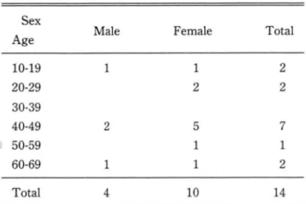
결 과환자의 연령 및 성벨분포는 주로 40대 이상에서 나 타났£켜, 10대에서는 낭, 여 각각 1영씩 말생하였으 며, 여자에서 10예로 남자보다 2_5배의 말생반도를 보 였다 (Table
1
).t날생부위 별로는 전두부 3예, 두정부 3예, 측두부
1
예 둥 7예로 궁흉부에 가장 않았고, 접형골 및 후두개 와에서도 각각 2예로 나타났으켜, 선반석인 뇌수막종 의 호발부위와 일치하였다 (Table2)
.전산화단층촬영상 중심부괴사를 보인 예가 4예, 중 심부 괴사 및 낭포형성한 예 가 3예, 광범위한 주변부
Table 1.
Age and Sex Distribution
Sex
Age Male Female Total
1 2
2 2
5 7
1 1
1 2
10 14
10-19
20-29 30-39 40-49 50-59 60-69
2
Total 4
Table 2. Site of Meningioma
Site Number
Convexity Sphenoid ridge Posterior fossa Suprasellar
Intraventr디icular
Sigmoid sinus
?I
nι ηι
1i 1i 1i
Total 14
종을 보인 예가 3예, 윤상증강 및 중삼성 저밀도를 보 이는 경우가 2예 있었 A며, 마발성무로 나타난 1예의 경우 약 2년간 추석검사한 결과 호기에 우측 후땅으로 경상구조에 연해서 생긴 종양。l 시간이 경파한후에는 좌측두정부에 마발성으로 발생했마. 그러고 두피,
뼈, 78 막을 동시에 침범한 예도 1예 등A로 닝|전형석 인 선산화단층촬영 소견을 보였다(Table 3) (Fig
1-3
).N.
고 잘대부분의 뇌수악종은 특징석인 전산화단층촬영 소 견을 보이고 있으나 적지 않은 예에서 매전형석인 전 산화단충촬영 소견을 보이 고 있 다. 전산화단충촬영 소견상 중심성 저농도를 보이는 경우는 주로 종양조직 괴사에 의해 나타냐며, 낭포화 형성 및 출혈 등에 의 해서도 나타날 수 있다2, 5, 6)
중성부 종양조직 괴사를 보이 는 경 우는 뇌 수악종 같 이 비교적 정적인( stati
c)
종양에서는 드물게 나타나 지만 원인으로 허혈에 의해 냐타난마고 보고되고 있 고 5, 6) 저자들의 경우 88예중 7예로 약 8% 에서 보였 으며, 마른 저자들에서는 약14%
발생율을 보였다고 보고되고 있마 5)낭포성 성분의 저농도를보이는 경우는얘우드물게 나타냐며, C
ushing and E isenhardt
등은 약 4% 에서,Sato
등은 420예 중 단지 5예 에 서 만 보였 다고 보고하 였고 7) 저자들은 88예중 5예로 약 5% 의 딸생율을 보 였다. 대개는 종양의 주변에 위치하고 있으며, 기계석 방추에 의해 뇌척수액이 국한 되었거나 반응석인 신경Table 3. Atypical CT Findings
Findings Number
Homogenously enhancing mass with irregular eccentric low density Mass with periphera l cyst fo rmation Mass with marked edema
Low densi ty mass with thick rim
enhancementMass in both intracranial and extracranial
43
3
2
areas
Multiplicity
A
B
-大韓放射線짧學會뜸 : 第
24
卷 第1
號1988-
증식증에 의해 2차적으로 생긴다고 보고하고 있다 5‘
6
.
7.8.
9)(F ig. l
-A).낭포가 중심부에 생기는 경우는 펀심성보다 드물 며, 중섬성 괴사나 종양의 알부가 부분적으로 지땅성 성분으로 대치되기 때운에 생긴다고 보고되고 있으 여, 선에 생겼던 출혈에 으l해서도 나타날 수 있마
Fig. 2 Meningioma with marked peritumoral edema Small enhanced lesion attached to falx with marked peritumoral edema is se en in right anterior frontal area.
(
Fig. l-B
). 이러한 낭포형성은 시상동안정, 겸상구조 또는 살바안열쿠어l 위치한 종양에 동반되어 잘 냐 타난마고 한마 5. 7)
주변부종은 대부분의 뇌수막종에서 다양한 정도로 나타나고 있으며, 혈뇌 장벽파괴에 의해 나타냐는 혈 판성 부종으로 부종정 도는 종괴 크기 와는 상판판계 가 없는 것으로 보고되고 있마. 부종정도는 주로 경도, 중등도의 부종을 보이며, 싱한 부종을 보이는 경우는
10%
미만에서 보고되고 있다 2-6.10-.11)Stevens 등에 의 하연 부종은 뇌 수막종이 전반부 시 상동 인접에 위치하거나 넓은 연석을 차지한 경우, 놔 수막 정맥동을 칩범했을혜 또는 뱅력이 짧은 경우에 잘동반되지만 종양의 크기, 석회화의 유무와는 상판 관계가 없다고 하였다 10) 저자들의 경우는 88예중
3
예로 약 4% 에서 심한 부종을 보였으며, 종양은 모우 웅륭부와 시상동인접에 위치하였다(Fi.g.2).
Fig. 1 Cystic meningiomas
뇌실 뇌수악종은 액락악종이냐 맥락조직에서 발생 하며, 성안인 경우 1.6% 의 딸생빈도를 보이며, 어린 이의 경우 17% 로 비교적 높은 발생벤도를 보안다 1. 12‘13) 이밖에 두피, 골 그러고 경막을 동시에 침엄한 예가 l 예 있었£며, 원말부위는 정확히 알 수 없£나
Jacob
등에 의하연 태생기의 선천성 기형으로 지주악A. Central necrotic areas and eccentric cystic le- sion in posterior aspect
(1)with peritumoral edema are seen in left frontal area
B. Central lo
\Vdensity with peripheral rim enhanc-
ed mass is seen in right deep parietal area
-임진우 외 . 뇌수악종의 "1 건형석 전산화단층촬영 소견-
A
C
조직이 골내에 갇혀 있다가 뇌수막종이 생걸 가능성이
있마고 보고하였마 1,14 )
대부분의 뇌수막종은 단일성이고 마발성인 경우는 매우 드물다. 간혹 신경섬유종증이 있는 환자에서 마 발성 뇌수막종을 동반하기도 하냐 판계없이 발생하기 도 한다 1 , 15) 저자들의 경우 1예에서 마딸성으로 냐타 났고 신경섬유종증과는 판계가 없었다.
v.
걸 료응 」B
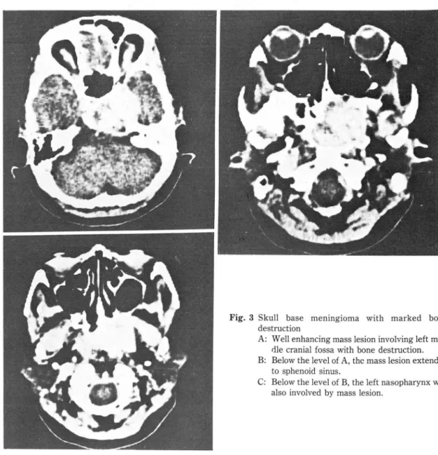
Fig. 3 Skull base meningioma with marked bone destruction
A: Well enhancing mass lesion involving left mid dle cranial fossa with bone destruction.
B: Below the level of A , thc mass lesion extended to
sphenoid
sinus.C: Below the level of B
,the left nasopharynx was also involved by mass
lesion.대부분의 뇌수막종은 특정석인 전산화단층촬영 소 견을 보이지얀드울지 않게 종괴괴사, 낭포성형성, 광 법위한 주변부종 등의 비전형적인 전산화단층촬영 소 견을 보일 수 있으므로 운헌 고찰과 함께 보고하는 바 이마.
REFERENCES
1. Seungho
Howard Lee
,Krishna
C.V.G
. Rao: Cranial com.puted tomography.
-大韓放射線훌훌學會誌 第24 卷 第 1 號 1988-
2. New PFJ, Aronow 5, Hesselink JR: National cancer institute study: Evaluation of computed tomography in the diagnosis of intracranial neoplasms. Radiology 136:665-675, 1980
3. Russel EF
,
George AE,
Kricheff 1 et al: A typical computed tomographic features of intracranial meningioma.Radiology 135:673-682, 1980
4 깅석천, 임정기, 이통호, 장기현 : 전산화단층촬영상
"1 전형석인 뇌수악종에 판한 고찰. 대한방사선의학회 지 18-4 : 683- 688
,
19825 서정호, 검영순, 이영태, 박창윤 Computed tomography에 의한 뇌수막종의 선난. 대한방사선의학 희지 16-1 :41-48
,
19806 이중식, 깅호진, 서수지, 최두석 : 뇌수악종에 대한 전산화단층촬영 소견. 대한방사션의학회지 16-1 :49 - 55
,
1980.7. Zagzag D, Gomori JN, Rappaport ZH, Shalt MN: Cystic meningioma presenting as a ring lesion. AjNR 7:911-912
,
19868. Nahser HC, Grole W, Löhr E et al: Multiple meningioms,
clinical and computed tomographic observation Neuroradiology 21:259-263, 1981
9. Sigel RM, Messina AV: Computed tomography: The anatomic basis of the zone of diminished density surroun- ding meningiomas. AjR 127:139-141, 1976
10. Becker D
,
Norman D,
Wison CB: Computed tomography and pathological correlation in cystic meningiomas, report of 2 cases. j. Neurosurgery 50:103-105, 197911. Ito J, Kade Karu T
,
Hyano M: Meningioma in tela cheroidea of 3rd ventricle: CT and angiographic correlations Neuroradiology 21:207-211, 198112. Dell 5, Ganti R, Steinberger A, McCartry J 111: Cystic men- ingiomas: a clinico-radiological study. j Neurosurgery 57:8-13, 1982
13. Taveras JM, Wood EH: Diagnostic neuroradiology. Williams and
‘ .villkins Co., 1976
14. Morrison G, Sobel DF, Kelley WM, Norman D: Intraven- tricular mass lesions. Radiology 153:435, 1984 15. Stevens JM, Ruiz JS, Kendall BE: Observation on peritumoral
edema in meningioma. Neuroradiology 25:125-131, 1983