http://dx.doi.org/10.4174/astr.2014.86.1.39 Annals of Surgical Treatment and Research
Risk factor analysis of new brain lesions associated with carotid endarterectmy
Jae Hoon Lee, Bo Yang Suh
Division of Vascular Surgery, Department of Surgery, Yeungnam University College of Medicine, Daegu, Korea
INTRODUCTION
Carotid artery stenosis, usually caused by atherosclerosis, is known to cause ischemic stroke [1]. Treatment for carotid artery stenosis, carotid endarterectomy (CEA) and carotid artery stenting (CAS) have been widely used [2]. CEA is the standard treatment for the primary and secondary prevention of stroke associated with carotid artery stenosis for its safety and durability [3]. CAS is less invasive and the use of embolic protection devices significantly reduces the frequency of adverse events, it is considered a therapeutic alternative to CEA, especially in patients at high risk for surgery [4,5].
New neurological deficit is a major concern associated with
these two procedures and finding new embolic lesions after the intervention is very important to both clinicians and patients [6]. Diffusion weighted imaging (DWI) is a good imaging modality for detecting new brain lesions (NBL) [7,8]. In the past, most lesions detected on DWI had not been considered clinically significant, but many data have shown that NBL on DWI should not be overlooked [9,10]. Bonati et al. [9] reported relationships between neurological complications and total DWI lesion volume. They suggest an increasing DWI lesion volume is associated with subsequent symptomatic strokes.
Vermeer et al. [10] suggested that patients with silent brain infarcts have a high risk of dementia and cognitive dysfunction.
As a result, silent DWI lesions might lead to future neurological Purpose: Carotid endarterectomy (CEA) is the standard treatment for carotid artery stenosis. New brain ischemia is a major concern associated with CEA and diffusion weighted imaging (DWI) is a good imaging modality for detecting early ischemic brain lesions. We aimed to investigate the surgical complications and identify the potential risk factors for the incidence of new brain lesions (NBL) on DWI after CEA.
Methods: From January 2006 to November 2011, 94 patients who had been studied by magnetic resonance imaging including DWI within 1 week after CEA were included in this study. Data were retrospectively investigated by review of vascular registry protocol. Seven clinical variables and three procedural variables were analyzed as risk factors for NBL after CEA.
Results: The incidence of periprocedural NBL on DWI was 27.7%. There were no fatal complications, such as ipsilateral disabling stroke, myocardial infarction or mortality. A significantly higher incidence of NBL was found in ulcer positive patients as opposed to ulcer negative patients (P = 0.029). The incidence of NBL after operation was significantly higher in patients treated with conventional technique than with eversion technique (P = 0.042).
Conclusion: Our data shows CEA has acceptable periprocedural complication rates and the existence of ulcerative plaque and conventional technique of endarterectomy are high risk factors for NBL development after CEA.
[Ann Surg Treat Res 2014;86(1):39-44]
Key Words: Catorid endarterectomy, Complications, New brain lesions, Risk factors
Received July 2, 2013, Revised September 3, 2013, Accepted September 24, 2013
Corresponding Author: Jae Hoon Lee
Division of Vascular Surgery, Department of Surgery, Yeungnam University College of Medicine, 170 Hyeonchung-ro, Nam-gu, Daegu 705-717, Korea.
Tel: +82-53-620-4032, Fax: +82-53-624-1213 E-mail: iami1124@hanmail.net
Copyright ⓒ 2014, the Korean Surgical Society
cc Annals of Surgical Treatment and Research is an Open Access Journal. All articles are distributed under the terms of the Creative Commons Attribution Non- Commercial License (http://creativecommons.org/licenses/by-nc/3.0/) which permits unrestricted non-commercial use, distribution, and reproduction in any medium, provided the original work is properly cited.
impairment.
Compared with CEA, CAS has a higher incidence of NBL. In 32 studies comprising 1,363 CAS and 754 CEA procedures, the incidence of NBL on DWI is 37% after CAS and 10% after CEA [11]. In the study by Bonati et al. [9], about three times more patients after CAS than CEA had at least one NBL detected on posttreatment scans. The number of patients with new DWI is 62 of 124 (50%) in CAS group, 8 of 107 (17%) in CEA group. Due to relatively low incidence of NBL, not much research on the risk factor for postoperative NBL after CEA has been reported.
In this study, we investigated surgical complications and identified the potential risk factors of clinical and procedural variables for the incidence of NBL on DWI after CEA.
METHODS
Study population
From January 2006 to November 2011, 94 patients who had been studied by magnetic resonance imaging (MRI) including DWI within 1 week after CEA were included in this study. The indication of CEA were symptomatic patients with stenosis over seventy percent according to North American Symptomatic Carotid Endarterectomy Trial (NASCET) criteria and asymptomatic patients were considered when they had a stenosis over eighty percent or over fifty percent if the lesion was ulcerative. The luminal area diameter by computed tomographic (CT) angiography was used for the assessment of carotid artery stenosis. Symptomatic patients were defined as those who had experienced amaurosis fugax, a transient ischemic attack (TIA), or a stroke in the territory of the ipsilateral carotid artery within 6 months before entry. Ulcer is defined as a concavity in the plaque that has a lower basal border compared to the adjacent plaque surface through CT angiography.
Surgical procedure
Patients had been given at least 1 antiplatelet agent (aspirin, 100 mg daily; cilostazol, 200 mg daily; or clopidogrel, 75 mg daily) for a minimum of 7 days before the operation. CEA was performed under general anesthesia with intraoperative monitoring. Intraoperative monitoring was done by transcranial doppler (TCD) or electroencephalography (EEG). Before the clamping of carotid artery, intravenous heparin (5,000 units) was administered to prevent the occurrence of acute thrombosis. When internal carotid artery (ICA) blood flow was reduced over 50% after clamping of ICA on TCD, or when significant EEG changes happened during operation, or the bones of the skull blocked the transmission of ultrasound, selective shunt (Pruitt-Inahara carotid shunt with T-port;
LeMaitre Vascular Inc., Burlington, MA, USA) was placed.
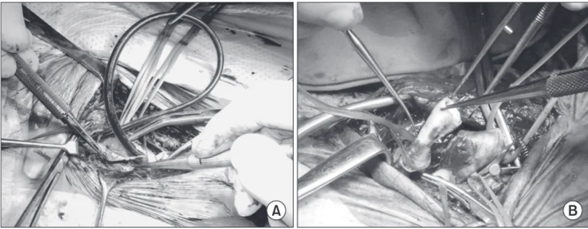
Surgical options were comprised of conventional and eversion CEA. The conventional CEA (cCEA) is a standard longitudinal carotid arteriotomy with or without patch angioplasty, whereas eversion CEA (eCEA) is done in the order of oblique transection, eversion of ICA and reimplantation of the latter into the common carotid artery (Fig. 1).
MRI technique
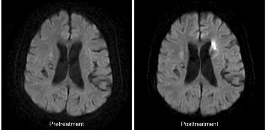
For evaluation of embolic events during procedure, MRI (Gyroscan Intera 1.5T, Philips Medical Systems, Best, The Netherlands) was performed in all patients within 1 week after treatment. The protocol included isotropic DWI sequence. MRI was analyzed by experienced neuroradiologists. The primary outcome was the occurrence of any new hyper-intense DWI lesion on the posttreatment scan that had not been present on the pretreatment scan (Fig. 2).
Patient assessment
Data were retrospectively investigated by review of vascular registry protocol. Seven clinical variables and three procedural variables were analyzed as potential risk factors for NBL on
Fig. 1. Conventional carotid endarterectomy with shunt (A) and eversion carotid endarterectomy (B).
DWI after CEA. The clinical variables considered were sex, age, side of lesion, neurologic symptom, severity of stenosis, level of lesion and ulcer on plaque. The procedural variables included the type of operation, the usage of shunt and the era (2006-2008 or 2009-2011). Preoperative and postoperative neurological examinations were investigated by a neurologist. Duplex scan (iU22 xMATRIX ultrasound system, Philips Medical Systems, Bothell, WA, USA) was performed on all the patients at 1, 3, 6, and 12 months for the first year and annually, thereafter. Early complication was defined as any stroke or death and other complication occurring within 30 days after treatment. Midterm complication was defined as any stroke or death occurring after
30 days after treatment.
Statistical analysis
Statistical analysis was performed using IBM SPSS ver. 18.0 (IBM Co., Armonk, NY, USA). Continuous data are reported as mean ± standard deviation. Nominal data were reported as number of subjects and percentage of individuals affected.
Univariate and multivariate analysis performed to determine the risk factor for NBL after CEA. The used univariate analysis was chi-square test, Fisher exact test and Student t-test.
Multivariate analysis was performed by the use of binary logistic regression analysis. A P-value < 0.05 was considered to Fig. 2. New brain lesions on magnetic resonance diffusion weighted imaging after carotid endarterectomy.
Table 1. Patient demographic and clinical data
Characteristic Total
(n = 94) Patients with NBL
(n = 26) Patients without NBL
(n = 68)
Age (yr) 66.64 ± 7.18 65.77 ± 7.67 66.97 ± 7.01
Male sex 81 (86.2) 22 (84.6) 59 (86.8)
Symptomatic ICA stenosis 72 (76.6) 19 (73.1) 53 (77.9)
TIA and amaurosis fugax 32 (34.0)
Stroke 40 (42.6)
Stenosis degree (%) 79.15 ± 12.53 78.69 ± 16.17 79.32 ± 10.96
Ulceration on plaque 24 (25.5) 10 (38.5) 14 (20.6)
Side of lesion, left 51 (54.3) 13 (50.0) 38 (55.9)
Level of lesion
C2 21 (22.3) 9 (34.6) 12 (17.6)
C3 58 (61.7) 15 (57.7) 43 (63.2)
C4 15 (16.0) 2 (7.7) 13 (19.1)
Comorbidities
Smoking 67 (71.3) 20 (76.9) 47 (69.1)
Hypertension 75 (79.8) 20 (76.9) 55 (80.9)
Diabetes mellitus 37 (39.4) 10 (38.5) 27 (39.7)
Hypercholesterolemia 75 (79.8) 21 (80.8) 54 (79.4)
Coronary artery disease 13 (13.8) 5 (19.2) 8 (11.8)
Values are presented as mean ± standard deviation or number (%)
NBL, new brain lesions; ICA, internal carotid artery; TIA, transient ischemic attack; C, cervical vertebra.
be statistically significant.
RESULTS
Demographic and clinical characteristics of total patients are presented in Table 1. A total of 94 patients were included this study. The mean age was 66.64 ± 7.18 years old and 86.2%
was male patient. Patients with symptomatic ICA stenosis were 72 (76.6%). Mean diameter of stenosis was 79.15% ± 12.53% in accordance with NASCET criteria. 24 patients (25.5%) had ulceration on plaque and 51 patients (54.3%) had left ICA stenotic lesion. Patients with the lesion located in the body of second cervical vertebra were 21 (22.3%). The common comorbidities were hypertension and hypercholesterolemia followed by smoking, diabetes, and coronary artery disease.
NBL occurred in 26 patients. The incidence of NBL in ulcer positive patients and ulcer negative patients was 10 (41.7%) and 16 (22.9%), respectively.
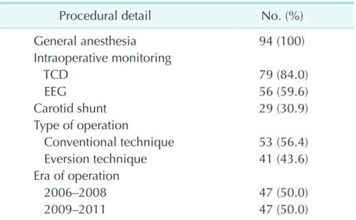
As shown in Table 2, all patients were treated under general anesthesia. Intraoperative monitoring was done by TCD in 79 cases, EEG in 56 cases and combined methods in 41 cases. Shunts were used in 29 cases for the prevention of ischemic events. The incidence of NBL in 29 shunted patients and 65 nonshunted patients was 12 (41.4%) and 14 (21.5%), respectively (P = 0.047). Fifty-three patients underwent CEA by conventional technique and 41 patients underwent eversion technique. NBL was found in 19 patients (35.8%) who underwent conventional technique and 7 patients (17.1%) who underwent eversion technique (P = 0.044). Fifty percent of patients had their operation performed before January 2009.
Sixteen patients (34.0%) had NBL from 2006 to 2008 and 10 patients (21.3%) had NBL from 2009 to 2011 (P = 0.167).
We examined the early and midterm postoperative complications. Table 3 shows the early complications within thirty days after CEA. There were 6 cases of nondisabling ipsilateral stroke such as amaurosis fugax and TIA. There
was no postoperative myocardiac infarction but 3 wound hematomas, 4 cases of cranial nerve palsies, and 1 case of hyperperfusion syndrome developed postoperatively. All of these events were treated conservatively without any complication. Thirty days after CEA, there were 3 cases of stroke and they were treated without any sequelae. And there were no deaths during the follow-up period (Table 4). Mean follow-up duration was 24 months.
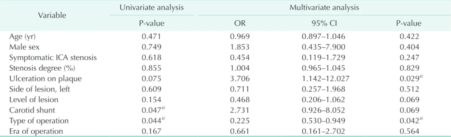
To identify the risk factors for NBL after CEA, we performed univariate and multivariate analysis (Table 5). In univariate analysis, the usage of carotid shunt and type of operation was significantly associated with the development of NLB (P = 0.047 and P = 0.044, respectively). In multivariate analysis by logistic regression analysis, the presence of ulcer on plaque (odds ration [OR], 3.706; 95% confidence interval [CI], 1.142 to 12.027; P = 0.029) and type of operation (OR, 0.225; 95% CI, 0.530 to 0.949; P = 0.042) were identified as significant risk factors for NBL after CEA. In our center, cCEA is preferred when shunt is needed or the level of lesion is high. In 2009- 2011, we attempted eCEA more regularly. Thus, in order to check for multicollinearity, we exclude confounding variables such as level of lesion, carotid shunt and era of operation and performed multivariate analysis. As a result, the significance of the presence of ulcer on plaque (OR, 3.112; 95% CI, 1.043 to 9.287;
P = 0.042) and type of operation (OR, 0.303; 95% CI, 0.104 to 0.884, P = 0.029) were retained. The usage of shunt and higher location of lesion were more frequently developed to NBL, but it failed to prove any statistical difference (P = 0.069). Age and gender were not associated with the incidence of NBL. No significant relationship was found between NBL and the degree
Table 3. Early (≤30 days) postoperative complications
Complication No. (%)
Ipsilateral stoke 6 (6.4)
Nodisabling 6 (6.4)
Disabling 0 (0)
Myocardial infarction 0 (0)
Wound hematoma 3 (3.1)
Cranial nerve palsya) 4 (4.3)
Hyperperfusion syndrome 1 (1.0)
Mortality 0 (0)
a)Transient hypoglossal nerve palsy.
Table 4. Midterm (>30 days) postoperative complications
Complication No. (%)
Ipsilateral stoke 3 (3.2)
Nodisabling 3 (3.3)
Disabling 0 (0)
Mortality 0 (0)
Table 2. The details of carotid endarterectomy
Procedural detail No. (%)
General anesthesia 94 (100)
Intraoperative monitoring
TCD 79 (84.0)
EEG 56 (59.6)
Carotid shunt 29 (30.9)
Type of operation
Conventional technique 53 (56.4)
Eversion technique 41 (43.6)
Era of operation
2006–2008 47 (50.0)
2009–2011 47 (50.0)
TCD, transcranial doppler; EEG, electroencephalography.
of stenosis.
DISCUSSION
In this study, CEA was performed with acceptable peripro- cedural complication rates. There were no fatal complications, such as ipsilateral disabling stroke, myocardial infarction and mortality. And we found that the presence of ulcer on plaque and the conventional type of operation are statistically significantly associated with the occurrence of NBL after CEA.
The incidence of NBL associated CEA on postoperative MRI has been variously reported from 0% to 34% [12]. In our data, periprocedural NBL was observed in 27.6% of patients.
This is compatible with previous results. But the incidence of NBL is rather high with cCEA compared to other studies. The reasons for the difference of incidence originated from regional differences, study method, patient characteristics, surgical technique and so on.
There are a few studies concerning the risk factor of NBL after treatment of carotid artery stenosis. The NASCET [13] examined 1,415 patients who underwent CEA and investigated surgical results. This trial reported that CEA is a durable procedure and five baseline variables were associated with a statistically significant increased risk of perioperative stroke and death: a hemispheric TIA compared with a retinal TIA as the qualifying event, a left-sided procedure, the presence of contralateral carotid occlusion, an ipsilateral ischemic lesion on the entry CT scan, and irregular or ulcerated plaque detected by angiography on the side of surgery. Lee et al. [14] investigated 233 CEAs performed on 222 patients with carotid artery stenosis. In this study, plaque with ulceration was a significant risk factor for the development of postoperative new brain infarction. In the European Carotid Surgery Trial [15], female sex, systolic hypertension and peripheral vascular disease were identified
Table 5. Risk factors analysis for new brain lesions after carotid endarterectomy on diffusionweighted MRI identified univariate and multivariate logistic regression analysis
Variable Univariate analysis Multivariate analysis
Pvalue OR 95% CI Pvalue
Age (yr) 0.471 0.969 0.897–1.046 0.422
Male sex 0.749 1.853 0.435–7.900 0.404
Symptomatic ICA stenosis 0.618 0.454 0.119–1.729 0.247
Stenosis degree (%) 0.855 1.004 0.965–1.045 0.829
Ulceration on plaque 0.075 3.706 1.142–12.027 0.029a)
Side of lesion, left 0.609 0.711 0.257–1.968 0.512
Level of lesion 0.154 0.468 0.206–1.062 0.069
Carotid shunt 0.047a) 2.731 0.926–8.052 0.069
Type of operation 0.044a) 0.225 0.530–0.949 0.042a)
Era of operation 0.167 0.661 0.161–2.702 0.564
MRI, magnetic resonance imaging; OR, odd ratio; CI, confidence interval; ICA, internal carotid artery.
a)P<0.05.
as independent risk factors for CEA related stroke and death in a multiple regression analysis. There was a trend towards an increased operative risk with irregular and ulcerated plaque in the ipsilateral carotid artery. The New York Carotid Artery Surgery, including 9,308 patients who underwent CEA, reported that the presence of a deep carotid ulcer was marginally associated with greater odds of perioperative death and stroke after CEA [16]. As you can see, plaque ulceration is highly associated with the occurrence of NBL after CEA.
In the aforementioned studies, type of operation was not investigated as a variable. In our study, there are two surgical options, cCEA and eCEA. Patients treated with conventional technique had a significantly higher incidence of NBL compared with patients treated with eversion technique. A large scaled meta-analysis of randomised and nonrandomised studies about the comparative results of cCEA and eCEA was performed by Antonopoulos et al. [17]. In this study, 8,530 eCEA and 7,721 cCEA procedures were investigated and eCEA compared to cCEA was associated with a lower incidence of perioperative stroke. Although perioperative stroke was not confirmed on MRI, this meta-analysis and our data showed the potential superiority of eCEA regarding perioperative stroke in common.
But, in order to conclude that the cCEA is a risk factor for NLB after CEA, a well-designed prospective study is needed. In our institute, shunt is inserted when decreased blood flow is confirmed by TCD during clamping. Reduced blood flow before surgery has a high risk of ischemia [18]. And, obligate shunt usage during procedure is reported to relate higher incidence of new ipsilateral DWI lesions compared to selective shunt usage [11]. The most common area of the carotid artery affected by atherosclerosis is the bifurcation of the common carotid artery [19]. Thus, the portion of patients with high-level lesion is relatively small and these patients are likely to have clotting disorders. And, a high level of lesion is difficult for
1. Petty GW, Brown RD Jr, Whisnant JP, Sicks JD, O'Fallon WM, Wiebers DO. Ischemic stroke subtypes: a population-based study of incidence and risk factors. Stroke 1999;30:2513-6.
2. Lanzino G, Rabinstein AA, Brown RD Jr. Treatment of carotid artery stenosis:
medical therapy, surgery, or stenting?
Mayo Clin Proc 2009;84:362-87.
3. Biller J, Feinberg WM, Castaldo JE, Whittemore AD, Harbaugh RE, Demp sey RJ, et al. Guidelines for carotid endar- terectomy: a statement for healthcare professionals from a special writing group of the Stroke Council, American Heart Association. Stroke 1998;29:554-62.
4. Furie KL, Kasner SE, Adams RJ, Albers GW, Bush RL, Fagan SC, et al. Guidelines for the prevention of stroke in patients with stroke or transient ischemic attack:
a guideline for healthcare professionals from the american heart association/
american stroke association. Stroke 2011;42:227-76.
5. Kastrup A, Nagele T, Groschel K, Schmidt F, Vogler E, Schulz J, et al. Incidence of new brain lesions after carotid stenting with and without cerebral protection.
Stroke 2006;37:2312-6.
6. Rosenthal D, Zeichner WD, Lamis PA, Stanton PE Jr. Neurologic deficit after carotid endarterectomy: pathogenesis and management. Surgery 1983;94:776-80.
7. van Everdingen KJ, van der Grond J, Kappelle LJ, Ramos LM, Mali WP.
Diffusion-weighted magnetic resonance imaging in acute stroke. Stroke 1998;29:
1783-90.
8. Bendszus M, Stoll G. Silent cerebral ischaemia: hidden fingerprints of inva- sive medical procedures. Lancet Neurol 2006;5:364-72.
9. Bonati LH, Jongen LM, Haller S, Flach HZ, Dobson J, Nederkoorn PJ, et al. New ischaemic brain lesions on MRI after stenting or endarterectomy for symp- tomatic carotid stenosis: a substudy of the International Carotid Stenting Study (ICSS). Lancet Neurol 2010;9:353-62.
10. Vermeer SE, Prins ND, den Heijer T, Hofman A, Koudstaal PJ, Breteler MM.
Silent brain infarcts and the risk of dementia and cognitive decline. N Engl J Med 2003;348:1215-22.
11. Schnaudigel S, Groschel K, Pilgram SM, Kastrup A. New brain lesions after carotid stenting versus carotid endarterectomy: a systematic review of the literature. Stroke 2008;39:1911-9.
12. Hebb MO, Heiserman JE, Forbes KP, Zabramski JM, Spetzler RF. Perioperative ischemic complications of the brain after carotid endarterectomy. Neurosurgery 2010;67:286-93.
13. Ferguson GG, Eliasziw M, Barr HW, Clagett GP, Barnes RW, Wallace MC, et al.
The North American Symptomatic Carotid Endarterectomy Trial: surgical results in 1415 patients. Stroke 1999;30:1751-8.
14. Lee KB, Lee KH, Chung CS, Kim GM, Byun HS, Jeon P, et al. Carotid endarterectomy:
analysis of early complications (<30 days) and risk factors for postoperative new brain infarction. J Korean Surg Soc
2009;77:195-201.
15. Bond R, Narayan SK, Rothwell PM, Warlow CP; European Carotid Surgery Trialists' Collaborative Group. Clinical and radiographic risk factors for operative stroke and death in the European carotid surgery trial. Eur J Vasc Endovasc Surg 2002;23:108-16.
16. Halm EA, Tuhrim S, Wang JJ, Rockman C, Riles TS, Chassin MR. Risk factors for perioperative death and stroke after carotid endarterectomy: results of the New York carotid artery surgery study.
Stroke 2009;40:221-9.
17. Antonopoulos CN, Kakisis JD, Sergentanis TN, Liapis CD. Eversion versus conven- tional carotid endarterectomy: a meta- analysis of randomised and non-rando- mised studies. Eur J Vasc Endovasc Surg 2011;42:751-65.
18. Parsons MW, Yang Q, Barber PA, Darby DG, Desmond PM, Gerraty RP, et al.
Perfusion magnetic resonance imaging maps in hyperacute stroke: relative cerebral blood flow most accurately identifies tissue destined to infarct.
Stroke 2001;32:1581-7.
19. Motomiya M, Karino T. Flow patterns in the human carotid artery bifurcation.
Stroke 1984;15:50-6.
20. Liapis CD, Bell PR, Mikhailidis D, Sivenius J, Nicolaides A, Fernandes e Fernandes J, et al. ESVS guidelines. Invasive treatment for carotid stenosis: indications, techni- ques. Eur J Vasc Endovasc Surg 2009;37(4 Suppl):1-19.
REFERENCES
manipulation. cCEA is preferred when shunt is required or the level of lesion is high [20] and in our multivariate analysis, the use of shunt and high level of lesion were marginally associated with the frequency of NBL (P = 0.069). Due to the influence of both shunt and level of lesion, the surgical technique may be one of the reasons for high incidence of NBL after cCEA. eCEA involves oblique transection and eversion of the ICA, eCEA may offer a great vision of entire parts of the interior ICA wall. So, the handling of embolic source can be easier in eCEA compared to cCEA.
In conclusion, our data shows CEA has acceptable peri- procedural complication rates and the existence of ulcerative plaque and conventional technique of endarterectomy are high risk factors for NBL development after CEA.
CONFLICTS OF INTEREST
No potential conflict of interest relevant to this article was reported.