165
Correspondence to: Byung Sik Kim, Division of Gastric Surgery, Department of Surgery, Asan Medical Center, University of Ulsan, Pungnap 2-dong, Songpa-gu, Seoul 138-736, Korea. Tel: 02-3010- 3491, Fax: 02-474-9027, E-mail: [email protected]
Received February 3, 2010, Accepted June 29, 2010
Surgical Treatment for Patients Who Underwent Endoscopic Mucosal Resection (EMR)/Endoscopic Submucosal Dissection (ESD) of
Early Gastric Cancer (EGC)
Department of Gastric Surgery, Asan Medical Center, University of Ulsan College of Medicine, Seoul, Korea
Min Gyu Kim, M.D., Beom Su Kim, M.D., Tae Hwan Kim, M.D., Kap Choong Kim, M.D.,
Jeong Hwan Yook, M.D., Ph.D., Sung Tae Oh, M.D., Ph.D., Byung Sik Kim, M.D., Ph.D.
Purpose: To evaluate the necessity for additional surgical treatment after Endoscopic Mucosal Resection (EMR)
and Endoscopic Submucosal Dissection (ESD), we analyzed the pathologic results of patients who underwent surgical treatment.Methods: 140 consecutive patients underwent additional surgical treatment after EMR/ESD with en bloc resection
between April 2005 and November 2009 at ASAN Medical Center. Additional surgical treatments were undergone for following conditions such as incomplete dissection (involvement of margin), undifferentiated-type histology (≥2 cm) and submucosal cancer.Results: One patient with deep margin involvement displayed advanced gastric cancer after gastrectomy. Three
of 74 patients with clear resection margin were confirmed to have residual cancer at ESD site and 2 of 3 patients displayed advanced gastric cancer after surgery. In univariate analysis for metastasis of lymph node, deep submucosal invasion (over sm2 or 500μm) and the presence of lymphovascular invasion showed significant differences for lymph node metastasis. Especially, lymphovascular invasion was an important predictive factor for lymph node metastasis in multivariate analysis. In analysis for residual cancer, lateral margin involvement and large tumor (>3 cm) were risk factors. And, only lateral margin involvement showed significant risk in multivariate analysis.Conclusion: Although EMR/ESD were fully accomplished for resection margin, gastrectomy and lymph node
dissection were positively necessary for patients with deepsubmucosal invasion (over sm2 or 500μm) and the presence of lymphovascular invasion to eliminate the possibility of residual cancer or more advanced gastric cancer or metastatic lymph nodes. (J Korean Surg Soc 2011;80:165-171)Key Words: Endoscopic mucosal resection (EMR), Endoscopic submucosal dissection (ESD), Early gastric cancer
(EGC)INTRODUCTION
Early gastric cancer (EGC) is defined as being confined to the mucosa or the submucosa, regardless of regional lymph-node metastasis.(1) If it has metastatic nodes, the curability depends on the dissection of metastatic lymph
nodes. But, it is hard to evaluate metastatic lymph nodes by either endoscopic ultrasound (EUS) or computed tomography (CT).(2,3) Recently, endoscopic mucosal resec- tion (EMR)/endoscopic submucosal dissection (ESD) has been widely accepted for standard and useful treatment of early gastric cancer as it is less invasive, conserving the whole stomach and improving postoperative quality of life.(4-7) Although several attempts have been made to predict metastatic lymph nodes for early gastric cancer, there have been some reports evaluating criteria for additional surgical treatment.(8-13) In general, both EMR and ESD were performed on the following conditions. 1)
differentiated (well- and/or moderately differentiated adenocarcinoma and/or papillary carcinoma) type, mucosal cancer without ulcer, and any size, 2) differentiated type, mucosal cancer with ulcer, and 3 cm or smaller, 3) undifferentiated type, mucosal cancer without ulcer, and 2 cm or smaller, 4) no lymphovascular invasion and 5) depth of tumor’ invasion, confined to the submucosal 1 (SM1;
∼500μm).(1,14,15)
Recently, in our institution, we extended indications of ESD for patients with mucosal cancer without ulcer findings irrespective of tumor size, mucosal cancer with ulcer findings (≤3 cm) and minute (<500 m from muscularis mucosae) submucosal invasive cancer (≤2 cm).
All patients with poorly differentiated type adenocarcinoma were excluded from ESD.
In our institution, before the ESD, EUS (Endoscopic Ultra-Sonography) and CT (Computed Tomography) gastrography were performed to evaluate the stage of patients.
In the present study, we therefore introduced the necessity of surgical treatment after EMR/ESD of EGC.
METHODS
1) Patient population and specimens
Between April 2005 and December 2009, 140 consecu- tive patients had received additional gastrectomy with lymph node dissection after the procedures of EMR/ESD in hospital. All EMR/ESD were performed by specialized enterologists. A single-channel endoscope (GIF-Q260, Olym- pus, Tokyo, Japan) was uses in patients under conscious sedation. Lesions were marked beyond a 5 mm margin using a conventional needle knife. A solution of epinephrine and methylene blue in normal saline was injected into the submucosal layer. Submucosal dissection with circumferential incision was performed using an insulated-tip diathermic (IT) knife. To confirm resection margin, the specimens containing tumors were sliced at intervals of 2 mm. In EMR procedures, the only difference was that dissection was performed by direct snaring using an oval device.
In this study, the criteria for surgical treatment were 1) mucosal cancer, with undifferentiated-type histology (>2 cm) or involvement of margin and 2) submucosal cancer.
In all patients, the extent of lymph node dissection was over D1+β. The surgically removed lymph nodes were histologically examined for metastasis.
2) Statistical analysis
Statistical analysis was performed using the "Statistical Package for Social Science (SPSS)," version 12.0 for Windows (SPSS, Inc, Chicago, IL). In analysis of metastatic lymph nodes, we excluded 3 patients who developed advanced gastric cancer after surgery. All parameters, considered tobe candidatesforrisk factorsin metastatic lymph nodes, were investigated by univariate and multi- variate analyses. The χ2 test was used for univariate analysis. Ap value ofless than 0.05 was considered as significant. Multivariate analysis was done with variables significant in univariate analysis using binary logistic multiple regression tests with backward elimination.
RESULTS 1) Demographics of patients
The clinical characteristics of 140 patients are presented in Table 1. The ESD was performed in 130 of the 140 patients. In mucosal lesion, 50 patients had additional surgery for undifferentiated-type histology (>2 cm) and lateral margin involvement. 105 of the 140 patients showed lesions at the lower part of the stomach. Laparoscopic gastrectomy was performed in 93 patients.
2) Incidence of residual cancer after EMR/ESD
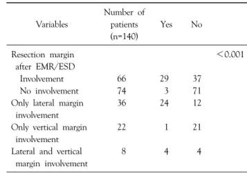
In the pathological results, 29 of the 66 patients with incomplete resection margin showed residual cancer after gastrectomy. Thirty-six patients with only lateral margin involvement showed high residual cancer compared with vertical margin. Also, patients who were diagnosed with incomplete vertical resection margin showed advanced gastric cancer in the final pathologic results. Especially, 3 of the 74 patients with clear resection margin showedTable 1. Demographics of patients who underwent surgical treat- ment after EMR/ESD
Variables
Age (years, mean±SD) 59.7±10.0
Gender (Male:Female) 91:49
EMR (number of patients) 10
ESD (number of patients) 130
Cause of surgery
Mucosal cancer (number of patients) 50 Undifferentiated-type histology (>2 cm) 20
Lateral margin involvement 30
Submucosal cancer 90
sm1 (<500μm) 26
sm2 (≥500μm) or deep margin involvement 64 Location of tumor
Upper 1/3 23
Middle 1/3 12
Lower 1/3 105
Type of operation
Distal gastrectomy 33
Total gastrectomy 14
Laparoscopic assisted distal gastrectomy 70 Laparoscopic assisted total gastrectomy 11 Totally laparoscopic distal gastrectomy 12
Table 2. Pathologic confirmation of residual cancer from surgical treatment
Variables
Number of patients (n=140)
Yes No
Resection margin <0.001
after EMR/ESD
Involvement 66 29 37
No involvement 74 3 71
Only lateral margin 36 24 12
involvement
Only vertical margin 22 1 21
involvement
Lateral and vertical 8 4 4
margin involvement
Table 3. Details of patients with residual cancer after clear EMR/ESD and advanced gastric cancer after surgery
Case Gender
/Age Location
Pathologic results after EMR/ESD Pathologic results after surgery Tumor’s
size (cm) Gross Depth (μm) Histology LVI RM Depth
(AJCC) RLNs MLNs
1 M/62 Lower 1/3 2.5 IIb 1,000 Differentiated Presence Clear Subserosa 26 0
2 M/76 Lower 1/3 1.7 IIa SM2 Differentiated Presence Clear Proper Muscle 20 0
Close to DRM
3 F/74 Lower 1/3 2.0 IIb 1,000 Differentiated Presence Clear SM3 19 0
4 M/72 Lower 1/3 4.5 IIb IIc DRM Differentiated Absent Involve Subserosa 10 1
involvement
LVI = lymphovascular invasion; RM = resection margin; RLNs = retrieved lymph nodes; MLNs = metastatic lymph nodes; DRM = deep resection margin.
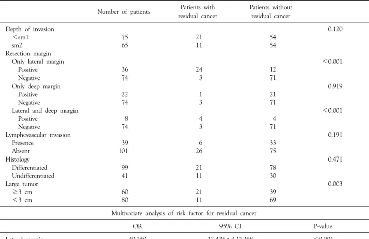
residual cancer after surgical treatment (Table 2). 3 of the 4 patients with deep submucosal invasion (Depth >1,000 μm or very close deep resection margin or deep margin involvement) developed advanced gastric cancer after surgery (Table 3). In univariate analysis for residual cancer, lateral margin, lateral and deep margin, and large tumor were significant predictive factors. Only lateral margin showed statistical significance in multivariate analysis (Table 4).
3) Incidence of lymph node metastasis
In univariate analysis, submucosal invasion with advanced depth (over sm2 or 500μm) and the presence of lymphovascular invasion had the statistical significance for metastatic lymph nodes. But in multivariate analysis, only the presence of lymphovascular invasion showed predictive factors for lymph node metastasis (Table 5).
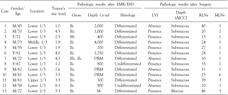
The details of patients with lymph node metastasis are described in Table 6. One patient with mucosal cancer had a metastatic lymph node. He had the longest diameter of 5.3 cm and the presence of lymphovascular invasion. Two patients with lymph node metastasis were in the sm1 group and had lymphovascular invasion.
Table 5. Univariate analysis of risk factors for metastatic lymph node
Number of patients Patients with metastatic lymph node
Patients without metastatic lymph node
Depth of invasion 0.021
<sm1 75 3 72
sm2 65 10 55
Confined to the submucosa 0.356
<sm1 25 2 23
sm2 65 10 55
Lymphovascular invasion <0.001
Presence 39 9 30
Absent 101 4 97
Histology 0.248
Differentiated 99 11 88
Undifferentiated 41 2 39
Large tumor 0.401
≥3 cm 60 7 53
<3 cm 80 6 74
Multivariate analysis of risk factor for metastatic lymph node
OR 95% CI P-value
Lymphovasular invasion 6.567 1.848∼23.335 0.004
Table 4. Univariate analysis of risk factors for residual cancer
Number of patients Patients with residual cancer
Patients without residual cancer
Depth of invasion 0.120
<sm1 75 21 54
sm2 65 11 54
Resection margin
Only lateral margin <0.001
Positive 36 24 12
Negative 74 3 71
Only deep margin 0.919
Positive 22 1 21
Negative 74 3 71
Lateral and deep margin <0.001
Positive 8 4 4
Negative 74 3 71
Lymphovascular invasion 0.191
Presence 39 6 33
Absent 101 26 75
Histology 0.471
Differentiated 99 21 78
Undifferentiated 41 11 30
Large tumor 0.003
≥3 cm 60 21 39
<3 cm 80 11 69
Multivariate analysis of risk factor for residual cancer
OR 95% CI P-value
Lateral margin 40.250 12.436∼130.268 <0.001
Table 6. Details of patients with metastatic lymph nodes
Case Gender/
Age Location Tumor’s size (cm)
Pathologic results after EMR/ESD Pathologic results after Surgery
Gross Depth (μm) Histology LVI Depth
(AJCC) RLNs MLNs
1 M/45 Lower 1/3 1.0 IIc 2,000 Differentiated Absence Submucosa 40 1
2 M/70 Lower 1/3 4.5 IIa 1,000 Differentiated Presence Submucosa 20 2
3 F/72 Lower 1/3 2.5 IIb 400 Differentiated Presence Submucosa 15 1
4 M/73 Middle 1/3 1.9 IIc 4,000 Differentiated Presence Submucosa 24 1
5 M/55 Lower 1/3 1.9 IIc 700 Differentiated Presence Submucosa 27 1
6 F/62 Lower 1/3 4.0 IIc 1,250 Differentiated Presence Submucosa 24 1
7 M/72 Lower 1/3 4.5 IIb, IIc DRM Differentiated Absence Subserosa 10 1
8 F/67 Lower 1/3 1.2 IIc 800 Undifferentiated Presence Submucosa 35 1
9 M/62 Lower 1/3 2.6 IIa DRM Differentiated Absence Submucosa 11 1
10 M/61 Lower 1/3 3.5 IIa DRM Differentiated Presence Submucosa 25 6
11 M/61 Upper 1/3 3.5 IIa 300 Differentiated Presence Submucosa 39 3
12 M/58 Lower 1/3 8.3 IIc 900 Undifferentiated Absence Submucosa 20 1
13 M/72 Lower 1/3 5.3 IIc M Differentiated Presence Mucosa 46 3
LVI = lymphovascular invasion; RLNs = retrieved lymph nodes; MLNs = metastatic lymph nodes.
DISCUSSION
EGC has high disease-specific five-year survival rate (over 95%) after surgical treatment.(16-20) EMR/ESD is the standard and preferred treatment for many patients with EGC without the risk of lymph node metastasis.(4,5) Also, ESD makes it possible to resect large lesions en bloc, permitting precise pathological results and decreasing the recurrence of cancer.(21) But after EMR/ESD, additional surgical treatment is also needed to remove residual cancer or metastatic lymph nodes in some cases. Surgical treat- ment was determined by the pathological results of specimens obtained from EMR/ESD. Resection margin involvement including lateral or deep margin was an important indication of additional surgical treatment to eliminate residual cancer.(22,23) The results of this study showed that the patients with lateral margin involvement needed additional surgical resection to remove residual cancer. But, although there was no significance statistically for deep margin involvement, additional surgical resection should be performed to eliminate the possibility of residual cancer. We speculate that the low ratio of residual cancer concerning deep margin involvement resulted from the singeing effects of the IT Knife during dissection. Also,
deep invasion of submucosal lesion (over sm2 or 500μm) also was an important indication for surgery to remove metastatic lymph nodes.(8,15,22) Some of the deep submucosal invasion with clear resection margin showed residual cancer or more advanced gastric cancer in the final pathological results. And, the presence of lymphovascular invasion was an important predictive factor for metastatic lymph nodes.
As stated above, the patients with lateral margin involvement showed high residual cancer rates. On the other hand, patients with deep margin involvement showed low residual cancer rates in the final pathologic results. But unlike lateral margin, one patient with deep resection margin involvement displayed advanced gastric cancer after gastrectomy. Besides this, 3 patients with clear resection margin showed residual cancer or more advanced gastric cancer. Especially, 2 of the 3 patients displayed advanced gastric cancer after gastrectomy. In terms of tumor invasion, it is conceivable that deep submucosal invasion (deep margin involvement or over sm2 group) had the possibility of more advanced gastric cancer compared with the tumor depth of ESD. Because the diathermic knife could provide a clear resection margin from the tumor, additional gastrectomy inevitable requires confirmation for more advanced gastric cancer in patients with deep
submucosal invasion.
In the present study, 13 of 140 patients displayed metastatic lymph nodes after gastrectomy. One patient with lymph node metastasis displayed advanced gastric cancer (depth of invasion, subserosa layer). Excepting 3 patients who were diagnosed with advanced gastric cancer after surgery, deepsubmucosal invasion (over sm2 or 500μm) and the presence of lymphovascular invasion showed significant differences for metastatic lymph nodes.
Especially in multivariate analysis, the presence of lymphovascular invasion was a predictive factor for metastatic lymph nodes. Therefore additional surgical treatment was positively necessary for patient with lymphovasular invasion or deepsubmucosal invasion.
In conclusion, because EMR/ESD of EGC is less traumatic than surgery, EMR/ESD should be considered as a first-line treatment in selected patients without risk of residual cancer or lymph node metastasis. But, additional surgical management should be done to eliminate the possibility of residual cancer or more advanced gastric cancer or metastatic lymph nodes for patients with deepsubmucosal invasion (over sm2 or 500μm) or presence of lymphovascular invasion.
REFERENCES
1) Japanese Gastric Cancer Association. Japanese Classification of Gastric Carcinoma - 2nd English Edition. Gastric Cancer 1998;
1:10-24.
2) Polkowski M, Palucki J, Wronska E, Szawlowski A, Nasierowska-Guttmejer A, Butruk E. Endosonography versus helical computed tomography for locoregional staging of gastric cancer. Endoscopy 2004;36:617-23.
3) Tsendsuren T, Jun SM, Mian XH. Usefulness of endoscopic ultrasonography in preoperative TNM staging of gastric cancer.
World J Gastroenterol 2006;12:43-7.
4) Furukawa H, Imamura H, Kodera Y. The role of surgery in the current treatment of gastric carcinoma. Gastric Cancer 2002;5 (Suppl 1):13-6.
5) Ono H. Endoscopic submucosal dissection for early gastric cancer. Chin J Dig Dis 2005;6:119-21.
6) Ono H, Hasuike N, Inui T, Takizawa K, Ikehara H, Yamaguchi Y, et al. Usefulness of a novel electrosurgical knife, the insulation-tipped diathermic knife-2, for endoscopic submu- cosal dissection of early gastric cancer. Gastric Cancer 2008;11:
47-52.
7) Ono S, Fujishiro M, Niimi K, Goto O, Kodashima S, Yama- michi N, et al. Technical feasibility of endoscopic submucosal dissection for early gastric cancer in patients taking anti- coagulants or anti-platelet agents. Dig Liver Dis 2009;41:725-8.
8) Gotoda T, Sasako M, Ono H, Katai H, Sano T, Shimoda T.
Evaluation of the necessity for gastrectomy with lymph node dissection for patients with submucosal invasive gastric cancer.
Br J Surg 2001;88:444-9.
9) Maehara Y, Orita H, Okuyama T, Moriguchi S, Tsujitani S, Korenaga D, et al. Predictors of lymph node metastasis in early gastric cancer. Br J Surg 1992;79:245-7.
10) Sano T, Kobori O, Muto T. Lymph node metastasis from early gastric cancer: endoscopic resection of tumour. Br J Surg 1992;79:241-4.
11) Ichikura T, Uefuji K, Tomimatsu S, Okusa Y, Yahara T, Tamakuma S. Surgical strategy for patients with gastric carcinoma with submucosal invasion. A multivariate analysis.
Cancer 1995;76:935-40.
12) Gotoda T, Yanagisawa A, Sasako M, Ono H, Nakanishi Y, Shimoda T, et al. Incidence of lymph node metastasis from early gastric cancer: estimation with a large number of cases at two large centers. Gastric Cancer 2000;3:219-25.
13) Abe N, Watanabe T, Suzuki K, Machida H, Toda H, Nakaya Y, et al. Risk factors predictive of lymph node metastasis in depressed early gastric cancer. Am J Surg 2002;183:168-72.
14) Yamao T, Shirao K, Ono H, Kondo H, Saito D, Yamaguchi H, et al. Risk factors for lymph node metastasis from intramucosal gastric carcinoma. Cancer 1996;77:602-6.
15) Abe N, Sugiyama M, Masaki T, Ueki H, Yanagida O, Mori T, et al. Predictive factors for lymph node metastasis of differentiated submucosally invasive gastric cancer. Gastrointest Endosc 2004;60:242-5.
16) Okamura T, Tsujitani S, Korenaga D, Haraguchi M, Baba H, Hiramoto Y, et al. Lymphadenectomy for cure in patients with early gastric cancer and lymph node metastasis. Am J Surg 1988;155:476-80.
17) Noguchi Y, Imada T, Matsumoto A, Coit DG, Brennan MF.
Radical surgery for gastric cancer. A review of the Japanese experience. Cancer 1989;64:2053-62.
18) Nakamura K, Ueyama T, Yao T, Xuan ZX, Ambe K, Adachi Y, et al. Pathology and prognosis of gastric carcinoma. Findings in 10,000 patients who underwent primary gastrectomy. Cancer 1992;70:1030-7.
19) Shimizu S, Tada M, Kawai K. Early gastric cancer: its surveillance and natural course. Endoscopy 1995;27:27-31.
20) Maruyama K, Kaminishi M, Hayashi K, Isobe Y, Honda I, Katai H, et al. Gastric cancer treated in 1991 in Japan: data analysis of nationwide registry. Gastric Cancer 2006;9:51-66.
21) Yokoi C, Gotoda T, Hamanaka H, Oda I. Endoscopic submucosal dissection allows curative resection of locally recurrent early gastric cancer after prior endoscopic mucosal
resection. Gastrointest Endosc 2006;64:212-8.
22) Ryu KW, Choi IJ, Doh YW, Kook MC, Kim CG, Park HJ, et al. Surgical indication for non-curative endoscopic resection in early gastric cancer. Ann Surg Oncol 2007;14:3428-34.
23) Chung YS, Park DJ, Lee HJ, Kim SG, Jung HC, Song IS, et al. The Role of Surgery After Incomplete Endoscopic Mucosal Resection for Early Gastric Cancer. Surg Today 2007;37:114-7.